[1] YOUNG WF, BROWN D, KENDLER A, et al. Delayed post-traumatic osteonecrosis of a vertebral body (Kummell’s disease). Acta Orthop Belg. 2002;68(1):13-19.
[2] NICKELL LT, SCHUCANY WG, OPATOWSKY MJ. Kummell disease. Proc (Bayl Univ Med Cent). 2013;26(3):300-301.
[3] LEE SH, KIM ES, EOH W. Cement augmented anterior reconstruction with short posterior instrumentation: a less invasive surgical option for Kummell disease with cord compression. J Clin Neurosci. 2011;18(4):509-514.
[4] 杨惠林,王根林,牛国旗,等. 骨质疏松性胸腰椎骨折不愈合的诊断与治疗[J]. 中华骨科杂志,2007,27(9):682-686.
[5] WANG HS, KIM HS, JU CI, et al. Delayed bone cement displacement following balloon kyphoplasty. Korean Neurosurg Soc. 2008;43(4):212-214.
[6] VAN DER SCHAAF I, FRANSEN H. Percutaneous vertebroplasty as treatment for Kummell’s disease. JBR-BTR. 2009;92(2):83-85.
[7] LI KC, WONG TU, KUNG FC, et al. Staging of Kümmell’s disease. J Musculoskelet Res. 2004;8(1):43-55.
[8] YEOM JS, KIM WJ, CHOY WS, et al. Leakage of cement in percutaneous transpedicular vertebroplasty for painful osteoporotic compression fractures. J Bone Joint Surg Br. 2003;85(1):83-89.
[9] YANG H, LIU H, WANG S, et al. Review of Percutaneous Kyphoplasty in China. Spine (Phila Pa 1976). 2016; 41 Suppl 19:B52-B58.
[10] PARK JW, PARK JH, JEON HJ, et al. Kummell’s Disease Treated with Percutaneous Vertebroplasty: Minimum 1 Year Follow-Up. Korean J Neurotrauma. 2017;13(2): 119-123.
[11] ZHANG J, FAN Y, HE X, et al. Is percutaneous kyphoplasty the better choice for minimally invasive treatment of neurologically intact osteoporotic Kummell’s disease? A comparison of two minimally invasive procedures. Int Orthop. 2018; 42(6):1321-1326.
[12] KONG LD, WANG P, WANG LF, et al. Comparison of vertebroplasty and kyphoplasty in the treatment of osteoporotic vertebral compression fractures with intravertebral clefts. Eur J Orthop Surg Traumatol. 2014;24 Suppl 1:S201-208.
[13] KRAUSS M, HIRSCHFELDER H, TOMANDL B, et al. Kyphosis reduction and the rate of cement leaks after vertebroplasty of intravertebral clefts. Eur Radiol. 2006;16(5):1015-1021.
[14] OH JS, DOH JW, SHIM JJ, et al. The Effectiveness of Gelfoam Technique before Percutaneous Vertebroplasy: Is It Helpful for Prevention of Cement Leakage? A Prospective Randomized Control Study. Korean J Spine. 2016;13(2):63-66.
[15] SUN HB, JING XS, LIU YZ, et al. The Optimal Volume Fraction in Percutaneous Vertebroplasty Evaluated by Pain Relief, Cement Dispersion, and Cement Leakage: A Prospective Cohort Study of 130 Patients with Painful Osteoporotic Vertebral Compression Fracture in the Thoracolumbar Vertebra. World Neurosurg. 2018; 114:e677-e688.
[16] HOPPE S, ELFIKY T, KEEL MJ, et al. Lavage prior to vertebral augmentation reduces the risk for cement leakage. Eur Spine J. 2016;25(11):3463-3469.
[17] HONG SJ, LEE S, YOON JS, et al. Analysis of intradiscal cement leakage during percutaneous vertebroplasty: multivariate study of risk factors emphasizing preoperative MR findings. J Neuroradiol. 2014;41(3):195-201.
[18] 唐永超,莫国业,张顺聪,等. 经皮椎体强化术治疗无神经症状Kümmell病的中长期疗效[J]. 中国脊柱脊髓杂志,2018,28(1):38-43.
[19] OHBA T, EBATA S, CLINTON D, et al. Instability of treated vertebrae after balloon kyphoplasty causing paraparesis in osteoporotic vertebral compression fracture: a report of two cases. Eur Spine J. 2013;22 Suppl 3:S341-345.
[20] HUANG KY, YAN JJ, LIN RM. Histopathologic Findings of Retrieved Specimens of Vertebroplasty with Polymethylmethacrylate Cement. Spine. 2005;30: E585-588.
[21] TSAI TT, CHEN WJ, LAI PL, et al. Polymethylmethacrylate cement dislodgment following percutaneous vertebroplasty: a case report. Spine. 2003;28(22):E457-460.
[22] 李鹏,高利峰,胡军华,等. 三柱强化经皮椎体成形术治疗Ⅰ、Ⅱ期Kummell病的疗效[J]. 临床骨科杂志,2020,23(3):322-324.
[23] 邓立明,黄凯,史建刚,等. 双侧穿刺骨水泥锚定椎体成形术治疗Kummell病临床观察[J]. 山东医药,2018,58(44):64-66.
[24] 张在青,陈波. 单侧穿刺骨水泥锚定椎体后凸成形术治疗Kummell病的疗效探讨[J]. 中国临床新医学,2020,13(5):512-515.
[25] 魏桂财,郑忠,李超雄,等. 骨水泥椎弓根附近锚定技术在Kummell病椎体成形术治疗的应用体会. 2019楚天骨科高峰论坛暨第二十六届中国中西医结合骨伤科学术年会论文集,2019:434-435.
[26] 杜心如,赵玲秀,张一模,等. 胸腰椎椎弓根内径的测量及其临床意义[J]. 中国脊柱脊髓杂志,2001,11(3):162-164.
|


 相反的是一项前瞻性的随机对照研究,发现在使用明胶海绵组有11处渗漏(22%),对照组有12处渗漏(26%);明胶海绵组椎管渗漏6例,椎旁静脉渗漏4例,软组织渗漏1例;对照组椎管渗漏4例,椎旁静脉渗漏8例,椎间盘间隙渗漏1例;提示明胶海绵技术与降低渗漏发生率无关(P=0.64)[14]。这可能因为该研究中选取的病例数少,手术方法为经皮椎体成形术,而且手术操作者是一位具有500例以上经皮椎体成形手术经验的医生,以上可能是导致与此次研究结果不一致的原因。
另外骨水泥量以及骨水泥椎体占比也是骨水泥渗漏的因素。SUN等[15]发现在单胸腰椎骨质疏松性椎体压缩性骨折中,轻、中度骨折时,椎体内骨水泥体积4-6 mL可迅速缓解疼痛,最佳的骨水泥椎体占比为19.78%,可以获得满意的骨水泥分布,随着骨水泥椎体占比的增加,骨水泥渗漏的发生率也随之增加。此次研究中骨水泥注入量为2-9 mL,平均为(4.32±1.72) mL,由于Kummell病中存在椎体内真空裂隙征,其骨水泥量有较大的波动性,故骨水泥注入量应个体化才能减少骨水泥的渗漏发生,切忌一概而论。
相反的是一项前瞻性的随机对照研究,发现在使用明胶海绵组有11处渗漏(22%),对照组有12处渗漏(26%);明胶海绵组椎管渗漏6例,椎旁静脉渗漏4例,软组织渗漏1例;对照组椎管渗漏4例,椎旁静脉渗漏8例,椎间盘间隙渗漏1例;提示明胶海绵技术与降低渗漏发生率无关(P=0.64)[14]。这可能因为该研究中选取的病例数少,手术方法为经皮椎体成形术,而且手术操作者是一位具有500例以上经皮椎体成形手术经验的医生,以上可能是导致与此次研究结果不一致的原因。
另外骨水泥量以及骨水泥椎体占比也是骨水泥渗漏的因素。SUN等[15]发现在单胸腰椎骨质疏松性椎体压缩性骨折中,轻、中度骨折时,椎体内骨水泥体积4-6 mL可迅速缓解疼痛,最佳的骨水泥椎体占比为19.78%,可以获得满意的骨水泥分布,随着骨水泥椎体占比的增加,骨水泥渗漏的发生率也随之增加。此次研究中骨水泥注入量为2-9 mL,平均为(4.32±1.72) mL,由于Kummell病中存在椎体内真空裂隙征,其骨水泥量有较大的波动性,故骨水泥注入量应个体化才能减少骨水泥的渗漏发生,切忌一概而论。 由此可见,虽然骨水泥团块移位的发生率并无骨水泥渗漏高,但一旦发生往往需二次手术,给医患双方都打来巨大压力及风险。结合既往文献,总结造成Kummell病术后骨水泥移位可能的原因有:①骨水泥注入椎体内后,在骨水泥周围可形成明显的骨坏死,骨水泥界面会形成纤维组织膜,而无新生血管形成[20],这破坏了骨水泥与周围组织的嵌合,从而导致机械性嵌合[21];②椎体内真空裂隙征的形成导致将骨水泥注射到空腔中与周围骨的嵌合要比注射到部分完整的骨小梁中少得多[21];③病椎前皮质的缺损增加骨水泥团块在负重条件下出现向前移位[21];④球囊扩张和骨水泥填充通常位于椎体的中前部,而椎体中后部往往缺少必要的骨水泥填充及加固[19]。
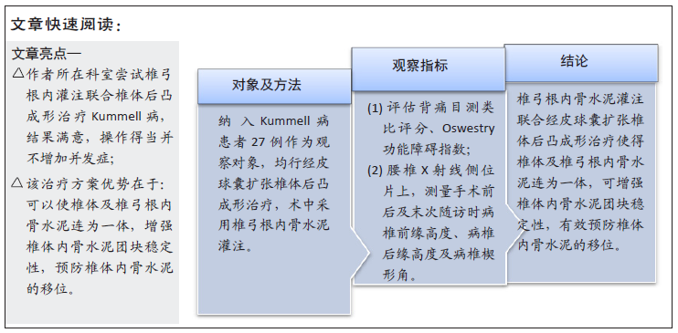
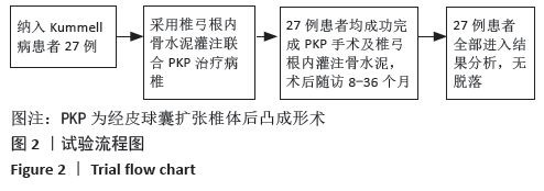
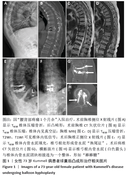
3.4 PKP术中联合椎弓根内骨水泥灌注预防术后骨水泥移位由于Kummell病的发生机制是椎体缺血性骨坏死,在椎体内形成裂隙,其周围形成硬化骨带,传统的PKP手术注入的骨水泥难以有效嵌入骨内而形成“鹅卵石”效应,当病椎负重后,骨水泥团块极有可能在椎体内自由滑动,导致患者再次出现疼痛,甚至二次手术。因此,若能将骨水泥团块稳定于椎体内,将有效降低其移位概率,提高手术长期的疗效。唐永超等[18]建议行椎体强化注入骨水泥结束退出穿刺针时,可边退边注入少量的骨水泥,使骨水泥沿工作通道弥散至椎弓根形成“拖尾征”,达到对前方裂隙内骨水泥团块锚定的作用,进而降低骨水泥团块松动的发生率。李鹏等[22]研究认为,对Ⅰ、Ⅱ期 Kummell 病可采用三柱强化经皮椎体成形治疗(即椎体前柱、中柱及椎弓根内骨水泥灌注),术后患者恢复快,疗效满意。邓立明等[23]也认为通过双侧穿刺骨水泥锚定椎体成形术治疗Kummell病患者,临床结果满意,能明显降低骨水泥松动或移位风险。另外,张在青等[24]采用单侧穿刺骨水泥锚定椎体后凸成形术治疗 Kummell 病也取得了满意疗效,并且并发症少,安全性高。这正是因为椎弓根相对于椎体是完整的,其内存在一定量的松质骨,有利于骨水泥更好地嵌合在椎弓根内,达到稳定椎体内骨水泥团块的作用。
由此可见,虽然骨水泥团块移位的发生率并无骨水泥渗漏高,但一旦发生往往需二次手术,给医患双方都打来巨大压力及风险。结合既往文献,总结造成Kummell病术后骨水泥移位可能的原因有:①骨水泥注入椎体内后,在骨水泥周围可形成明显的骨坏死,骨水泥界面会形成纤维组织膜,而无新生血管形成[20],这破坏了骨水泥与周围组织的嵌合,从而导致机械性嵌合[21];②椎体内真空裂隙征的形成导致将骨水泥注射到空腔中与周围骨的嵌合要比注射到部分完整的骨小梁中少得多[21];③病椎前皮质的缺损增加骨水泥团块在负重条件下出现向前移位[21];④球囊扩张和骨水泥填充通常位于椎体的中前部,而椎体中后部往往缺少必要的骨水泥填充及加固[19]。
3.4 PKP术中联合椎弓根内骨水泥灌注预防术后骨水泥移位由于Kummell病的发生机制是椎体缺血性骨坏死,在椎体内形成裂隙,其周围形成硬化骨带,传统的PKP手术注入的骨水泥难以有效嵌入骨内而形成“鹅卵石”效应,当病椎负重后,骨水泥团块极有可能在椎体内自由滑动,导致患者再次出现疼痛,甚至二次手术。因此,若能将骨水泥团块稳定于椎体内,将有效降低其移位概率,提高手术长期的疗效。唐永超等[18]建议行椎体强化注入骨水泥结束退出穿刺针时,可边退边注入少量的骨水泥,使骨水泥沿工作通道弥散至椎弓根形成“拖尾征”,达到对前方裂隙内骨水泥团块锚定的作用,进而降低骨水泥团块松动的发生率。李鹏等[22]研究认为,对Ⅰ、Ⅱ期 Kummell 病可采用三柱强化经皮椎体成形治疗(即椎体前柱、中柱及椎弓根内骨水泥灌注),术后患者恢复快,疗效满意。邓立明等[23]也认为通过双侧穿刺骨水泥锚定椎体成形术治疗Kummell病患者,临床结果满意,能明显降低骨水泥松动或移位风险。另外,张在青等[24]采用单侧穿刺骨水泥锚定椎体后凸成形术治疗 Kummell 病也取得了满意疗效,并且并发症少,安全性高。这正是因为椎弓根相对于椎体是完整的,其内存在一定量的松质骨,有利于骨水泥更好地嵌合在椎弓根内,达到稳定椎体内骨水泥团块的作用。