[1] AGARWAL R, GARCIA AJ. Biomaterial strategies for engineering implants for enhanced osseointegration and bone repair.Adv Drug Deliver Rev.2015; 94:53-62.
[2] WOODARD LN, KMETZ KT, ROTH AA, et al. Porous poly (ε-caprolactone)–poly (L-lactic acid) semi-interpenetrating networks as superior, defect-specific scaffolds with potential for cranial bone defect repair.Biomacromolecules. 2017;18(12): 4075-4083.
[3] CUNNIFFE GM, DÍAZ-PAYNO PJ, RAMEY JS, et al. Growth plate extracellular matrix-derived scaffolds for large bone defect healing. Eur Cell Mater.2017;33:130-142.
[4] EL-RASHIDY AA, ROETHER JA, HARHAUS L, et al. Regenerating bone with bioactive glass scaffolds: A review of in vivo studies in bone defect models.Acta Biomater. 2017;62:1-28.
[5] SOUNDARYA SP, SANJAY V, MENON AH, et al. Effects of flavonoids incorporated biological macromolecules based scaffolds in bone tissue engineering.Int J Biol Macromol 2018; 110:74-87.
[6] LI JJ, EBIED M, XU J, et al.Current approaches to bone tissue engineering: the interface between biology and engineering. Adv Healthc Mater.2018;7(6):e1701061.
[7] HENDRIKSON W, VAN BLITTERSWIJK C, ROUWKEMA J, et al. The use of finite element analyses to design and fabricate three-dimensional scaffolds for skeletal tissue engineering.Front Bioeng Biotech.2017;5:30.
[8] JIN L, FENG ZQ, WANG T, et al. A novel fluffy hydroxylapatite fiber scaffold with deep interconnected pores designed for three-dimensional cell culture.J Mater Chem B.2014;2(1): 129-136.
[9] 唐丽宇,龚飞飞,庄劭玉,等.珊瑚羟基磷灰石用于上颌前牙区位点保存的锥体束 CT 研究[J].医学研究生学报, 2018,31(6):641-643.
[10] 刘冬,秦虎,汪永新,等.3D打印羟基磷灰石/聚乳酸网状复合物修复颅骨缺损[J].中国组织工程研究,2019,23(6):833-837.
[11] SHORT AR, KORALLA D, DESHMUKH A, et al. Hydrogels that allow and facilitate bone repair, remodeling, and regeneration. J Mater Chem B.2015;3(40):7818-7830.
[12] RYAN AJ, GLEESON JP, MATSIKO A, et al. Effect of different hydroxyapatite incorporation methods on the structural and biological properties of porous collagen scaffolds for bone repair.J Anat.2015;227(6):732-745.
[13] ZHANG H, MAO X, DU Z, et al. Three dimensional printed macroporous polylactic acid/hydroxyapatite composite scaffolds for promoting bone formation in a critical-size rat calvarial defect model. Sci Technol Adv Mat.2016;17(1):136-148.
[14] WAN T, STYLIOS GK, GIANNOUDI M, et al. Investigating a new drug delivery nano composite membrane system based on PVA/PCL and PVA/HA (PEG) for the controlled release of biopharmaceuticals for bone infections.Injury.2015;46:S39-S43.
[15] ISIKLI C, HASIRCI V, HASIRCI N. Development of porous chitosan-gelatin/hydroxyapatite composite scaffolds for hard tissue-engineering applications.J Tissue Eng Regen M.2012; 6(2):135-143.
[16] TORSTRICK FB, LIN ASP, POTTER D, et al. Porous PEEK improves the bone-implant interface compared to plasma-sprayed titanium coating on PEEK.Biomaterials. 2018;185:106-116.
[17] GEORGOPOULOU A, KALIVA M, VAMVAKAKI M, et al. Osteogenic potential of pre-osteoblastic cells on a chitosan-graft- polycaprolactone copolymer.Mater. 2018;11(4):490.
[18] CUERVO-LOZANO CE, SOTO-DOMÍNGUEZ A, SAUCEDO- CÁRDENAS O, et al. Osteogenesis induced by a three-dimensional bioimplant composed of demineralised bone matrix, collagen, hydroxyapatite, and bone marrow-derived cells in massive bone defects: An experimental study.Tissue Cell.2018; 50:69-78.
[19] LI Z, CHU D, GAO Y, et al. Biomimicry, biomineralization, and bioregeneration of bone using advanced three-dimensional fibrous hydroxyapatite scaffold.Mater Today Adv.2019;3:100014.
[20] HEIMANN RB. Plasma-sprayed hydroxylapatite-based coatings: chemical, mechanical, microstructural, and biomedical properties. J Therm Spray Techn. 2016;25(5):827-850.
[21] ISLAMI M,MORTAZAVI Y,SOLEIMANI M,et al.In vitro expansion of CD 133+ cells derived from umbilical cord blood in poly-L-lactic acid (PLLA) scaffold coated with fibronectin and collagen.Artif Cell Blood Sub.2018;46(5):1025-1033.
[22] HANAS T, KUMAR TSS, PERUMAL G, et al. Electrospun PCL/HA coated friction stir processed AZ31/HA composites for degradable implant applications.J Mater Process Tech. 2018;252:398-406.
[23] GHASSEMI T, SHAHROODI A, EBRAHIMZADEH MH, et al. Current concepts in scaffolding for bone tissue engineering. Arch Bone Joint Surg.2018;6(2):90.
[24] JI K, WANG Y, WEI Q, et al. Application of 3D printing technology in bone tissue engineering. Bio-Design Manuf. 2018;1(3):203-210.
[25] SHADJOU N, HASANZADEH M, KHALILZADEH B. Graphene based scaffolds on bone tissue engineering. Bioengineered. 2018;9(1):38-47.
[26] LEE H, YANG GH, KIM M, et al. Fabrication of micro/nanoporous collagen/dECM/silk-fibroin biocomposite scaffolds using a low temperature 3D printing process for bone tissue regeneration.Mat Sci Eng C.2018;84:140-147.
[27] SUBUKI I, ADNAN N, SHARUDIN RW. Biodegradable scaffold of natural polymer and hydroxyapatite for bone tissue engineering: A short review.AIP Conf Proc. 2018;2031(1):020019.
[28] SIDDIQUI N, ASAWA S, BIRRU B, et al.PCL-based composite scaffold matrices for tissue engineering applications.Mol Biotechnol. 2018;60(7):506-532.
[29] ZHAO W, LI J, JIN K, et al.F abrication of functional PLGA-based electrospun scaffolds and their applications in biomedical engineering.Mat Sci Eng C.2016;59: 1181-1194.
[30] TOOSI S, NADERI-MESHKIN H, KALALINIA F, et al. PGA-incorporated collagen: Toward a biodegradable composite scaffold for bone-tissue engineering.J Biomed Mater Res A. 2016;104(8): 2020-2028.
[31] SCAFFARO R, LOPRESTI F, BOTTA L, et al. Preparation of three-layered porous PLA/PEG scaffold: relationship between morphology, mechanical behavior and cell permeability.J Mech Behav Biomed.2016;54:8-20.
[32] LUO X, KULIG KM, FINKELSTEIN EB, et al. In vitro evaluation of decellularized ECM-derived surgical scaffold biomaterials. J Biomed Mater Res B.2017;105(3):585-593.
[33] DEARTH CL, SLIVKA PF, STEWART SA, et al. Inhibition of COX1/2 alters the host response and reduces ECM scaffold mediated constructive tissue remodeling in a rodent model of skeletal muscle injury.Acta Biomater.2016;31:50-60.
[34] LIN S, CUI L, CHEN G, et al. PLGA/β-TCP composite scaffold incorporating salvianolic acid B promotes bone fusion by angiogenesis and osteogenesis in a rat spinal fusion model. Biomaterials.2019;196:109-121.
[35] KHOJASTEH A, FAHIMIPOUR F, ESLAMINEJAD MB, et al. Development of PLGA-coated β-TCP scaffolds containing VEGF for bone tissue engineering.Mat Sci Eng.2016;69: 780-788.
[36] WU W, FANG J, LIU W, et al. Preparation and properties of BMPLGA/NBAG-β-TCP composite scaffold materials.Int J Polym Anal Ch.2018;23(8):710-720.
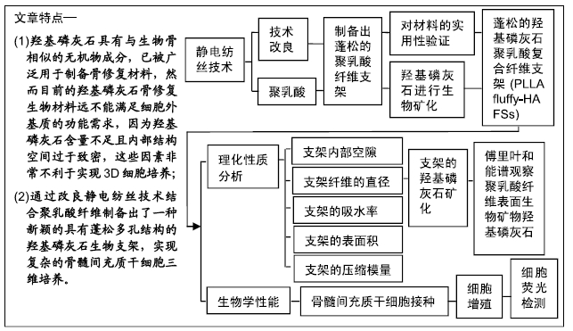
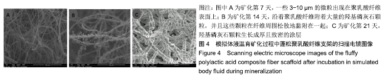
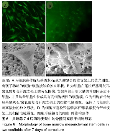
|