中国组织工程研究 ›› 2017, Vol. 21 ›› Issue (7): 1086-1091.doi: 10.3969/j.issn.2095-4344.2017.07.019

• 骨与关节图像与影像 bone and joint imaging • 上一篇    下一篇

基于影像学技术股骨头坏死病灶范围、血液供应与病理变化关系:诊断性动物实验方案

傅维民,王本杰   

  1. 大连大学附属中山医院,辽宁省大连市 116001
  • 修回日期:2017-01-16 出版日期:2017-03-08 发布日期:2017-04-11
  • 通讯作者: 王本杰,博士,副主任医师,大连大学附属中山医院,辽宁省大连市 116001
  • 作者简介:傅维民,男,1982年生,汉族,2014年南方医科大学毕业,博士,主治医师,主要从事骨与关节创伤方面的研究。
  • 基金资助:

    2015年辽宁省科学事业公益研究基金【辽财教便字2015(13)号】


Assessing the degree of necrotic femoral head, and association of blood supply with pathlogical changes: study protocol for a diagnostic animal trial  

Fu Wei-min, Wang Ben-jie   

  1. Affiliated Zhongshan Hospital of Dalian University, Dalian 116001, Liaoning Province, China
  • Revised:2017-01-16 Online:2017-03-08 Published:2017-04-11
  • Contact: Wang Ben-jie, M.D., Associate chief physician, Affiliated Zhongshan Hospital of Dalian University, Dalian 116001, Liaoning Province, China
  • About author:Fu Wei-min, M.D., Attending physician, Affiliated Zhongshan Hospital of Dalian University, Dalian 116001, Liaoning Province, China
  • Supported by:

    the Science Public Welfare Foundation of Liaoning Province in 2015, No. 2015(13)

摘要:

文章快速阅读:


文题释义:
激素性股骨头坏死动物模型的建立:采用香港中文大学Qin等介绍的的方法,静脉注射一次低剂量的脂多糖(10 μg/kg),之后以24 h为间隔给予肌肉注射3次高剂量的甲基强的松龙(20 mg/kg),最后一次给药后6周能成功建立股骨头坏死的动物模型。该模型的特点是造模成功率高,模型死亡率低,已经被证实有较好的安全性和有效性。
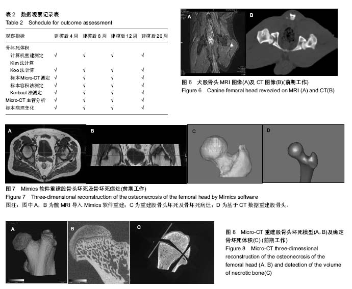
股骨头标本的Micro-CT检测:可以使用微型CT对股骨头样本进行数据采集与三维重建,以获得矢状面、冠状面和横断面的组织结构,并可对股骨头坏死区进行构建,确定骨坏死体积。Micro-CT检测结合显微对比剂灌注能够评价股骨头内血供情况,明确股骨头内主干血管的分布、走行及供血范围。
 
摘要
背景:目前评价股骨头坏死病灶体积大小的影像学方法有基于X射线的Kerboul角、坏死指数,基于CT、MRI图像三维重建计算股骨头坏死体积等,但各方法的准确度报道不一,尚无一种被广泛接受;同时,并没有研究明确揭示骨坏死病变范围、程度与股骨头血供损害的直接联系。
目的:评估各种常用影像技术在不同时期骨坏死病灶评价中的准确性,尝试确立一种更加简便易用的骨坏死病灶临床计算方法。
方法:实验为诊断性动物体内实验,在中国大连,大连大学附属中山医院完成。制备激素性股骨头坏死犬模型,通过MRI、CT检查,结合Micro-CT离体标本检测对坏死股骨头病变进行精确评估,结合血管灌注造影及病理检测评价骨坏死发生范围、骨破坏程度与血供变化的关联;同时将结果与临床常用仅基于影像学的骨坏死分期分级标准相比较,以期验证此精确测量方法与传统方法的一致性。实验过程遵循了国际兽医学编辑协会《关于动物伦理与福利的作者指南共识》和本地及国家法规,并遵守《动物实验体内实验研究报告规范指南》(ARRIVE指南)。
结果与结论:课题拟建立针对骨坏死不同分期采用不同影像学技术测定以获得准确结果的新理念,将使不同技术能力的医疗机构能够应用获得的影像资料给出相对更准确的骨坏死评估结果,使骨坏死病变分期评价更准确,进而患者有望得到针对评估结果的标准化治疗方案。
 
中国组织工程研究杂志出版内容重点:人工关节;骨植入物;脊柱骨折;内固定;数字化骨科;组织工程
ORCID: 0000-0003-1716-0409(王本杰)

关键词: 骨科植入物, 人工关节, 股骨头坏死, X射线片, MRI成像, CT

Abstract:

BACKGROUND: The imaging methods of evaluating the volume of necrotic femoral head are based on Kerboul angle and necrosis index revealed on X-ray, as well as CT and MRI three-dimensional reconstruction, but the accuracy of each method is different, and none is widely accepted. Additionally, the association of the volum and degree of necrotic femoral head with the status of blood supply to the femoral head remain unclear.

OBJECTIVE: To evaluate the accuracy of each imaing method for assessing different stages of femoral head necrosis, and to confirm a convenient method used for measuring the volum of necrotic femoral head.
METHODS: This diagnostic in vivo animal trial was finished in the Affiliated Zhongshan Hospital of Dalian University, Liaoning Province, China. The canine models of steroid-induced necrosis of femoral head were prepared, and received CT and MRI examinations, followed by three-dimensional reconstruction combined with Micro-CT to make accurate diagnosis; the degree of necrotic femoral head and its association with the status of blood supply were evaluated through angiography and histopathological examination. All above outcome measures were compared with the Association Research Circulation Osseous to testify the consistency of this precise method with traditional methods. The experiment follows the national guidelines for the Care and Use of Laboratory Animals, and “Consensus author guidelines on animal ethics and welfare” by the international Association for Veterinary Editors (IAVE). The article was prepared in accordance with the “Animal Research: Reporting of In Vivo Experiments Guidelines” (ARRIVE Guidelines).
RESULTS AND CONCLUSION: The aim of the study is to obtain a accurate measurement method by detecting different stages of osteonecrosis using different imaging techniques, which enables surgeons make exact diagnosis and clinical stage of osteonecrosis, thus desiging standardized treatment program.

中国组织工程研究杂志出版内容重点:人工关节;骨植入物;脊柱骨折;内固定;数字化骨科;组织工程

Key words: Femur Head Necrosis, Radiography, Tomography, X-ray Computed, Magnetic Resonance Imaging, Tissue Engineering

中图分类号: