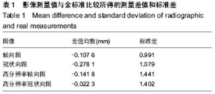
| [1] Mozzo P, Procacci C, Tacconi A, et al.A new volumetric CT machine for dental imaging based on the cone-beam technique: preliminary results.Eur Radiol. 1998;8(9):1558-1564.
[2] Sukovic P.Cone beam computed tomography in craniofacial imaging.Orthod Craniofac Res. 2003;6 Suppl 1:31-36.
[3] 马绪臣.口腔颌面锥形束CT的临床应用[M].北京:人民卫生出版社,2011:110-111.
[4] Brown AA, Scarfe WC, Scheetz JP,et al.Linear accuracy of cone beam CT derived 3D images.Angle Orthod. 2009;79(1):150-157.
[5] El-Beialy AR, Fayed MS, El-Bialy AM, et al. Accuracy and reliability of cone-beam computed tomography measurements: Influence of head orientation.Am J Orthod Dentofacial Orthop. 2011;140(2):157-165.
[6] Loubele M, Van Assche N, Carpentier K,et al.Comparative localized linear accuracy of small-field cone-beam CT and multislice CT for alveolar bone measurements.Oral Surg Oral Med Oral Pathol Oral Radiol Endod. 2008;105(4):512-518.
[7] van Daatselaar AN, Dunn SM, Spoelder HJ,et al. Feasibility of local CT of dental tissues.Dentomaxillofac Radiol. 2003;32(3): 173-180.
[8] Araki K, Maki K, Seki K,et al.Characteristics of a newly developed dentomaxillofacial X-ray cone beam CT scanner (CB MercuRay): system configuration and physical properties.Dentomaxillofac Radiol. 2004;33(1):51-59.
[9] Fatemitabar SA, Nikgoo A.Multichannel computed tomography versus cone-beam computed tomography: linear accuracy of in vitro measurements of the maxilla for implant placement.Int J Oral Maxillofac Implants. 2010 ;25(3):499- 505.
[10] Tsiklakis K, Donta C, Gavala S,et al.Dose reduction in maxillofacial imaging using low dose Cone Beam CT.Eur J Radiol. 2005;56(3):413-417.
[11] Lascala CA, Panella J, Marques MM.Analysis of the accuracy of linear measurements obtained by cone beam computed tomography (CBCT-NewTom).Dentomaxillofac Radiol. 2004 ; 33(5):291-294.
[12] Stratemann SA, Huang JC, Maki K,et al.Comparison of cone beam computed tomography imaging with physical measures.Dentomaxillofac Radiol. 2008;37(2):80-93.
[13] van Vlijmen OJ, Maal TJ, Bergé SJ, et al.A comparison between two-dimensional and three-dimensional cephalometry on frontal radiographs and on cone beam computed tomography scans of human skulls.Eur J Oral Sci. 2009;117(3):300-305.
[14] Draenert FG, Coppenrath E, Herzog P, et al. Beam hardening artifacts occur in dental implant scans with the NesTom cone beam CT but not with the dental 4-row multidetector CT. Dentomaxillofac Radiol.2007;36:198-203.
[15] Jacobs R, Lambrichts I, Liang X,et al.Neurovascularization of the anterior jaw bones revisited using high-resolution magnetic resonance imaging.Oral Surg Oral Med Oral Pathol Oral Radiol Endod. 2007;103(5):683-693.
[16] Van Assche N, van Steenberghe D, Guerrero ME,et al. Accuracy of implant placement based on pre-surgical planning of three-dimensional cone-beam images: a pilot study.J Clin Periodontol. 2007;34(9):816-821.
[17] Hassan B, vd Stelt PF, Sanderink G. Accuracy of three-dimensional measurements of mandibular anatomy in cone beam computed tomography images. Oral Surg Oral Med Oral Pathol Oral Radiol Endod.2007;103:534-542.
[18] Alsaadi G, Quirynen M, Komárek A,et al.Impact of local and systemic factors on the incidence of oral implant failures, up to abutment connection.J Clin Periodontol. 2007;34(7):610- 617.
[19] Roberts JA, Drage NA, Davies J,et al.Effective dose from cone beam CT examinations in dentistry.Br J Radiol. 2009; 82(973):35-40.
[20] Silva MA, Wolf U, Heinicke F,et al.Cone-beam computed tomography for routine orthodontic treatment planning: a radiation dose evaluation.Am J Orthod Dentofacial Orthop. 2008;133(5):640.e1-5.
[21] Ludlow JB, Davies-Ludlow LE, Brooks SL, et al. Dosimetry of 3 CBCT devices for oral and maxillofacial radiology: CB Mercuray, NewTom 3G and i-CAT. Dento maxilla facial radiology. 2006;35(4):219-226.
[22] Pinsky HM, Dyda S, Pinsky RW,et al. Accuracy of three-dimensional measurements using cone-beam CT.Dentomaxillofac Radiol. 2006;35(6):410-416.
[23] Dalessandri D, Bracco P, Paganelli C,et al.Ex vivo measurement reliability using two different cbct scanners for orthodontic purposes.Int J Med Robot. 2012;8(2):230-242.
[24] De Vos W, Casselman J, Swennen GR. Cone-beam computerized tomography(CBCT) imaging of the oral and maxillofacial region: A systematic review of the literature. Int J Oral Maxillofac Surg.2009;38:609-625. |