Chinese Journal of Tissue Engineering Research ›› 2018, Vol. 22 ›› Issue (2): 204-209.doi: 10.3969/j.issn.2095-4344.0007
Previous Articles Next Articles
Platelet-rich fibrin combined with tooth ash promotes bone repair
- 1Mudanjiang Medical University, Mudanjiang 157011, Heilongjiang Province, China; 2Department of Stomatology, Hongqi Hospital of Mudanjiang, Mudanjiang 157011, Heilongjiang Province, China
-
Received:2017-08-29Online:2018-01-18Published:2018-01-18 -
Contact:Luan Hai-rong, Associate professor, Mudanjiang Medical University, Mudanjiang 157011, Heilongjiang Province, China -
About author:Wang De-li, Master, Associate professor, Mudanjiang Medical University, Mudanjiang 157011, Heilongjiang Province, China -
Supported by:the Scientific and Technological Plan Project of Mudanjiang City, No. Z2016s0077; the Scientific Plan of Mudanjiang City, No. G2007d2368; the Fundamental Rearch Foundation for Heilongjiang Provincial Universities in 2017
CLC Number:
Cite this article
Wang De-li, Xu Wen-xiu, Lin Na, Wang Hai-yan, Shi Yi, Yu Xue-gang, Li Qiao-ling, Zhou Yang,Luan Hai-rong.
share this article
Add to citation manager EndNote|Reference Manager|ProCite|BibTeX|RefWorks
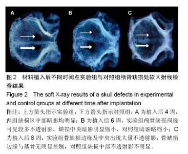
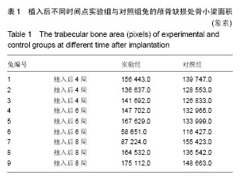
2.1 实验动物数量分析 9只新西兰兔造模后全部成活,进入结果分析,中途无脱失。 2.2 影像学检查结果 见图2。"
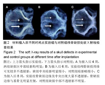
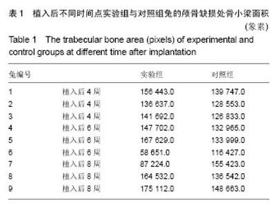
植入后4周,实验组(上方箭头)与对照组(下方箭头)缺损区无明显变化,只是边缘有散在不透射影,缺损区中部暗影均明显,实验组略小,实验组骨小梁面积较对照组稍大,但差异无显著性意义(P > 0.05)。植入后6周,实验组颅骨缺损周缘可见较多不透射影,缺损中央暗影明显缩小;对照组暗影略缩小,骨小梁面积较实验组明显减少(P < 0.05)。植入后8周时,实验组骨缺损边缘及中央出现大量不透射影,骨缺损边缘与基骨无明显差别;对照组缺损中部不透射影不明显,骨小梁面积较实验组小(P < 0.05),见表1,2。"
"
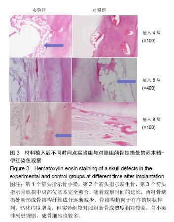
2.3 苏木精-伊红染色结果 见图3。 植入后4周:两组缺损处中心部位大部分未长合,实验组缺损处可见牙齿煅烧颗粒分散在缺损区,骨小梁形态不规则,但较对照侧骨小梁粗大、致密(箭头所示),纤维成分相对较少;对照组骨小梁相对稀疏细小。 植入后6周:与对照组相比,实验侧骨缺损处中心部位牙齿煅烧颗粒被大部分保留在缺损区,围绕在牙齿煅烧颗粒周围有更多的新骨长成;对照组缺隙处有纤维组织长入,新骨形成较少(箭头所示),未长合。两组缺损区表面新骨均不连续,但实验组骨小梁形态趋于规则,骨小梁更粗大、致密,骨结构出现层次,缺损区边缘及中央均有大量新骨形成;对照组相对无序,新骨形成较少;实验组成骨细胞含量较多,数量明显多于对照组。 植入后8周:实验组骨缺损中央部位基本完全愈合(箭头所示),对照侧尚未完全长合;实验组有较多的牙齿煅烧颗粒保留,颗粒周围新骨形成较多,矿化程度也较高,成骨细胞较对照组偏多,新骨形成较厚,表面也较对照组光滑,骨小梁排列更规则、粗大,骨质更致密;对照组新骨虽有形成,但较薄不连续,骨小梁细小、稀疏。 随着观察时间的延长,两组骨缺损处新形成骨结构纤维成分逐渐减少,骨结构趋向于有序的层状排列,钙化程度增高,但实验组较对照组新骨成熟度相对较高,骨小梁排列更规则,成骨细胞也较多。如果延长实验动物观察时间,实验组与对照组的骨缺损修复程度相差会越来越小,直至最终完全愈合。"
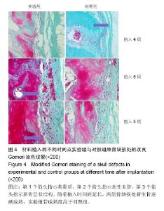
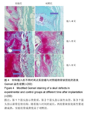
2.4 改良Gomori染色结果 见图4。 植入后4周,两组新生骨成熟程度均较低、形态不规则,实验组较对照组纤维成分相对较少、骨基质偏多,对照组纤维成分较多,类骨质偏多(箭头所示)。植入后6周,两组骨成熟程度均较4周组高,实验组骨形态更趋于规则,并含有丰富的新生血管(箭头所示),红染区域面积即新骨形成量较4周时显著增多,并且显著多于对照组。植入后8周,骨成熟程度较6周时高,实验组新骨层状结构相对明显(箭头所示),骨结构较对照组规则,骨成熟程度相对较高。"
| [1]Kim WB,Kim SG,Lim SC,et al.Effect of Tisseel on bone healing with particulate dentin and plaster of Paris mixture.Oral Surg Oral Med Oral Pathol Oral Radiol Endod. 2010;109(2):34-40.[2]Hara S,Mitsugi M,Kanno T,et al.Bone Transport and Bone Graft Using Auto-Tooth Bone for Alveolar Cleft Repair.J Craniofac Surg.2013;24(1):65-68.[3]Reena AB,Tamara R.Bone Graft Substitutes.Hand Clin.2012; 28(4):457-468.[4]李琦.Choukroun富血小板纤维蛋白修复兔颅骨骨缺损的实验研究[D].吉林大学口腔医学院,2009.[5]MiyataY,Ozawa S,Kojima N,et al.An experimental study of bone grafting using rat milled tooth.Int J Oral Maxil lofac Implants.2011;26(6):1210-1216.[6]郭津源,仲维剑,柴松岭,等.牙齿煅烧颗粒结合富血小板纤维蛋白修复骨缺损的实验研究[J].口腔医学研究, 2015,31(11): 1069-1072.[7]中华人民共和国科学技术部.关于善待实验动物的指导性意见. 2006-09-30.[8]郭慧媛,张录达,郑丽敏,等.X射线显微CT用于大鼠骨小梁结构分析的研究[J].光谱学与光谱分析,2009,29(8):2276-2280.[9]Dohan DM,Del Corso M,Charrier JB.Cytotoxicity analyses of-Choukroun's platelet-rich fibrin( PRF) on a wide range of hnm R-cells:the answer to a commercial controversy.Oral Surg O Metl Orai Pathol Oral Radiol Ended.2007;103(3): 587-593.[10]Braecini F,Dohnn DM.Relevance of Choukroun'8 plate-let-rich fibrin(PRF) duringfacial aesthetic lipostrueture (Colem 's tech-nique):prcliminary results.Revue de Laryngol Otol Rhinol.2007;128(4) : 255-260.[11]Dohan DM,Choukroun J, Diss A,et al.Platelet-rich fibrin(PRF): a second-generation platelet concentrate. Part II: platelet-related biologic features. Oral Surg Oral Med Oral Pathol Oral Radiol Endod.2006;101(3):e45-50.[12]Choukroun J, Diss A,Simonpieri A,et al.Platelet-rich fibrin (PRF):a second-generation platelet concentrate. Part V: histologic evaluations of PRF effects on bone allograft maturation in sinus lift. Oral Surg Oral Med Oral Pathol Oral Radiol Endod.2006;101(3):299-303. [13]Sunitha RV, Munirathnam NE.Platelet-rich fibrin: Evolution of a second-generation platelet concentrate. Indian J Dent Res. 2008;19(1):42-46.[14]Weibrich G,Kleis WK,Hafner G.Growth factor levels in the platelet-rich plasma produced by 2 different methods: curasan-type PRP kit versus PCCS PRP system.Int J Oral Maxillofac Implants.2002;17(2):184-190.[15]Kushida S,Kakudo N,Morimoto N,et al.Platelet and growth factor concentrations in activated platelet-rich plasma:a comparison of seven commercial separation systems.J Artif Organs.2014;17(2):186-192.[16]Kim TH,Kim SH,Sándor GK,et al. Comparison of platelet-rich plasma (PRP), platelet-rich fibrin (PRF), and concentrated growth factor (CGF) in rabbit-skull defect healing. Arch Oral Biol.2014;59(5):550-558.[17]Appel TR,P tzsch B,Müller J,et al.Comparison of three different preparations of platelet concentrates for growth factor enrichment.Clin Oral Implants Res. 2002;13(5): 522-528.[18]Jeong KI,Kim SG,Kim YK,et al.Clinicl study of graft materials using autogenous teeth in maxillary sinus augmentation. Implant Dent.2011;20(6):471-475.[19]Dohan DM,Choukroun J,Diss A,et al.Platelet-rich fibrin(PRF):A Second-generation platelet concentrart.Part II Platelet-related biologic features.Oral Surg Oral Med Oral Pathol Oral Radiol Endod. 2006;101(3):45-50.[20]Dohan Ehrenfest DM,Doglioli P,de Peppo GM,et al. Choukroun’s platelet-rich fibrin,(PRF) stimulates in vitro proliferation and differentiation of human oral bone mesenchymal stem cell in a dose-dependent way.Arch oral Biol.2010;55(3):185-194.[21]Lee JW,Kim SG,Kim JY,et al. Restoration of a peri-implant defect by platelet-rich fibrin.Oral Surg Oral Med Oral Pathol Oral Radiol Endod.2012;113(4):469-463.[22]Marrelli M,Tatullo M.Influence of PRF in the healing of bone and gingival tissues. Clinical and histological evaluations.Eur Rev Med Pharmacol Sci.2013;17(14):1958-1962.[23]Louis PJ,Gutta R,Said-Al-Naief N,et al.Reconstruction of the maxilla and mandible with particulate bone graft and titanium mesh for implant placement.J Oral Maxillofac Surg. 2008; 66(2):235-245.[24]Yassibag-Berkman Z,Tuncer O,Subasioglu T,et al. Combined use of platelet-rich plasma and bone grafting with or without guided tissue regeneration in the treatment of anterior interproximal defects.J Periodontol. 2007;78(5): 801-809.[25]Chiapasco M,Zaniboni M,Rimondini L.Autogenous onlay bone grafts vs.alveolar distraction osteogenesis for the correction of vertically deficient edentulous ridges:a 2-4-year prospective study on humans.Clin Oral Implants Res.2007;18(4):432-440.[26]Scarano A,Orsini G,Pecora G,et al.Peri-implant bone regeneration with calcium sulfate:a light and transmission electron microscopy case report.Implant Dent. 2007;16(2): 195-203.[27]Donos N,Kostopoulos L,Karring T.Augmentation of the rat jaw with autogeneic cortico-cancellous bone grafts and guided tissue regeneration.Clin Oral Implants Res. 2002;13(2): 192-202.[28]Araújo MG,Sonohara M,Hayacibara R,et al.Lateral ridge augmentation by the use of grafts comprised of autologous bone or a biomaterial.An experiment in the dog.J Clin Periodontol 2002;29(12):1122-1131[29]付冬梅,肖琼,杨琴秋,等.富血小板纤维蛋白新生诱导骨的组织学观察[J].中国组织工程研究,2016,20(7):933-939.[30]Bonucci E,Marini E,Valdinucci F,et al.Osteogenic response to hydroxyapatite-fibrin implants in maxillofacial bone defects. Eur J Oral Sci.1997;105:557-561. |
| [1] | Zhang Tongtong, Wang Zhonghua, Wen Jie, Song Yuxin, Liu Lin. Application of three-dimensional printing model in surgical resection and reconstruction of cervical tumor [J]. Chinese Journal of Tissue Engineering Research, 2021, 25(9): 1335-1339. |
| [2] | Zeng Yanhua, Hao Yanlei. In vitro culture and purification of Schwann cells: a systematic review [J]. Chinese Journal of Tissue Engineering Research, 2021, 25(7): 1135-1141. |
| [3] | Xu Dongzi, Zhang Ting, Ouyang Zhaolian. The global competitive situation of cardiac tissue engineering based on patent analysis [J]. Chinese Journal of Tissue Engineering Research, 2021, 25(5): 807-812. |
| [4] | Wu Zijian, Hu Zhaoduan, Xie Youqiong, Wang Feng, Li Jia, Li Bocun, Cai Guowei, Peng Rui. Three-dimensional printing technology and bone tissue engineering research: literature metrology and visual analysis of research hotspots [J]. Chinese Journal of Tissue Engineering Research, 2021, 25(4): 564-569. |
| [5] | Chang Wenliao, Zhao Jie, Sun Xiaoliang, Wang Kun, Wu Guofeng, Zhou Jian, Li Shuxiang, Sun Han. Material selection, theoretical design and biomimetic function of artificial periosteum [J]. Chinese Journal of Tissue Engineering Research, 2021, 25(4): 600-606. |
| [6] | Liu Fei, Cui Yutao, Liu He. Advantages and problems of local antibiotic delivery system in the treatment of osteomyelitis [J]. Chinese Journal of Tissue Engineering Research, 2021, 25(4): 614-620. |
| [7] | Li Xiaozhuang, Duan Hao, Wang Weizhou, Tang Zhihong, Wang Yanghao, He Fei. Application of bone tissue engineering materials in the treatment of bone defect diseases in vivo [J]. Chinese Journal of Tissue Engineering Research, 2021, 25(4): 626-631. |
| [8] | Zhang Zhenkun, Li Zhe, Li Ya, Wang Yingying, Wang Yaping, Zhou Xinkui, Ma Shanshan, Guan Fangxia. Application of alginate based hydrogels/dressings in wound healing: sustained, dynamic and sequential release [J]. Chinese Journal of Tissue Engineering Research, 2021, 25(4): 638-643. |
| [9] | Chen Jiana, Qiu Yanling, Nie Minhai, Liu Xuqian. Tissue engineering scaffolds in repairing oral and maxillofacial soft tissue defects [J]. Chinese Journal of Tissue Engineering Research, 2021, 25(4): 644-650. |
| [10] | Xing Hao, Zhang Yonghong, Wang Dong. Advantages and disadvantages of repairing large-segment bone defect [J]. Chinese Journal of Tissue Engineering Research, 2021, 25(3): 426-430. |
| [11] | Chen Siqi, Xian Debin, Xu Rongsheng, Qin Zhongjie, Zhang Lei, Xia Delin. Effects of bone marrow mesenchymal stem cells and human umbilical vein endothelial cells combined with hydroxyapatite-tricalcium phosphate scaffolds on early angiogenesis in skull defect repair in rats [J]. Chinese Journal of Tissue Engineering Research, 2021, 25(22): 3458-3465. |
| [12] | Wang Hao, Chen Mingxue, Li Junkang, Luo Xujiang, Peng Liqing, Li Huo, Huang Bo, Tian Guangzhao, Liu Shuyun, Sui Xiang, Huang Jingxiang, Guo Quanyi, Lu Xiaobo. Decellularized porcine skin matrix for tissue-engineered meniscus scaffold [J]. Chinese Journal of Tissue Engineering Research, 2021, 25(22): 3473-3478. |
| [13] | Mo Jianling, He Shaoru, Feng Bowen, Jian Minqiao, Zhang Xiaohui, Liu Caisheng, Liang Yijing, Liu Yumei, Chen Liang, Zhou Haiyu, Liu Yanhui. Forming prevascularized cell sheets and the expression of angiogenesis-related factors [J]. Chinese Journal of Tissue Engineering Research, 2021, 25(22): 3479-3486. |
| [14] | Liu Chang, Li Datong, Liu Yuan, Kong Lingbo, Guo Rui, Yang Lixue, Hao Dingjun, He Baorong. Poor efficacy after vertebral augmentation surgery of acute symptomatic thoracolumbar osteoporotic compression fracture: relationship with bone cement, bone mineral density, and adjacent fractures [J]. Chinese Journal of Tissue Engineering Research, 2021, 25(22): 3510-3516. |
| [15] | Liu Liyong, Zhou Lei. Research and development status and development trend of hydrogel in tissue engineering based on patent information [J]. Chinese Journal of Tissue Engineering Research, 2021, 25(22): 3527-3533. |
| Viewed | ||||||
|
Full text |
|
|||||
|
Abstract |
|
|||||