[1] PAE HC, KANG JH, CHA JK, et al. 3D-printed polycaprolactone scaffold mixed with beta-tricalcium phosphate as a bone regenerative material in rabbit calvarial defects.J Biomed Mater Res B Appl Biomater. 2019; 107(4):1254-1263.
[2] ZHU W, MA X, GOU M, et al. 3D printing of functional biomaterials for tissue engineering.Curr Opin Biotechnol.2016;40:103-112.
[3] 赵胤杰.3D打印骨组织工程支架前沿进展[J].通讯世界, 2018(10): 277-278.
[4] 王凯,郑爽,潘肃,等.制备3D打印骨组织工程支架修复骨缺损的特征[J].中国组织工程研究,2019,23(34):5516-5522.
[5] SHAH AM, JUNG H, SKIRBOLL S. Materials used in cranioplasty: a history and analysis.Neurosurg Focus.2014;36(4):E19.
[6] OKUMUS A, GUVEN E, ERMIS I, et al. Use of Muscle Graft Versus Fascia Flap as Interpositional Material in Prevention of Reossification in a Rat Model of Skull Bone Defect.Turk Neurosurg.2020;30(1):119-123.
[7] SASAKI T, NIIZUMA K, KANOKE A, et al. Octacalcium phosphate collagen composite (OCP/Col) enhance bone regeneration in a rat model of skull defect with dural defect. Heliyon. 2020;6(2):e03347.
[8] 马保金.基于羟基磷灰石纳米晶功能材料的制备及其在骨组织工程中的应用[D].济南:山东大学,2019.
[9] 王宁宁.3D打印PDA/PLGA/β-TCP复合组织工程支架的制备及相关研究[D].吉林:吉林大学,2019.
[10] SINGH BN, VEERESH V, MALLICK SP, et al. Generation of scaffold incorporated with nanobioglass encapsulated in chitosan/chondroitin sulfate complex for bone tissue engineering.Int J Biol Macromol. 2020; 153:1-16.
[11] 徐史兴.自体下颌骨外板和纳米羟基磷灰石/胶原复合材料修复颅骨缺损的研究[D].北京:北京协和医学院,2017.
[12] HAMIDABADI HG, SHAFAROUDI MM, SEIFI M, et al. Repair of Critical-Sized Rat Calvarial Defects With Three-Dimensional Hydroxyapatite-Gelatin Scaffolds and Bone Marrow Stromal Stem Cells.Med Arch. 2018;72(2):88-93.
[13] CRUZ-NEVES S, RIBEIRO N, GRACA I, et al. Behavior of prostate cancer cells in a nanohydroxyapatite/collagen bone scaffold.J Biomed Mater Res A.2017;105(7):2035-2046.
[14] 樊园.3D打印纳米羟基磷灰石增强水凝胶软骨支架研究[D].杭州:杭州电子科技大学,2019.
[15] 弓勋,云升. 间充质干细胞的生物学特性及应用[J].中国医学工程, 2018,26(12):40-44.
[16] ABAZARI MF, HOSSEINI Z, ZARE KARIZI S, et al. Different osteogenic differentiation potential of mesenchymal stem cells on three different polymeric substrates.Gene.2020;740:144534.
[17] 先德彬. BMSCs和HUVECs复合HA-TCP支架对大鼠颅骨缺损修复的成血管研究[D].泸州:西南医科大学,2018.
[18] KIM JH, KIM GH, KIM JW, et al. In Vivo Angiogenic Capacity of Stem Cells from Human Exfoliated Deciduous Teeth with Human Umbilical Vein Endothelial Cells.Mol Cells.2016;39(11):790-796.
[19] 先德彬,明华伟,徐荣胜,等.骨髓间充质干细胞与血管内皮生长因子165基因转染人脐静脉内皮细胞共移植促进缺血皮瓣血管的生成[J].中国组织工程研究,2018,22(1):46-52.
[20] 徐荣胜.BMSCs和HUVECs复合HA-TCP支架共培养对大鼠颅骨缺损修复的成骨能力影响[D].泸州:西南医科大学,2018.
[21] CHEN W, LIU X, CHEN Q, et al. Angiogenic and osteogenic regeneration in rats via calcium phosphate scaffold and endothelial cell co-culture with human bone marrow mesenchymal stem cells (MSCs), human umbilical cord MSCs, human induced pluripotent stem cell-derived MSCs and human embryonic stem cell-derived MSCs.J Tissue Eng Regen M. 2018;12(1):191-203.
[22] EL-GHANNAM A. Bone reconstruction: from bioceramics to tissue engineering. Expert Rev Med Dev.2005;2(1):87-101.
[23] IZAL I, ARANDA P, SANZ-RAMOS P, et al. Culture of human bone marrow-derived mesenchymal stem cells on of poly(L-lactic acid) scaffolds: potential application for the tissue engineering of cartilage.Knee Surg Sports Traumatol Arthrosc.2013;21(8):1737-1750.
[24] TOHMA Y, DOHI Y, OHGUSHI H, et al. Osteogenic activity of bone marrow-derived mesenchymal stem cells (BMSCs) seeded on irradiated allogenic bone.J Tissue Eng Regen Med.2012;6(2):96-102.
[25] DULCHAVSKY D, GAO X, LIU YB, et al. Bone marrow-derived stromal cells (BMSCs) interact with fibroblasts in accelerating wound healing.J Invest Surg.2008;21(5):270-279.
[26] 金永利.生物陶瓷复合材料结合外源性血管内皮生长因子对骨缺损的修复作用[J].基因组学与应用生物学,2019,38(10):4780-4784.
[27] 陈琦,李石岩,禹鑫,等.CGF复合丝素蛋白-羟基磷灰石支架对兔下颌骨缺损的修复作用[J].河北医学,2020,26(4):698-702.
[28] 董少杰,王旭东,沈国芳,等.生物陶瓷支架的功能改性及应用研究进展[J].无机材料学报,2020(8):1-15.
[29] RUNYAN CM, TAYLOR JA. Clinical applications of stem cells in craniofacial surgery.Facial Plast Surg.2010;26(5):385-395.
[30] 李冬梅,刘新晖,李庆星.纳米级细胞型组织工程人工骨的构建:修复下颌骨损[J].中国组织工程研究,2018,22(14):2146-2151.
[31] HE S, LIN KF, SUN Z, et al. Effects of Nano-hydroxyapatite/Poly(DL-lactic-co-glycolic acid) Microsphere-Based Composite Scaffolds on Repair of Bone Defects: Evaluating the Role of Nano-hydroxyapatite Content.Artif Organs.2016;40(7):E128-135.
[32] LIAO J, LI Y, LI H, et al. Preparation, bioactivity and mechanism of nano-hydroxyapatite/sodium alginate/chitosan bone repair material.J Appl Biomater Func.2018;16(1):28-35.
[33] KIM HW, KIM HE, SALIH V. Stimulation of osteoblast responses to biomimetic nanocomposites of gelatin-hydroxyapatite for tissue engineering scaffolds.Biomaterials.2005;26(25):5221-5230.
[34] 耿海霞,Ko C-c,郭秀娟,等.羟基磷灰石-凝胶支架复合成骨细胞修复兔颅骨缺损的影像学评价[J].临床口腔医学杂志,2014,30(10): 610-612.
[35] WONG TM, JIN J, LAU TW, et al. The use of three-dimensional printing technology in orthopaedic surgery.Journal of orthopaedic surgery (Hong Kong).2017;25(1):2309499016684077.
[36] 赵靖,王笛,刘继全,等.3D打印技术在医学领域应用的现状及问题[J].中国现代医学杂志,2017,27(12):71-74.
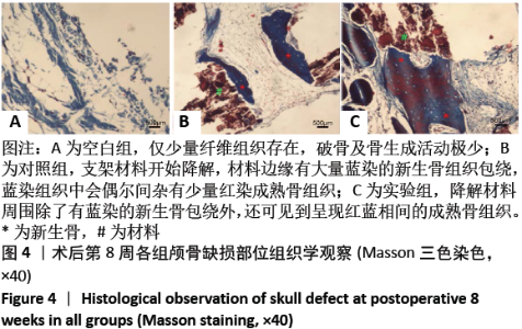
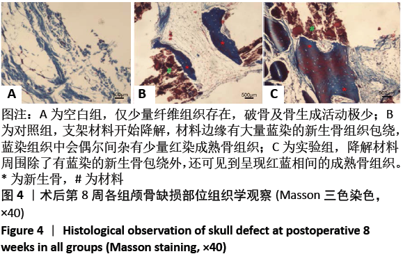
|