Chinese Journal of Tissue Engineering Research ›› 2017, Vol. 21 ›› Issue (36): 5793-5798.doi: 10.3969/j.issn.2095-4344.2017.36.010
Previous Articles Next Articles
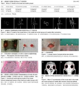
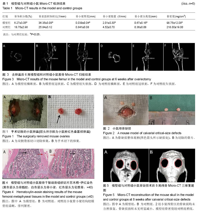
Establishment of a C57 mouse model of osteoporosis with calvarial critical-size defects
Luo Dao-wen1, Huang Kui1, Wang Lei1, Luo Shi-hong1, Rao Peng-cheng1, Xiao Jin-gang1, 2
- 1Orofacial Reconstruction and Regeneration Laboratory, 2Department of Oral and Maxillofacial Surgery, Hospital of Stomatology, Southwest Medical University, Luzhou 646000, Sichuan Province, China
-
Received:2017-07-26Online:2017-12-28Published:2018-01-04 -
Contact:Xiao Jin-gang, M.D., Professor, Chief physician, Master’s supervisor, Orofacial Reconstruction and Regeneration Laboratory, Hospital of Stomatology, Southwest Medical University, Luzhou 646000, Sichuan Province, China -
About author:Luo Dao-wen, Studying for master’s degree, Orofacial Reconstruction and Regeneration Laboratory, Hospital of Stomatology, Southwest Medical University, Luzhou 646000, Sichuan Province, China -
Supported by:the National Natural Science Foundation of China, No. 813671125; the Program of Science & Technology Department of Sichuan Province, No. 2014JY0044; the Project of Education Department of Sichuan Province, No. 10-ZB030; the Project of Health Department of Sichuan Province, No. 80170; the Key Project of Luzhou Medical College, No. 201207; the Luzhou Municipal Government-Southwest Medical University Science and Technology Strategic Cooperation Project, No. 2015LZCYD-S05(2/12)
Cite this article
Luo Dao-wen1, Huang Kui1, Wang Lei1, Luo Shi-hong1, Rao Peng-cheng1, Xiao Jin-gang1, 2. Establishment of a C57 mouse model of osteoporosis with calvarial critical-size defects[J]. Chinese Journal of Tissue Engineering Research, 2017, 21(36): 5793-5798.
share this article
| [1] Fonseca H,Moreira-Goncalves D,Coriolano HJ,et al.Bone quality: the determinants of bone strength and fragility. Sports Med.2014;44(1):37-53.[2] Raisz LG. Pathogenesis of osteoporosis: concepts, conflicts, and prospects.J Clin Invest.2005;115(12):3318-3325.[3] Coipeau P,Rosset P,Langonne A,et al.Impaired differentiation potential of human trabecular bone mesenchymal stromal cells from elderly patients.Cytotherapy.2009;11(5): 584-594.[4] Lill CA,Hesseln J,Schlege lU,etal.Biomechanical evaluation of healing in a non-critical defect in a large animal model of osteoporosis.JOrthop Res.2003;21(5):836-842.[5] Nikolaou VS, Efstathopoulos N, Kontakis G, et al.The influence of osteoporosis in femoral fracture healing time. Injury.2009;40(6):663-668.[6] Rachner TD, Khosla S, Hofbauer LC.et al.Osteoporosis: now and the future. Lancet. 2011;377(9773):1276-1287.[7] Eldesoqi K,Henrich D, El-Kady AM,e tal.Safety Evaluation of a Bioglass–Polylactic Acid Ctrlmposite Scaffold Seeded with Progenitor Cells in a Rat Skull Critical-Size Bone Defect. PLoS One.2014;9(2):1-13.[8] 毕志伟,黄东,欧阳海洋,等.同种异体骨移植治疗骨缺损的应用研究进展[J].中国临床解剖学杂志,2014,32(5):623-625.[9] Kwan MD,Sellmyer MA,Quarto N, et al.Chemical control of FGF-2 release forpromotingcalvarial healing with adipose stem cells. J Biol Chem.2011;286(13): 11307-11313.[10] Pigossi SC,de Oliveira GJ,Finoti LS,et al.Bacterial cellulose- hydroxyapatite composites with osteogenic growth peptide (OGP) or pentapeptide OGP on bone regeneration in critical-size calvarial defect model. J Biomed Mater Res A.2015;103(10): 3397-3406.[11] Liu YS, Ou ME, Liu H, et al. The effect of simvastatin on chemotactic capability of SDF-1alpha and the promotion of bone regeneration. Biomaterials.2014;35(15): 4489-4498.[12] 徐伟,谢杨丽,翁土军,等.Wnt信号参与小鼠颅骨缺损后骨形成过程的初步研究[J].第三军医大学学报, 2014,36(8): 785-788.[13] Li S, Huang KJ, Wu JC,et al. Peripheral blood-derived mesenchymal stem cells: candidate cells responsible for healing critical-sizedcalvarial bone defects. Stem Cells Transl Med. 2015;4(4):359-368.[14] Yi Liu, Ruili Yang, Songtao Shi, et al. Systemic Infusion of Mesenchymal Stem Cells Improves Cell-Based Bone Regeneration via Upregulation of Regulatory T Cells. Tissue Eng Part A. 2015;21(3-4): 498-509.[15] Zheng RC, Park YK, Kim SK,et al. Bone Regeneration of Blood-derived Stem Cells within Dental Implants. J Dent Res. 2015;94(9):1318-1325.[16] Gupta DM, Kwan M D, Slater BJ, et al. Applications of an athymic nude mouse model of nonhealing critical-sized calvarial defects. J Craniofac Surg.2008;19(1):192-197.[17] SavillePD.Changes in skeletal mass and fragility with castration in the rat:a model of osteoporosis.J Am Geriatr Soc.1969;17(2):155-166.[18] 何柳,王然,王晓文,等.低强度脉冲超声对骨质疏松症小鼠模型的作用[J].中国骨质疏松杂志,2015,21(6):657-660.[19] Bliuc D, NguyenND, Nguyen TV, et al. Compound risk of high mortality following osteoporotic fracture and refracture in elderly women and men. J Bone Miner Res.2013;28(11): 2317-2324.[20] Rokn AR, Shakeri AS, Etemad-Moghadam S, et al. Regenerative Effects of Three Types of Allografts on Rabbit Calvarium: An Animal Study.J Dent (Tehran).2015;12(11): 823-834.[21] Felsenberg D,Boonen S.The bone quality framework:determinants of bone strength and their interrelationships and implications for osteoporosis management.Clin Ther.2005; 27(1):1-11.[22] 杨其芬,邵秉一,杨德琴,等. OP小鼠模型的建立及其骨髓贴壁基质细胞体外增殖活性的初步研究[J]. 第三军医大学学报, 2015, 19(1),1966-1971.[23] Li Y, Fan L, Hu J,et.al. MiR-26a Rescues Bone Regeneration Deficiency of Mesenchymal Stem Cells Derived From Osteoporotic. Mol Ther.2015;23(8):1349-57.[24] Azuma K, Furuzawa M, Fujiwara S.et al.Effects of Active Mastication on Chronic Stress-Induced Bone Loss in Mice. Int J Med Sci. 2015; 12(12): 952-957.[25] Riggs BL, Khosla S, Melton LJ 3rd.Sex steroids andthe construction and conservation of the adult skeleton. Endocr Rev. 2002;23(3):279-302.[26] Wang L, Huang C, Li Q, et al.Osteogenic differentiation potential of adipose-derived stem cells from ovariectomized mice.Cell Prolif. 2017 Apr;50(2). doi: 10.1111/cpr.12328.[27] Zhang Y, Wang L, Deng F, et al. Determination of a critical size calvarial defect in senile osteoporotic mice model based on in vivo micro-computed tomography and histological evaluation. Arch Gerontol Geriatr.2015; 61(1): 44-55.[28] 胡宜成,刘鑫,沈际佳,等.犬骨髓间充质干细胞复合磷酸钙骨水泥修复骨缺损的实验研究 [J].上海口腔医学, 2014, 23(4): 402-408.[29] Wang X, Ackermann M, Wang S, et al. Amorphous polyphosphate/amorphous calcium carbonate implant material with enhanced bone healing efficacy in a critical-size defect in rats. Biomed Mater.2016;11(3): 035005.[30] Wang Z, Hu H, Li Z, et al. Sheet of osteoblastic cells combined with platelet-rich fibrin improves the formation of bone in critical-size calvarial defects in rabbits. Br J Oral Maxillofac Surg.2016;54(3): 316-321.[31] 甘宁,郁雪松,陈军,等.杨梅素减轻去卵巢大鼠OP [J].第三军医大学学报,2016, 38(6): 614-618.[32] 唐宇星,赵庆,杨中萌,等.音猬因子修饰纳米晶胶原基骨复合骨髓间充质干细胞修复股骨缺损[J]. 中国组织工程研究,2017,21(14):2180-2185[33] Chen J, Yu J, He Q, et al. A novel injectable porous surface modified bioactive bone cement for vertebroplasty: an in vivo biomechanical and osteogenic study in a rabbit osteoporosis model. Am J Transl Res.2015;7(3): 548.[34] Costa L, Lopes B, Lanis A, et al. Bone demineralization in the lumbar spine of dogs submitted to prednisone therapy. J Vet PharmacolTher. 2010; 33(6): 583-586.[35] Schmitz JP, Hollinger JO. The critical size defect as an experimental model for craniomandibulofacialnonunions . ClinOrthopRelat Res.1986;2(05): 299-308.[36] Choi H,Jeong BC,Hur SW, et al.The Angiopoietin-1 Variant COMP-Ang1 Enhances BMP2-Induced Bone Regeneration with Recruiting Pericytes in Critical Sized Calvarial Defects. PLoS One. 2015; 10(10): e0140502.[37] Qureshi AT,Doyle A,Chen C,et al.Photoactivated miR-148b-nanoparticle conjugates improve closure of critical size mouse calvarial defects.Acta Biomater. 2015;12: 166-173.[38] Das A, Segar CE, Hughley BB, et al. The promotion of mandibular defect healing by the targeting of S1P receptors and the recruitment of alternatively activated macrophages. Biomaterials.2013;34(38): 9853-9862.[39] 靳慧勇,侯天勇,罗飞,等.小鼠股骨临界骨缺损模型的构建及评估 [J].第三军医大学学报, 2011, 33(22): 2331-2334. |
| [1] | Yao Xiaoling, Peng Jiancheng, Xu Yuerong, Yang Zhidong, Zhang Shuncong. Variable-angle zero-notch anterior interbody fusion system in the treatment of cervical spondylotic myelopathy: 30-month follow-up [J]. Chinese Journal of Tissue Engineering Research, 2022, 26(9): 1377-1382. |
| [2] | Jiang Huanchang, Zhang Zhaofei, Liang De, Jiang Xiaobing, Yang Xiaodong, Liu Zhixiang. Comparison of advantages between unilateral multidirectional curved and straight vertebroplasty in the treatment of thoracolumbar osteoporotic vertebral compression fracture [J]. Chinese Journal of Tissue Engineering Research, 2022, 26(9): 1407-1411. |
| [3] | Zhu Chan, Han Xuke, Yao Chengjiao, Zhou Qian, Zhang Qiang, Chen Qiu. Human salivary components and osteoporosis/osteopenia [J]. Chinese Journal of Tissue Engineering Research, 2022, 26(9): 1439-1444. |
| [4] | Li Wei, Zhu Hanmin, Wang Xin, Gao Xue, Cui Jing, Liu Yuxin, Huang Shuming. Effect of Zuogui Wan on bone morphogenetic protein 2 signaling pathway in ovariectomized osteoporosis mice [J]. Chinese Journal of Tissue Engineering Research, 2022, 26(8): 1173-1179. |
| [5] | Xiao Hao, Liu Jing, Zhou Jun. Research progress of pulsed electromagnetic field in the treatment of postmenopausal osteoporosis [J]. Chinese Journal of Tissue Engineering Research, 2022, 26(8): 1266-1271. |
| [6] | Gao Yujin, Peng Shuanglin, Ma Zhichao, Lu Shi, Cao Huayue, Wang Lang, Xiao Jingang. Osteogenic ability of adipose stem cells in diabetic osteoporosis mice [J]. Chinese Journal of Tissue Engineering Research, 2022, 26(7): 999-1004. |
| [7] | Zhang Jinglin, Leng Min, Zhu Boheng, Wang Hong. Mechanism and application of stem cell-derived exosomes in promoting diabetic wound healing [J]. Chinese Journal of Tissue Engineering Research, 2022, 26(7): 1113-1118. |
| [8] | An Weizheng, He Xiao, Ren Shuai, Liu Jianyu. Potential of muscle-derived stem cells in peripheral nerve regeneration [J]. Chinese Journal of Tissue Engineering Research, 2022, 26(7): 1130-1136. |
| [9] | Peng Kun. Improvement of the treatment effect of osteoporotic fractures: research status and strategy analysis [J]. Chinese Journal of Tissue Engineering Research, 2022, 26(6): 980-984. |
| [10] | Shen Song, Xu Bin. Diffuse distribution of bone cement in percutaneous vertebroplasty reduces the incidence of refracture of adjacent vertebral bodies [J]. Chinese Journal of Tissue Engineering Research, 2022, 26(4): 499-503. |
| [11] | He Yunying, Li Lingjie, Zhang Shuqi, Li Yuzhou, Yang Sheng, Ji Ping. Method of constructing cell spheroids based on agarose and polyacrylic molds [J]. Chinese Journal of Tissue Engineering Research, 2022, 26(4): 553-559. |
| [12] | He Guanyu, Xu Baoshan, Du Lilong, Zhang Tongxing, Huo Zhenxin, Shen Li. Biomimetic orientated microchannel annulus fibrosus scaffold constructed by silk fibroin [J]. Chinese Journal of Tissue Engineering Research, 2022, 26(4): 560-566. |
| [13] | Chen Xiaoxu, Luo Yaxin, Bi Haoran, Yang Kun. Preparation and application of acellular scaffold in tissue engineering and regenerative medicine [J]. Chinese Journal of Tissue Engineering Research, 2022, 26(4): 591-596. |
| [14] | Kang Kunlong, Wang Xintao. Research hotspot of biological scaffold materials promoting osteogenic differentiation of bone marrow mesenchymal stem cells [J]. Chinese Journal of Tissue Engineering Research, 2022, 26(4): 597-603. |
| [15] | Shen Jiahua, Fu Yong. Application of graphene-based nanomaterials in stem cells [J]. Chinese Journal of Tissue Engineering Research, 2022, 26(4): 604-609. |
| Viewed | ||||||
|
Full text |
|
|||||
|
Abstract |
|
|||||