Chinese Journal of Tissue Engineering Research ›› 2016, Vol. 20 ›› Issue (20): 2899-2906.doi: 10.3969/j.issn.2095-4344.2016.20.002
Previous Articles Next Articles
Trypsin plus type II collagenase digestion for isolation of nucleus pulposus cells: the optimal glucose concentration in complete medium
Zhang Cun-xin, Ma Jin-feng, Wang De-chun
- Department of Spine Surgery, the Affiliated Hospital of Qingdao University, Qingdao 266000, Shandong Province, China
-
Received:2016-03-30Online:2016-05-13Published:2016-05-13 -
Contact:Wang De-chun, M.D., Professor, Department of Spine Surgery, the Affiliated Hospital of Qingdao University, Qingdao 266000, Shandong Province, China -
About author:Zhang Cun-xin, Studying for master’s degree, Department of Spine Surgery, the Affiliated Hospital of Qingdao University, Qingdao 266000, Shandong Province, China -
Supported by:the National Natural Science Foundation of China, No. 21472104
Cite this article
Zhang Cun-xin, Ma Jin-feng, Wang De-chun. Trypsin plus type II collagenase digestion for isolation of nucleus pulposus cells: the optimal glucose concentration in complete medium[J]. Chinese Journal of Tissue Engineering Research, 2016, 20(20): 2899-2906.
share this article
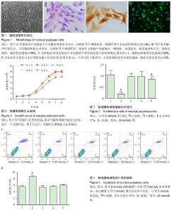
2.1 髓核细胞体外分离培养形态学观察及鉴定结果 胰蛋白酶联合Ⅱ型胶原酶消化分离髓核组织后,可收获较多髓核细胞。光学显微镜下观察发现髓核细胞从网状的纤维组织中释放出来。原代髓核细胞接种后倒置显微镜下观察可见细胞呈球形,大小不等,悬浮在培养液中,三四天后可见有细胞贴壁。细胞贴壁后呈圆形并且变大,伸出伪足后呈三角形、多角形或短梭形,细胞核较大,有时可见双核,核仁明显,胞浆中可见分泌颗粒,细胞折光性好。培养约1周后,髓核细胞完全贴壁,细胞伸出的伪足相互连接成片。培养约15 d可见大部分细胞汇合,细胞周围可见基质样物质沉淀。培养约3周时,原代髓核细胞汇合可达85%以上。苏木精-伊红染色后,可见髓核细胞呈多角形、短梭形等不规则形状,胞核位于细胞中央或偏向一侧胞壁,染成蓝色,胞浆染成粉红色,越靠近胞核,胞浆着色越深。细胞免疫组化染色后可见胞质中Ⅱ型胶原蛋白染成黄褐色,越靠近细胞核着色越深。免疫荧光染色可见Ⅱ型胶原蛋白呈绿色荧光,大部分位于胞质中,且靠近胞核处荧光强度越强,胞核呈蓝色荧光。见图1。 2.2 髓核细胞生长曲线变化 通过对1-5代髓核细胞计数观察发现,细胞呈“S”型曲线生长,见图2。传代后第1天,细胞生长缓慢,从第2-4天细胞生长迅速,第五天细胞生长进入停滞期,以后细胞生长更为缓慢,几乎不再增殖。且随着细胞传代次数的增加,细胞的生长活性逐渐降低。经观察发现,第1-3代细胞生长活性较强,细胞形态相对维持不变。第4代髓核细胞开始发生退变,属于过渡阶段。第5代之后,细胞生长增殖能力显著减退,细胞形态发生变化,由多角形、短梭形逐渐变成长梭形。 2.3 葡萄糖浓度筛选结果 与17.5 mmol/L组相比, 25 mmol/L组细胞增殖受抑制(P < 0.05);6.25, 12.5 mmol/L组细胞增殖比率与对照组(17.5 mmol/L)相比差异无显著性意义;0 mmol/L组细胞增殖受抑制显著(P < 0.01),见图3。 与对照组(17.5 mmol/L)比较,当培养液中葡萄糖浓度为6.25,12.5,25 mmol/L时,细胞凋亡差异无显著性意义(P > 0.05),当培养液中葡萄糖浓度0 mmol/L时,细胞凋亡率明显增加(P < 0.05)。见图4。"

| [1] Tsuji T, Watanabe K, Hosogane N, et al. Risk factors of radiological adjacent disc degeneration with lumbar interbody fusion for degenerative spondylolisthesis. J Orthop Sci. 2015.[2] Ma JF, Zang LN, Xi YM, et al. MiR-125a Rs12976445 Polymorphism is Associated with the Apoptosis Status of Nucleus Pulposus Cells and the Risk of Intervertebral Disc Degeneration. Cell Physiol Biochem. 2016;38(1): 295-305.[3] Battie MC, Videman T. Lumbar disc degeneration: epidemiology and genetics. J Bone Joint Surg Am. 2006;88 Suppl 2:3-9. [4] Watanabe T, Sakai D, Yamamoto Y, et al. Human nucleus pulposus cells significantly enhanced biological properties in a coculture system with direct cell-to-cell contact with autologous mesenchymal stem cells. J Orthop Res. 2010;28(5):623-630.[5] Erwin WM, Islam D, Inman RD, et al. Notochordal cells protect nucleus pulposus cells from degradation and apoptosis: implications for the mechanisms of intervertebral disc degeneration. Arthritis Res Ther. 2011;13(6):R215.[6] Gebhard H, Bowles R, Dyke J, et al. Total disc replacement using a tissue-engineered intervertebral disc in vivo: new animal model and initial results. Evid Based Spine Care J. 2010;1(2):62-66.[7] 李树文,武海军,银和平,等.Ⅱ型胶原酶消化结合组织块贴壁法分离培养兔髓核细胞[J].中国组织工程研究, 2013, 17(39):6861-6866.[8] 李全修,陈伯华,刘勇,等.构建兔髓核细胞诱导人骨髓间充质干细胞向类髓核细胞分化的体外模型[J].中国组织工程研究与临床康复,2009,13(45):8961-8965.[9] 于占革,杨威,温莹,等.兔不同节段椎间盘髓核细胞培养特性的比较[J].中国脊柱脊髓杂志,2010,20(8):689-693.[10] Kong JG, Park JB, Lee D, et al. Effect of high glucose on stress-induced senescence of nucleus pulposus cells of adult rats. Asian Spine J. 2015;9(2):155-161.[11] Neidlinger-Wilke C, Wurtz K, Urban JP, et al. Regulation of gene expression in intervertebral disc cells by low and high hydrostatic pressure. Eur Spine J. 2006;15 Suppl 3:S372-S378.[12] Zheng CJ, Chen J. Disc degeneration implies low back pain. Theor Biol Med Model. 2015;12:24.[13] Sambrook PN, Macgregor AJ, Spector TD. Genetic influences on cervical and lumbar disc degeneration: a magnetic resonance imaging study in twins. Arthritis Rheum. 1999;42(2):366-372.[14] Wang F, Cai F, Shi R, et al. Aging and age related stresses: a senescence mechanism of intervertebral disc degeneration. Osteoarthritis Cartilage. 2016; 24(3):398-408.[15] Bali T, Kumar MN. Relative Contribution of Upper and Lower Lumbar Spinal Segments to Flexion/Extension: comparison between Normal Spines and Spines with Disc Disease in Asian Patients. Asian Spine J. 2015; 9(5):770-775.[16] Ding F, Shao ZW, Xiong LM. Cell death in intervertebral disc degeneration. Apoptosis. 2013; 18(7):777-785.[17] Xu TT, Liao F, Jin HT, et al. Research advance on intervertebral disc degeneration and cell death. Zhongguo Gu Shang. 2015;28(7):673-678.[18] Yu X, Li Z, Shen J, et al. MicroRNA-10b promotes nucleus pulposus cell proliferation through RhoC-Akt pathway by targeting HOXD10 in intervetebral disc degeneration. PLoS One. 2013;8(12):e83080.[19] Shi L, Teng H, Zhu M, et al. Paeoniflorin inhibits nucleus pulposus cell apoptosis by regulating the expression of Bcl-2 family proteins and caspase-9 in a rabbit model of intervertebral disc degeneration. Exp Ther Med. 2015;10(1):257-262.[20] Woiciechowsky C, Abbushi A, Zenclussen ML, et al. Regeneration of nucleus pulposus tissue in an ovine intervertebral disc degeneration model by cell-free resorbable polymer scaffolds. J Tissue Eng Regen Med. 2014;8(10):811-820.[21] 王新光,叶伟,郭汉明,等.白细胞介素-1β诱导人椎间盘髓核细胞凋亡[J].中国医药导报,2011,8(21):20-24.[22] 丁树伟,徐学振.椎间盘组织工程种子细胞的选取、鉴定及培养难点[J].中国医药指南,2013,11(11):470-472.[23] Mern DS, Thome C. Identification and characterization of human nucleus pulposus cell specific serotypes of adeno-associated virus for gene therapeutic approaches of intervertebral disc disorders. BMC Musculoskelet Disord. 2015;16:341.[24] Kadow T, Sowa G, Vo N, et al. Molecular basis of intervertebral disc degeneration and herniations: what are the important translational questions? Clin Orthop Relat Res. 2015;473(6):1903-1912.[25] 肖剑,赵剑,潘欣,等.兔腰椎间盘髓核细胞的培养及形态观察[J].中国矫形外科杂志,2001,8(8):65-66.[26] 张传志,周跃,李长青,等.兔髓核细胞体外培养和重组人转化生长因子-β1对其代谢影响的研究[J].中国脊柱脊髓杂志,2006,16(4):280-283.[27] Gu W, Zhu Q, Gao X, et al. Simulation of the progression of intervertebral disc degeneration due to decreased nutritional supply. Spine (Phila Pa 1976). 2014;39(24):E1411-E1417.[28] Chen JW, Ni BB, Zheng XF, et al. Hypoxia facilitates the survival of nucleus pulposus cells in serum deprivation by down-regulating excessive autophagy through restricting ROS generation. Int J Biochem Cell Biol. 2015;59:1-10.[29] Kong JG, Park JB, Lee D, et al. Effect of high glucose on stress-induced senescence of nucleus pulposus cells of adult rats. Asian Spine J. 2015;9(2):155-161.[30] Park EY, Park JB. Dose- and time-dependent effect of high glucose concentration on viability of notochordal cells and expression of matrix degrading and fibrotic enzymes. Int Orthop. 2013;37(6): 1179-1186.[31] Naqvi SM, Buckley CT. Extracellular matrix production by nucleus pulposus and bone marrow stem cells in response to altered oxygen and glucose microenvironments. J Anat. 2015;227(6):757-766.[32] Elmore S. Apoptosis: a review of programmed cell death. Toxicol Pathol. 2007;35(4):495-516.[33] Zhang X, Chen Y, Jenkins LW, et al. Bench-to-bedside review: Apoptosis/programmed cell death triggered by traumatic brain injury. Crit Care. 2005;9(1):66-75. |
| [1] | Zhang Tongtong, Wang Zhonghua, Wen Jie, Song Yuxin, Liu Lin. Application of three-dimensional printing model in surgical resection and reconstruction of cervical tumor [J]. Chinese Journal of Tissue Engineering Research, 2021, 25(9): 1335-1339. |
| [2] | Geng Qiudong, Ge Haiya, Wang Heming, Li Nan. Role and mechanism of Guilu Erxianjiao in treatment of osteoarthritis based on network pharmacology [J]. Chinese Journal of Tissue Engineering Research, 2021, 25(8): 1229-1236. |
| [3] | Pei Lili, Sun Guicai, Wang Di. Salvianolic acid B inhibits oxidative damage of bone marrow mesenchymal stem cells and promotes differentiation into cardiomyocytes [J]. Chinese Journal of Tissue Engineering Research, 2021, 25(7): 1032-1036. |
| [4] | Zeng Yanhua, Hao Yanlei. In vitro culture and purification of Schwann cells: a systematic review [J]. Chinese Journal of Tissue Engineering Research, 2021, 25(7): 1135-1141. |
| [5] | Li Shibin, Lai Yu, Zhou Yi, Liao Jianzhao, Zhang Xiaoyun, Zhang Xuan. Pathogenesis of hormonal osteonecrosis of the femoral head and the target effect of related signaling pathways [J]. Chinese Journal of Tissue Engineering Research, 2021, 25(6): 935-941. |
| [6] | Xu Yinqin, Shi Hongmei, Wang Guangyi. Effects of Tongbi prescription hot compress combined with acupuncture on mRNA expressions of apoptosis-related genes,Caspase-3 and Bcl-2, in degenerative intervertebral discs [J]. Chinese Journal of Tissue Engineering Research, 2021, 25(5): 713-718. |
| [7] | Zhang Wenwen, Jin Songfeng, Zhao Guoliang, Gong Lihong. Mechanism by which Wenban Decoction reduces homocysteine-induced apoptosis of myocardial microvascular endothelial cells in rats [J]. Chinese Journal of Tissue Engineering Research, 2021, 25(5): 723-728. |
| [8] | Liu Qing, Wan Bijiang. Effect of acupotomy therapy on the expression of Bcl-2/Bax in synovial tissue of collagen-induced arthritis rats [J]. Chinese Journal of Tissue Engineering Research, 2021, 25(5): 729-734. |
| [9] | Xie Chongxin, Zhang Lei. Comparison of knee degeneration after anterior cruciate ligament reconstruction with or without remnant preservation [J]. Chinese Journal of Tissue Engineering Research, 2021, 25(5): 735-740. |
| [10] | Xu Dongzi, Zhang Ting, Ouyang Zhaolian. The global competitive situation of cardiac tissue engineering based on patent analysis [J]. Chinese Journal of Tissue Engineering Research, 2021, 25(5): 807-812. |
| [11] | Wu Zijian, Hu Zhaoduan, Xie Youqiong, Wang Feng, Li Jia, Li Bocun, Cai Guowei, Peng Rui. Three-dimensional printing technology and bone tissue engineering research: literature metrology and visual analysis of research hotspots [J]. Chinese Journal of Tissue Engineering Research, 2021, 25(4): 564-569. |
| [12] | Chang Wenliao, Zhao Jie, Sun Xiaoliang, Wang Kun, Wu Guofeng, Zhou Jian, Li Shuxiang, Sun Han. Material selection, theoretical design and biomimetic function of artificial periosteum [J]. Chinese Journal of Tissue Engineering Research, 2021, 25(4): 600-606. |
| [13] | Liu Fei, Cui Yutao, Liu He. Advantages and problems of local antibiotic delivery system in the treatment of osteomyelitis [J]. Chinese Journal of Tissue Engineering Research, 2021, 25(4): 614-620. |
| [14] | Li Xiaozhuang, Duan Hao, Wang Weizhou, Tang Zhihong, Wang Yanghao, He Fei. Application of bone tissue engineering materials in the treatment of bone defect diseases in vivo [J]. Chinese Journal of Tissue Engineering Research, 2021, 25(4): 626-631. |
| [15] | Zhang Zhenkun, Li Zhe, Li Ya, Wang Yingying, Wang Yaping, Zhou Xinkui, Ma Shanshan, Guan Fangxia. Application of alginate based hydrogels/dressings in wound healing: sustained, dynamic and sequential release [J]. Chinese Journal of Tissue Engineering Research, 2021, 25(4): 638-643. |
| Viewed | ||||||
|
Full text |
|
|||||
|
Abstract |
|
|||||