Chinese Journal of Tissue Engineering Research ›› 2014, Vol. 18 ›› Issue (51): 8286-8291.doi: 10.3969/j.issn.2095-4344.2014.51.016
Previous Articles Next Articles
Two arsenicals induce apoptosis of islet cells
Yao Xiao-feng, Wang Fang-fang, Jiang Li-ping, Geng Cheng-yan, Zhong Lai-fu, Zheng Bai-lu, Yang Guang, Sun Xian-ce
- Liaoning Natural Products Engineering Research Center for Anti-Degenerative Diseases, Department of Occupational and Environmental Health, Dalian Medical University, Dalian 116044, Liaoning Province, China
-
Online:2014-12-10Published:2014-12-10 -
Contact:Sun Xian-ce, M.D., Professor, Liaoning Natural Products Engineering Research Center for Anti-Degenerative Diseases, Department of Occupational and Environmental Health, Dalian Medical University, Dalian 116044, Liaoning Province, China -
About author:Yao Xiao-feng, Master, Lecturer, Liaoning Natural Products Engineering Research Center for Anti-Degenerative Diseases, Department of Occupational and Environmental Health, Dalian Medical University, Dalian 116044, Liaoning Province, China -
Supported by:the National Natural Science Foundation of China, No. 30972562; the Natural Science Foundation of Liaoning Province, No. 2014023050
CLC Number:
Cite this article
Yao Xiao-feng, Wang Fang-fang, Jiang Li-ping, Geng Cheng-yan, Zhong Lai-fu, Zheng Bai-lu, Yang Guang, Sun Xian-ce. Two arsenicals induce apoptosis of islet cells[J]. Chinese Journal of Tissue Engineering Research, 2014, 18(51): 8286-8291.
share this article

2.1 两种砷化物对INS-1细胞生存率的影响 两种砷化物处理INS-1细胞48 h后,与对照组比较,Na2HAsO4·7H2O (iAs5+)和C2H6AsNaO2·3H2O (DMA5+)引起INS-1细胞的存活率降低(P < 0.05,P < 0.01),且呈剂量依赖性下降(表1)。"

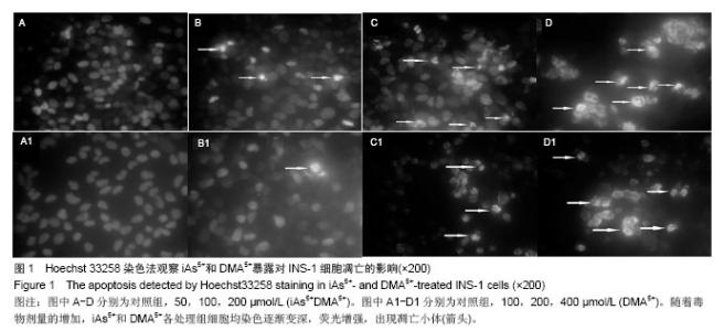
2.2 两种砷化物对INS-1细胞凋亡的影响 两种砷化物处理INS-1细胞48 h后,Hoechst 33258染色结果表明对照组INS-1细胞核形态均一无边聚, 胞内染色均匀一致,形状规则。而随着毒物剂量的增加,iAs5+和DMA5+各处理组细胞边缘渐不平整,染色变深,染色质浓集,荧光增强,部分染色质出现浓缩状态,高度凝聚、边缘化,出现凋亡小体(图1)。"

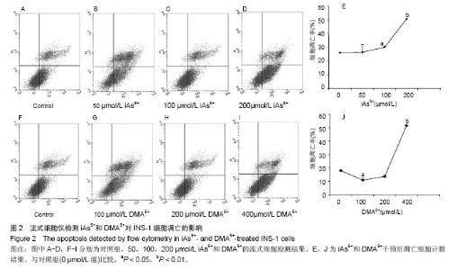
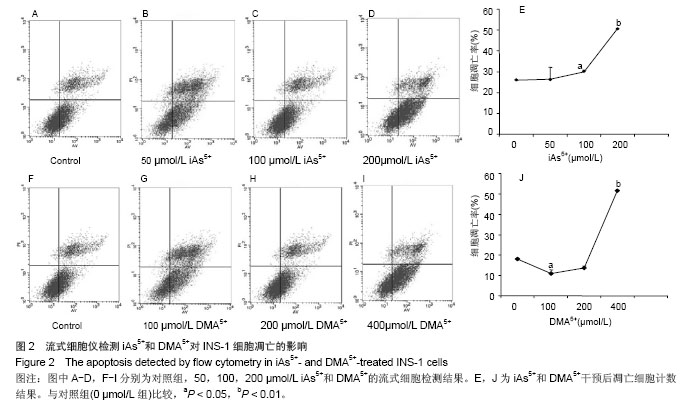
Annexin V/PI染色结果显示,iAs5+和DMA5+暴露均引起INS-1细胞凋亡。与对照组比较,200 µmol/L的iAs5+暴露组凋亡细胞显著增多(P < 0.05)(图2A-E)。100 µmol/L的DMA5+暴露组凋亡细胞减少(P < 0.05);400 µmol/L的DMA5+暴露组凋亡细胞显著增加(P < 0.01)(图2F-J)。"

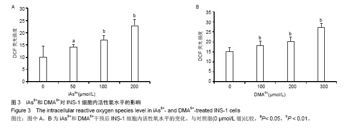
2.3 两种砷化物对INS-1细胞内活性氧水平的影响 两种砷化物处理INS-1细胞24 h后,细胞内DCF荧光强度见图3。iAs5+和DMA5+作用INS-1细胞24 h后,细胞内活性氧水平显著升高,并且与iAs5+浓度存在剂量依赖关系(P < 0.05, P < 0.01)。"
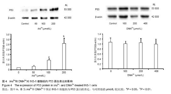
2.4 两种砷化物对INS-1细胞内P53蛋白表达的影响 iAs5+作用INS-1细胞48 h后,Western blot检测核内P53蛋白表达,结果显示随iAs5+剂量增加,P53的表达量逐渐增加(图4A)。100,200 µmol/L iAs5+处理细胞后,核内P53蛋白呈剂量依赖性增加(P < 0.05)。不同浓度DMA5+作用INS-1细胞48 h后,与对照组比较,染毒组P53蛋白表达差异无显著性意义 (图4B)。"
| [1]Yang W, Lu J, Weng J, et al. Prevalence of diabetes among men and women in China. N Engl J Med. 2010;362(12):1090-1101. [2]Sciandrello G, Barbaro R, Caradonna F, et al. Early induction of genetic instability and apoptosis by arsenic in cultured Chinese hamster cells. Mutagenesis. 2002;17(2):99-103. [3]孙贵范.深入研究慢性砷中毒的分子作用机制[J].中国地方病学杂志,2004,23(1):1-2. [4]洪峰.无机砷的肾脏损伤作用研究进展[J].国外医学卫生学分册, 2002,29(1):7-30. [5]Wang Y, Zhao FD, Liao YJ, et al. Arsenic exposure and glutamate-induced gliotransmitter release from astrocytes. Neural Regen Res. 2012; 7(31): 2439-2445. [6]Das AK, Sahu R, Dua TK, et al. Arsenic-induced myocardial injury:protective role of Corchorus olitorius leaves. Food Chem Toxicol. 2010;48(5):1210-1217. [7]Tseng CH, Tai TY, Chong CK, et al. Long-term arsenic exposure and incidence of non-insulin-dependent diabetes mellitus:a cohort study in arseniasis hyperendemic villages in Taiwan. Environ Health Perspect. 2000;108(9):847-851. [8]Chui HF, Chang CC, Tsai SS, et al. Does arsenic exposure increase the risk for diabetes mellitus. J Occup Environ Med. 2006;48(1):63-67. [9]Wang JP, Wang SL, Lin Q, et al. Association of arsenic and kidney dysfunction in people with diabetes and validation of its effects in rats. Environ Int. 2009;35(3):507-511. [10]Lam DW, Leroith D. The worldwide diabetes epidemic. Curr Opin Endocrinol Diabetes Obes. 2012;19(2):93-96. [11]Cheng XB, Hsieh YT, Tu ST, et al. Obesity and low target attainment rates in Chinese with type 2 diabetes. Eur J Inter Med. 2012;23(4):101-105. [12]Navas-Acien A, Silbergeld EK, Pastor-Barriuso R, et al. Arsenic exposure and prevalence of type 2 diabetes in US adults. JAMA. 2008;300(7):814-822. [13]Wang JP, Wang SL, Lin Q, et al. Association of arsenic and kidney dysfunction in people with diabetes and validation of its effects in rats. Environ Int. 2009;35(3):507-511. [14]Tseng CH. Arsenic Exposure and Diabetes Mellitus in the United. JAMA. 2008;300(23):2728. [15]Gong Z, Lu X, Ma M, et al. Arsenic speciation analysis. Talanta. 2002;58(1):77-96. [16]彭珊茁,蔡原.砷的生物学指标研究进展[J].中国工业医学杂志, 2006,19(3):168-171. [17]Pan X, Jiang L, Zhong L, et al. Arsenic induces apoptosis by the lysosomal-mitochondrial pathway in INS-1 cells. Environ Toxicol. 2014[Epub ahead of print]. [18]Martín-Pardillos A, Sosa C, Sorribas V. Arsenic increases Pi-mediated vascular calcification and induces premature senescence in vascular smooth muscle cells. Toxicol Sci. 2013;131(2):641-653. [19]Calatayud M, Devesa V, Vélez D. Differential toxicity and gene expression in Caco-2 cells exposed to arsenic species. Toxicol Lett. 2013;218(1):70-80. [20]Ding H, Shi J, Wang Y, et al. Neferine inhibits cultured hepatic stellate cell activation and facilitates apoptosis. Eur J Pharmacol. 2011;650(1):163-169. [21]Ninsontia C, Pongjit K, Chaotham C, et al. Silymarin selectively protects human renal cells from cisplatin-induced cell death. Pharm Biol. 2011;49(10):1082-1090. [22]Wongtongtair S, Chanvorachote P, Hutamekalin P, et al. Barakol-induced apoptosis in P19 cells through generation of reactive oxygen species and activation of caspase-9. Ethnopharmacol. 2011;137(2):971-978. [23]Bedner E, Li X, Gorczyca W, et al. Analysis of apoptosis by laser scanning cytometry. Cytometry. 1999;35(3):181-195. [24]Vermes I, Haanen C, Reutelingsperger C. Flow cytometry of apoptotic cell death. J Immunol Methods. 2000;243(1): 167-190. [25]LeBel CP, Ischiropoulos H, Bondy SC. Evaluation of the probe 2',7'-dichlorofluorescin as an indicator of reactive oxygen species formation and oxidative stress. Chem Res Toxicol. 1992;5(2):227-231. [26]Sohn JH, Han KL, Lee SH, et al. Protective effects of panduratin A against oxidative damage of tert-butylhydroperoxide in human HepG2 cells. Biol Pharm Bull. 2005;28(6):1083-1086. [27]IARC. IARC Monographs on the evaluation of the carcinogenic risk to humans. Some Drinking-Water Disinfectants and Contaminants,Including arsenic. IARC Monogr Eval Carcinog Risks Hum. 2004;84:1-477. [28]陆景坤,陈朝军,翟小涵,等.不同砷化合物对人皮肤成纤维细胞系的毒性研究[J].中国地方病防治杂志,2011,26(5):328-331. [29]李冰,陆春伟,孙贵范.不同砷化合物对血管内皮细胞毒性的研究[J]卫生研究,2006,35(6):700-702. [30]陆景坤,陈朝军,刘小雷.不同砷化合物对人胚肾细胞系HEK-293细胞的毒性研究[J].环境与健康杂志,2011,28(1): 25-28. [31]Afanasyev VN, Korol BA, Matylevich NP, et al. The use of flow cytometry for the investigation of cell death. Cytometry. 1993;14(6):603-609. [32]徐跃飞,任凤,赵宝昌.Adinbitor对人胃癌MGC803细胞增殖及凋亡作用[J].实用癌症杂志,2009,24(3):233-236. [33]Epe B. Genotoxicity of singlet oxygen. Chem Biol Interact. 1991;80(3):239-260. [34]Martindale JL, Holbrook NJ. Cellular response to oxidative strees: signaling for suicide and survival. J Cell Physiol. 2002;192(1):1-15. [35]Yamanaka K, Hoshino M, Okamoto M, et al. Induction of DNA damage by dimethylarsine,ametabolite of inorganic, is for the major part likely due to its peroxyl radical. Biochem Biophys Res Commum. 1990;168(1):58-64. [36]Kitchin KT. Recent advances in arsenic carcinogenesis: Modes of aetion,animal model systems, and methylated arsenic metabolites. Toxicol Appl Pharmacol. 2001;172(3): 249-261. [37]Pietsch EC, Sykes SM, McMahon SB, et al. The P53 family and programmed cell death. Oncogene. 2008;27(50):6507-6521. [38]杨从容,王雅棣.活性氧、锰超氧化物歧化酶和P53的相互作用及对肿瘤的影响[J].癌变•畸变•突变,2011,23(3):238-241. [39]李玉祥,罗小平,廖玲洁,等.高氧对新生大鼠肺caspase-3和P53基因表达及肺细胞凋亡的影响[J].中华儿科杂志,2005,43(8): 585-590. [40]Diaz-Villasenor A, Sanchez-Soto MC, Cebrian ME. Sodium arsenite impairs insulin secretion and transcription in pancreatic beta-cells. Toxico Appl Pharmacol. 2006;214(1): 30-34. |
| [1] | Zhang Tongtong, Wang Zhonghua, Wen Jie, Song Yuxin, Liu Lin. Application of three-dimensional printing model in surgical resection and reconstruction of cervical tumor [J]. Chinese Journal of Tissue Engineering Research, 2021, 25(9): 1335-1339. |
| [2] | Geng Qiudong, Ge Haiya, Wang Heming, Li Nan. Role and mechanism of Guilu Erxianjiao in treatment of osteoarthritis based on network pharmacology [J]. Chinese Journal of Tissue Engineering Research, 2021, 25(8): 1229-1236. |
| [3] | Pei Lili, Sun Guicai, Wang Di. Salvianolic acid B inhibits oxidative damage of bone marrow mesenchymal stem cells and promotes differentiation into cardiomyocytes [J]. Chinese Journal of Tissue Engineering Research, 2021, 25(7): 1032-1036. |
| [4] | Zeng Yanhua, Hao Yanlei. In vitro culture and purification of Schwann cells: a systematic review [J]. Chinese Journal of Tissue Engineering Research, 2021, 25(7): 1135-1141. |
| [5] | Li Shibin, Lai Yu, Zhou Yi, Liao Jianzhao, Zhang Xiaoyun, Zhang Xuan. Pathogenesis of hormonal osteonecrosis of the femoral head and the target effect of related signaling pathways [J]. Chinese Journal of Tissue Engineering Research, 2021, 25(6): 935-941. |
| [6] | Xu Yinqin, Shi Hongmei, Wang Guangyi. Effects of Tongbi prescription hot compress combined with acupuncture on mRNA expressions of apoptosis-related genes,Caspase-3 and Bcl-2, in degenerative intervertebral discs [J]. Chinese Journal of Tissue Engineering Research, 2021, 25(5): 713-718. |
| [7] | Zhang Wenwen, Jin Songfeng, Zhao Guoliang, Gong Lihong. Mechanism by which Wenban Decoction reduces homocysteine-induced apoptosis of myocardial microvascular endothelial cells in rats [J]. Chinese Journal of Tissue Engineering Research, 2021, 25(5): 723-728. |
| [8] | Liu Qing, Wan Bijiang. Effect of acupotomy therapy on the expression of Bcl-2/Bax in synovial tissue of collagen-induced arthritis rats [J]. Chinese Journal of Tissue Engineering Research, 2021, 25(5): 729-734. |
| [9] | Xie Chongxin, Zhang Lei. Comparison of knee degeneration after anterior cruciate ligament reconstruction with or without remnant preservation [J]. Chinese Journal of Tissue Engineering Research, 2021, 25(5): 735-740. |
| [10] | Xu Dongzi, Zhang Ting, Ouyang Zhaolian. The global competitive situation of cardiac tissue engineering based on patent analysis [J]. Chinese Journal of Tissue Engineering Research, 2021, 25(5): 807-812. |
| [11] | Wu Zijian, Hu Zhaoduan, Xie Youqiong, Wang Feng, Li Jia, Li Bocun, Cai Guowei, Peng Rui. Three-dimensional printing technology and bone tissue engineering research: literature metrology and visual analysis of research hotspots [J]. Chinese Journal of Tissue Engineering Research, 2021, 25(4): 564-569. |
| [12] | Chang Wenliao, Zhao Jie, Sun Xiaoliang, Wang Kun, Wu Guofeng, Zhou Jian, Li Shuxiang, Sun Han. Material selection, theoretical design and biomimetic function of artificial periosteum [J]. Chinese Journal of Tissue Engineering Research, 2021, 25(4): 600-606. |
| [13] | Liu Fei, Cui Yutao, Liu He. Advantages and problems of local antibiotic delivery system in the treatment of osteomyelitis [J]. Chinese Journal of Tissue Engineering Research, 2021, 25(4): 614-620. |
| [14] | Li Xiaozhuang, Duan Hao, Wang Weizhou, Tang Zhihong, Wang Yanghao, He Fei. Application of bone tissue engineering materials in the treatment of bone defect diseases in vivo [J]. Chinese Journal of Tissue Engineering Research, 2021, 25(4): 626-631. |
| [15] | Zhang Zhenkun, Li Zhe, Li Ya, Wang Yingying, Wang Yaping, Zhou Xinkui, Ma Shanshan, Guan Fangxia. Application of alginate based hydrogels/dressings in wound healing: sustained, dynamic and sequential release [J]. Chinese Journal of Tissue Engineering Research, 2021, 25(4): 638-643. |
| Viewed | ||||||
|
Full text |
|
|||||
|
Abstract |
|
|||||