Chinese Journal of Tissue Engineering Research ›› 2014, Vol. 18 ›› Issue (18): 2848-2854.doi: 10.3969/j.issn.2095-4344.2014.18.011
Previous Articles Next Articles
Apoptosis of optic nerve cells after retinal contusion injury in rabbits
Wang Zhi-yu1, Shi Ai-yun2
- 1Department of Ophthalmology, 2Department of Neurology, Mindong Hospital of Ningde, Fuan 355000, Fujian Province, China
-
Received:2014-03-08Online:2014-04-30Published:2014-04-30 -
About author:Wang Zhi-yu, Master, Associate chief physician, Department of Ophthalmology, Mindong Hospital of Ningde, Fuan 355000, Fujian Province, China -
Supported by:Key Program of Fujian Provincial Science and Technology Bureau, No. 2008Y01010605
CLC Number:
Cite this article
Wang Zhi-yu, Shi Ai-yun. Apoptosis of optic nerve cells after retinal contusion injury in rabbits[J]. Chinese Journal of Tissue Engineering Research, 2014, 18(18): 2848-2854.
share this article
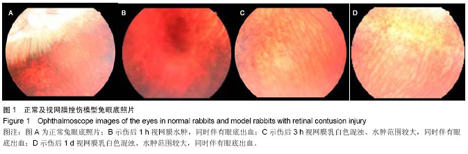
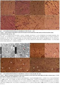
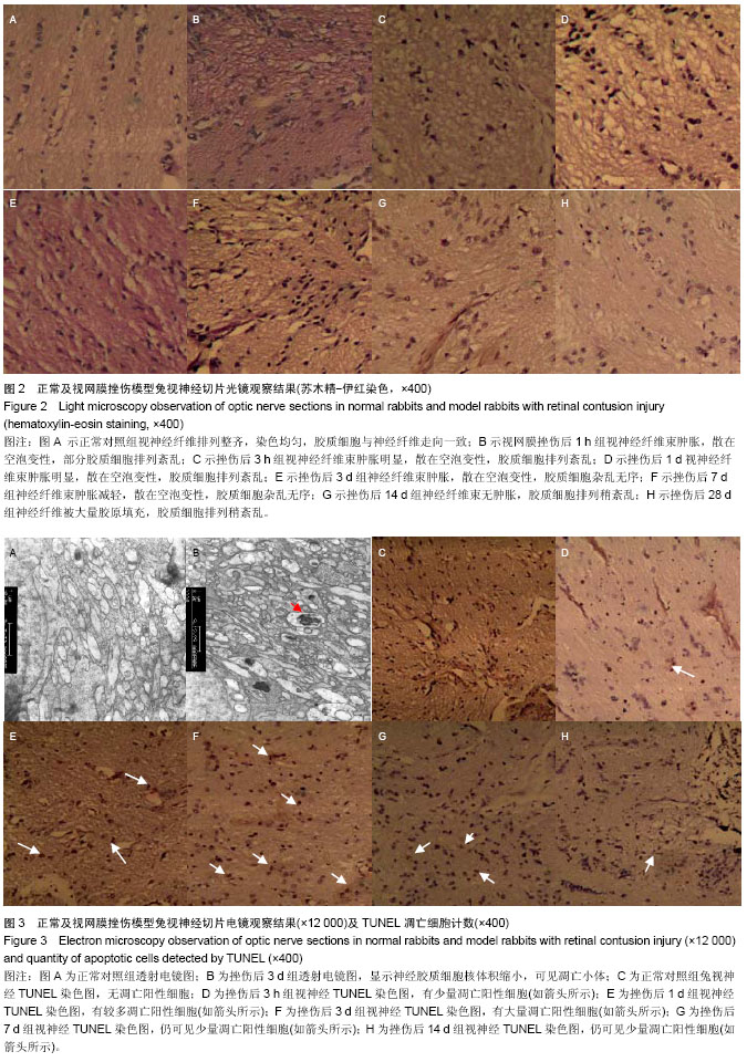
2.1 实验动物数量分析 纳入青紫蓝兔48只分为8组,每组6只,全部进入结果分析,无脱失。 2.2 光镜观察结果 挫伤模型建立后不同时间点的神经胶质细胞光镜下计数:正常对照组80.60±15.46,挫伤后1 h组71.70±12.83,挫伤后3 h组59.00±13.37,挫伤后1 d组45.60±15.43,挫伤后3 d组56.10±17.79,挫伤后7 d组67.90±14.75,挫伤后14 d组72.80±12.91,挫伤后28 d组74.71±13.83。 光镜所见正常对照组视神经纤维排列整齐,染色均匀,其间有散在的胶质细胞与神经纤维走向一致,呈极性排列。挫伤后1,3 h,挫伤后1,3,7 d视神经纤维排列紊乱,纤维束肿胀,散在空泡变性,视神经胶质细胞杂乱无序。挫伤后1,3 h,挫伤后1,3,7,14,28 d细胞计数减少,与正常对照组相比,差异有非常显著性意义(P < 0.01)。挫伤后14, 28 d组可见神经胶质细胞增生,神经纤维被大量胶原填充(图2)。 2.3 透射电镜观察结果 透射电镜下见正常对照组视神经髓鞘板层状排列致密,轴突内布满空心环状结构的微管和实心圆的神经微丝,并可见线粒体等细胞器(图3A)。挫伤后1 h组:部分神经胶质细胞排列稀疏、间隙增宽,线粒体、轴突轻度肿胀。挫伤后3 h组:神经胶质细胞胞体空泡变性,线粒体水肿透明,次级溶酶体轻度增多,核染色质浓缩,核膜厚薄不均,轴突轻度肿胀。挫伤后1 d组:神经胶质细胞髓鞘样变化,核体积轻度缩小,染色质浓缩、边集,可见凋亡小体。挫伤后3 d组:神经胶质细胞电子密度"
"
| [1] 刘家琦,李凤鸣.实用眼科学[M].2版.北京:人民卫生出版社,2003: 583. [2] Sipperley JQ, Quigley HA, Hass JDM. Traumatic retinopathy of prematurity in primates; the explanation of commotio retinae.Arch Ophthalmol. 1978;96: 2267-2273. [3] Chakravarthy U, Douglas AJ, Bailie R, et al. Immnoreactive endothelin distribution in ocular tissues. Invest Opthhalmol Vis Sci. 1994;35:2448. [4] Chen CS, Odel JG, Miller JS, et al.Multifocal visual evoked potentials and multifocal electroretinograms in papillorenal syndrome. Arch Ophthalmol. 2002; 120(6): 870-871. [5] 窦宏亮,宋琛.眼球挫伤后眼血流动力学改变[J].中华眼科杂志, 1992,28(4): 243-245. [6] 安美霞,张效房,张金嵩.挫伤性视网膜病变中光感受器细胞凋亡与氧化损伤的实验研究[J].中华眼科杂志,2004,40(2): 118-121. [7] Yang L, Bula D, Arroyo JG, et al. Preventing retinal detachment- associated photoreceptor cell loss in Bax-deficient mice. Invest Ophthalmol Vis Sci. 2004;45(2): 648-654. [8] Seddon JM, Cote J, Page WF, et al. The US twin study of age-related macular degeneration: relative roles of genetic and environmental influences. Arch Ophthalmol. 2005;123: 321-327. [9] Tomany SC, Cruickshanks KJ, Klein R, et al. Sunlight and the 10-year incidence of age-related maculopathy: the beaver dam eye study. Arch Ophthalmol. 2004;122: 750-757. [10] 李凤鸣.眼科全书下册[M].2版.北京:人民卫生出版社,1996: 3281-3285. [11] 王志玉,付群,史爱云.视网膜挫伤后神经感觉层细胞凋亡的实验研究[J].眼科新进展, 2008,28(8):590-594. [12] Wenzel A, Grimm C, Samardzija M, et al. Molecular mechanisms of light-induced photoreceptor apoptosis and neuroprotection for retinal degeneration. Prog Retin Eye Res. 2005;24(2):275-306. [13] Zacks DN, Hanninen V, Pantcheva M, et al. Caspase activation in an experimental model of retinal detachment. Invest Ophthalmol Vis Sci. 2003;44(3):1262-1267. [14] Peter ME, Heufelder AE, Hengartner MO. Advances in apoptosis research. Proc Natl Acad Sci USA. 1997;94(24): 12736-12737. [15] 欧阳科,袁援生,李燕.大鼠青光眼模型视网膜神经节细胞凋亡的研究[J].眼科研究, 2008,26(10): 725-725. [16] 张琳琳,吕瀛娟,于荣国,等.标准化大鼠外伤性视神经损伤动物模型建立[J].眼外伤职业眼病杂志,2008, 30(12): 913-917. [17] Hisatomi T, Sakamoto T, Murata T, et al. Relocalization of apoptosis-inducing factor in photoreceptor apoptosis induced by retinal detachment in vivo. Am J Pathol. 2001;158(4): 1271-1278. [18] Nakano Y, Oyamada M, Dai P, et al. Connexin43 knockdown accelerates wound healing but inhibits mesenchymal transition after corneal endothelial injury in vivo. Invest Ophthalmol Vis Sci. 2008;49(1):93-104. [19] Zhang Y, Wang W. Effects of bone marrow mesenchymal stem cell transplantation on light damaged retina. Invest Ophthalmol Vis Sci. 2010;51(7): 3742-3748. [20] Calera MR,Wang Z,Sanchez-Olea R,et al.Depression of intraocular pressure following inactivation of connexin43 in the nonpigmented epithelium of theciliary body. Invest Ophthalmol Vis Sci.2009;50(5):2185-2193. [21] Bak M, Silahtaroglu A, Moller M, et al. MicroRNA expression in the adult mouse central nervous system. RNA. 2008;14(3): 432-444. [22] 徐格致,李维英,曹安民.老年黄斑变性中感光细胞的凋亡[J].中华眼科杂志,1998,34(1):59-61. [23] Monteiro BG, Serafim RC, Melo GB, et al. Human immature dental pulp stem cells share key characteristic features with limbal stem cells. Cell Prolif. 2009;42(5):587-594. [24] Navaratna D, Menicucci G, Maestas J, et al. A peptideinhibitor of the uPA / uPAR system inhibits alteration of the blood retinal barrier in diabetes. FASEB J. 2008;22(9) : 3310-3317. [25] Leal EC, Aveleira CA, Castilho AF, et al. High glucose and oxidative /nitrosative stress conditions induce apoptosis in retinal endothelial cells by a caspase-independent pathway. Exp Eye Res. 2009;88(5) : 983-991. [26] Romeo G, Liu WH, Asnaghi V, et al. Activation of nuclear factor kappa B induced by diabetes and high glucose regulates a proapoptotic program in retinal pericytes. Diabetes. 2002; 51(7) : 2241-2248. [27] Ejaz S, Chekarova I, Ejaz A, et al. Importance of pericytes and mechanisms of pericyte loss during diabetes retinopathy. Diabetes Obes Metab. 2008;10(1): 53-63. [28] Loscher CJ, Hokamp K, Wilson JH, et al. A common microRNA signature in mouse models of retina l degeneration. Exp Eye Res. 2008;87 (6) : 5292534. [29] Damiani D, Alexander JJ, O ’Rourke JR, et al. Dicer inactivation leads to progressive functional and structural degeneration of the mouse retina. Neuroscience. 2008; 28(19) : 4878-4887. [30] Ueki Y,Wang J, Chollangi S, et al. STAT3 activation in photoreceptors by leukemia inhibitory factor is associated with protection from light damage. J Neurochem. 2008;105 (3): 784-796. [31] Cong LD, Sun DW, Zhang ZY, et al. A novel rabbit model for studying RPE transplantation. Invest Ophthalmol Vis Sci. 2008; 49(9):4115-4125. [32] Lee JK, Jin HK, Bae JS. Bone marrow derived mesenchymal stem cells attenuate amyloid beta2 induced memoryimpa irmentand apoptosis by inhibiting neuronal cell death. Curr Alzheimer Res. 2010;7(6):540-548. [33] Mu H, Zhang XM, Liu JJ, et al.Effect of high glucose concentration on VEGF and PEDF express ion in cultured retinal Müller cells. Mol Biol Rep. 2009;36(8): 2147-2151. [34] Joly S, Lange C, Thiersch M, et al. Leukemia inhiitory factor extends the life span of injured photoreceptors in vivo. J Neurosci. 2008;28 (51) : 13765-13774. [35] Petrovic MG, Korosec P, Kosnik M, et al. Local and genetic determinants of vascular endothelial growth factor expression in advanced proliferative diabetic retinopathy. Mol Vis. 2008; 14: 1382-1387. [36] Loewen N, Chen J, Dudley VJ, et al.Genomic response of hypoxic Müller cells involves the very low density lipoprote in receptor as part of an angiogenic network. Exp Eye Res. 2009; 88(5):928-937. [37] Loscher CJ, Hokamp K, Wilson JH, et al. A common microRNA signature in mouse models of retina l degeneration. Exp Eye Res. 2008;87(6):529-534. [38] 王雨生,李蓉.胶质细胞在眼内疾病发生和视网膜功能重建中的作用[J].眼科国际纵览,2010,34(6):361-363. [39] Nakazawa T, Takeda M, Lewis GP, et al. Attenuated glial reactions and photoreceptor degeneration after retina l detachment in mice deficient in glial fibrillary acidic protein and vimentin. Invest Ophthalmol Vis Sci. 2007;48(6) : 2760-2768. [40] Yan F, Hui YN, Li YJ. Epidermal growth factor receptor in cultured human retinal pigment epithelial cells. Ophthalmologica. 2007;221(4):244-250. [41] Shen J, Yang X, Xie B, et al. MicroRNAs regulate ocular neovascularization. Mol Ther. 2008;16(7) : 1208-1216. [42] Hisatomi T, Sakamoto T, Goto Y, et al. Critical role of photoreceptor apoptosis in functional damage after retinaldetachment. Curr Eye Res. 2002;24:161-172. [43] Chang CJ, Lai WW, Edward DP, et al. Apoptotic photoreceptor cell death after traumatic retinal detachment in humans. Arch Ophthalmol. 1995;113(7):880-886. [44] Nakazawa T, Takeda M, Lewis GP, et al. Attenuated glia l reactions and photoreceptor degeneration after retina l detachment in mice deficient in glial fibrillary acidic protein and vimentin. Invest Ophthalmol Vis Sci. 2007;48(6) : 2760-2768. [45] Zhao Y, Hong DH, Pawlyk B, et al. The retinitis pigmentosa GTPase regulator (RPGR)-interacting protein: subserving RPGR function1 and participating in disk morphogenesis. Proc Natl Acad Sci USA. 2003;100:3965-3970. [46] Lam TT, Abler AS, Tso MM, et al. Apoptosis and caspases after ischemia-reperfusion injury in rat retina. Invest Ophthalmol Vis Sci. 1999;40(5):967-975. |
| [1] | Geng Qiudong, Ge Haiya, Wang Heming, Li Nan. Role and mechanism of Guilu Erxianjiao in treatment of osteoarthritis based on network pharmacology [J]. Chinese Journal of Tissue Engineering Research, 2021, 25(8): 1229-1236. |
| [2] | Pei Lili, Sun Guicai, Wang Di. Salvianolic acid B inhibits oxidative damage of bone marrow mesenchymal stem cells and promotes differentiation into cardiomyocytes [J]. Chinese Journal of Tissue Engineering Research, 2021, 25(7): 1032-1036. |
| [3] | Li Shanshan, Guo Xiaoxiao, You Ran, Yang Xiufen, Zhao Lu, Chen Xi, Wang Yanling. Photoreceptor cell replacement therapy for retinal degeneration diseases [J]. Chinese Journal of Tissue Engineering Research, 2021, 25(7): 1116-1121. |
| [4] | Li Shibin, Lai Yu, Zhou Yi, Liao Jianzhao, Zhang Xiaoyun, Zhang Xuan. Pathogenesis of hormonal osteonecrosis of the femoral head and the target effect of related signaling pathways [J]. Chinese Journal of Tissue Engineering Research, 2021, 25(6): 935-941. |
| [5] | Xu Yinqin, Shi Hongmei, Wang Guangyi. Effects of Tongbi prescription hot compress combined with acupuncture on mRNA expressions of apoptosis-related genes,Caspase-3 and Bcl-2, in degenerative intervertebral discs [J]. Chinese Journal of Tissue Engineering Research, 2021, 25(5): 713-718. |
| [6] | Zhang Wenwen, Jin Songfeng, Zhao Guoliang, Gong Lihong. Mechanism by which Wenban Decoction reduces homocysteine-induced apoptosis of myocardial microvascular endothelial cells in rats [J]. Chinese Journal of Tissue Engineering Research, 2021, 25(5): 723-728. |
| [7] | Liu Qing, Wan Bijiang. Effect of acupotomy therapy on the expression of Bcl-2/Bax in synovial tissue of collagen-induced arthritis rats [J]. Chinese Journal of Tissue Engineering Research, 2021, 25(5): 729-734. |
| [8] | Xie Chongxin, Zhang Lei. Comparison of knee degeneration after anterior cruciate ligament reconstruction with or without remnant preservation [J]. Chinese Journal of Tissue Engineering Research, 2021, 25(5): 735-740. |
| [9] | Su Liping, Lu Ziyang, Liu Li, Zhang Wei, Su Tianyuan, Hu Xiayun, Pu Hongwei, Han Dengfeng. C-jun, Cytc and Caspase-9 in the apoptosis of cerebellar granule neurons induced by diacetylmorphine in rats [J]. Chinese Journal of Tissue Engineering Research, 2021, 25(25): 3943-3948. |
| [10] | Zuo Zhenkui, Han Jiarui, Ji Shuling, He Lulu. Pretreatment with ginkgo biloba extract 50 alleviates radiation-induced acute intestinal injury in mice [J]. Chinese Journal of Tissue Engineering Research, 2021, 25(23): 3666-3671. |
| [11] | Zhang Liang, Ma Xiaoyan, Wang Jiahong. Regulatory mechanism of Shenshuai Yin on cell apoptosis in the kidney of chronic renal failure rats [J]. Chinese Journal of Tissue Engineering Research, 2021, 25(23): 3672-3677. |
| [12] | Xie Yang, Lü Zhiyu, Zhang Shujiang, Long Ting, Li Zuoxiao. Effects of recombinant adeno-associated virus mediated nerve growth factor gene transfection on oligodendrocyte apoptosis and myelination in experimental autoimmune encephalomyelitis mice [J]. Chinese Journal of Tissue Engineering Research, 2021, 25(23): 3678-3683. |
| [13] | Li Shanshan, You Ran, Guo Xiaoxiao, Zhao Lu, Wang Yanling, Chen Xi. Advances in the mechanisms of optic nerve regeneration [J]. Chinese Journal of Tissue Engineering Research, 2021, 25(23): 3740-3745. |
| [14] | Xu Bin, Yang Xiushu, Liu Xuan, Wang Zhenxing. Changes of intestinal epithelial cells and their apoptotic factors Caspase-3, Bax and Bcl-2 under urinary environment [J]. Chinese Journal of Tissue Engineering Research, 2021, 25(20): 3173-3177. |
| [15] | Song Shilei, Chen Yueping, Zhang Xiaoyun, Li Shibin, Lai Yu, Zhou Yi. Potential molecular mechanism of Wuling powder in treating osteoarthritis based on network pharmacology and molecular docking [J]. Chinese Journal of Tissue Engineering Research, 2021, 25(20): 3185-3193. |
| Viewed | ||||||
|
Full text |
|
|||||
|
Abstract |
|
|||||