Chinese Journal of Tissue Engineering Research ›› 2013, Vol. 17 ›› Issue (16): 2981-2988.doi: 10.3969/j.issn.2095-4344.2013.16.019
Previous Articles Next Articles
Inflammatory factor release from leukocyte- and platelet-rich plasma gel
Wen Tian-yang1, Wang Ai-hong2, Xu Zhang-rong2
- 1 Department of Endocrinology, the 306th Teaching Hospital of PLA, Peking University, Beijing 100101, China
2 Department of Endocrinology, Diabetes Center, the 306th Hospital of PLA, Beijing 100101, China
-
Received:2013-02-04Revised:2013-03-26Online:2013-04-16Published:2013-04-16 -
Contact:Xu Zhang-rong, Master, Chief physician, Department of Endocrinology, Diabetes Center, the 306th Hospital of PLA, Beijing 100101, China xzr1021@vip.sina.com -
About author:Wen Tian-yang★, Studying for master’s degree, Department of Endocrinology, the 306th Teaching Hospital of PLA, Peking University, Beijing 100101, China pekingkw@163.com -
Supported by:the Military Clinical High and New Technology in 2010, No. 2010gxjs054*
CLC Number:
Cite this article
Wen Tian-yang, Wang Ai-hong, Xu Zhang-rong. Inflammatory factor release from leukocyte- and platelet-rich plasma gel[J]. Chinese Journal of Tissue Engineering Research, 2013, 17(16): 2981-2988.
share this article

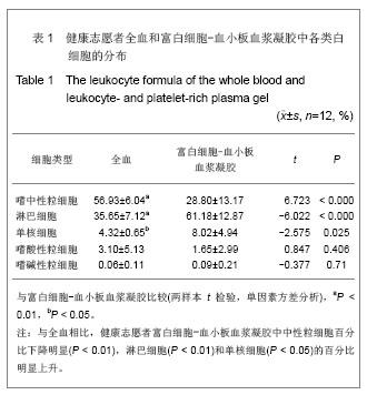
2.1 健康志愿者数量分析 共纳入对象12例,无中途退出者,最终均进入结果分析。 2.2 健康志愿者富白细胞-血小板血浆凝胶中血小板计数结果 12 例志愿者全血样本的血小板计数为(190.00±41.91)×109 L-1,富白细胞-血小板血浆凝胶的血小板计数为(812.96±334.53)×109 /L-1,两者比较差异有非常显著性意义(P < 0.01)。富白细胞-血小板血浆凝胶血小板浓缩倍数为(4.28±1.51)倍,富白细胞-血小板血浆凝胶血小板回收率为(62.43±19.87)%。 2.3 健康志愿者富白细胞-血小板血浆凝胶中白细胞计数及分类 12 例全血样本的白细胞总数为(5.42±1.43)×109 L-1,富白细胞血小板血浆凝胶的白细胞总数为(10.48±3.12)×109 L-1,两者比较差异有显著性意义(P < 0.01)。富白细胞-血小板血浆凝胶白细胞浓缩倍数为(1.93±0.63)倍,富白细胞-血小板血浆凝胶白细胞回收率为(28.92±7.83)%。富白细胞-血小板血浆凝胶中淋巴细胞所占比例最大,达到(61.18±12.87)%,与全血相比,健康志愿者富白细胞-血小板血浆凝胶中的嗜中性粒细胞百分比下降明显(P < 0.01),淋巴细胞 (P < 0.01)和单核细胞(P < 0.05)的百分比明显上升。见表1。"
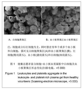
2.4 健康志愿者富白细胞-血小板血浆凝胶的超微结构 扫描电镜下观察可见,富白细胞-血小板血浆凝胶中除了纤维蛋白形成的粗细不等的网状结构外,细胞成分以红细胞为主,同时散在有单个或多个血小板和白细胞,偶可见白细胞聚集区和血小板聚集区,其中大部分白细胞为淋巴细胞。见图1。"
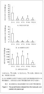
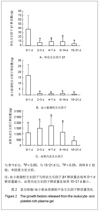
2.5 富白细胞-血小板血浆凝胶生长因子的释放量变化 富白细胞-血小板血浆凝胶的血小板源性生长因子与转化生长因子β1释放量相似,在培养0-1 d释放量最大,与其他时间段相比差异有显著性意义(P < 0.05),随后迅速下降,维持在较低水平。血管内皮生长因子表现为先逐渐上升,后缓慢下降,在培养4-7 d时达到峰值,但各时间段释放量比较,只有培养15-21 d与其他时间段差异有显著性意义(P < 0.05)。见图2。"
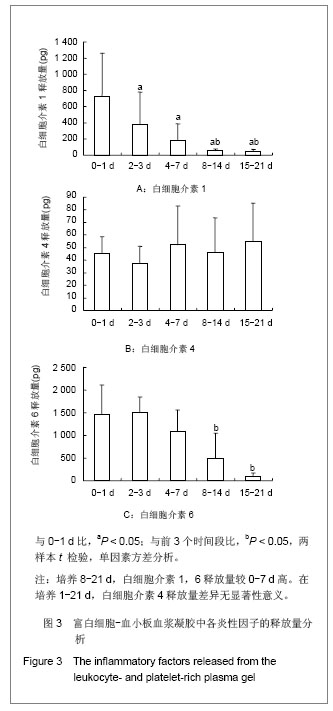
2.6 富白细胞-血小板血浆凝胶各时间段炎性因子的释放量 白细胞介素1释放量在0-1 d即达到峰值(P < 0.05),之后逐渐下降。白细胞介素6的释放量在培养0- 7 d均保持较高水平,培养8-21 d释放量明显下降,与培养0-7 d比较差异有显著性意义(P < 0.05)。白细胞介素1于培养0-1 d达到峰值(P < 0.05),之后逐渐下降;白细胞介素4各时间段释放量差异无显著性意义;白细胞介素6在培养0-7 d大量释放,随后明显下降(P < 0.05)。见图3。"
| [1]Uggeri J. Dose-dependent effects of platelet gel releasate on activities of human osteoblasts. Periodontol. 2007;78(10): 1985-1991.[2]Lindeboom JA. Influence of the application of platelet-enriched plasma in oral mucosal wound healing. Clin Oral Implants Res. 2007;18(1):133-139.[3]Rozman P, Bolta Z. Use of platelet growth factors in treating wounds and soft-tissue injuries. Acta Dermatovenerol Alp Panonica Adriat. 2007;16(4):156-165.[4]Thor A. Early bone formation in human bone grafts treated with platelet-rich plasma: preliminary histomorphometric results. Oral Maxillofac Surg. 2007;36(12):1164-1171.[5]Everts PA. Autologous platelet gel and fibrin sealant enhance the efficacy of total knee arthroplasty: improved range of motion, decreased length of stay and a reduced incidence of arthrofibrosis. Knee Surg Sports Traumatol Arthrosc. 2007; 15(7):888-894.[6]Gardner MJ, Demetrakopoulos D, Klepchick PR, et al. The efficacy of autologous platelet gel in pain control and blood loss in total knee arthroplasty. An analysis of the haemoglobin, narcotic requirement and range of motion. Int Orthop. 2007; 31(3):309-313.[7]Yuan NB, Long Y, Zhang XX, et al. Sichuan Daxue Xuebao: Yixueban. 2009;40(2):292-294.袁南兵,龙洋,张祥迅,等.自体富血小板凝胶治疗糖尿病难治性皮肤溃疡作用机制的初步探讨[J].四川大学学报:医学版, 2009, 40(2):292-294.[8]Eppley BL, Woodell JE, Higgins J. Platelet quantification and growth factor analysis from platelet-rich plasma: implications for wound healing. Plast Reconstr Surg. 2004;114(6): 1502-1508. [9]Yuan NB, Wang C, Wang Y, et al. Zhongguo Xiufu Chongjian Waike Zazhi. 2008;22(4):468-471.袁南兵,王椿,王艳,等.自体富血小板凝胶的制备及其生长因子分析[J].中国修复重建外科杂志,2008,22(4):468-471.[10]Bielecki TM, Gazdzik TS, Arendt J, et al. Antibacterial effect of autologous platelet gel enriched with growth factors and other active substances: an in vitro study. J Bone Joint Surg Br. 2007; 89(3):417-420.[11]Moojen DJ, Everts PA, Schure RM, et al. Antimicrobial activity of platelet-leukocyte gel against Staphylococcus aureus. J Orthop Res. 2008;26(3):404-410.[12]National People's Congress of the People's Republic of China. Blood Donation Law of the People's Republic of China.1997.[13]State Council of the People's Republic of China. Administrative Regulations on Medical Institution. 1994-09-01.[14]Sonnleitner D, Huemer P, Sullivan DY. A simplified technique for producing platelet-rich plasma and platelet concentrate for intraoral bone grafting techniques: a technical note. Int J Oral Maxillofac Implants. 2000;15(6):879-882.[15]Weibrich G. Comparison of platelet, leukocyte, and growth factor levels in point-of-care platelet-enriched plasma, prepared using a modified Curasan kit, with preparations received from a local blood bank. Clin Oral Implants Res. 2003;14(3):357-362.[16]Mazzucco L. Platelet-rich plasma and platelet gel preparation using Plateltex. Vox Sang. 2008;94(3):202-208.[17]Conaway JE. New trends in analytical technology and methods for pesticide residue analysis. J Assoc Off Anal Chem. 1991;74(5):715-717.[18]Wei D, Huang ZH, Zhao YP, et al. Anhui Nongye Kexue. 2009,37(6):2357-2358.魏东,黄智鸿,赵月平,等.浅析ELISA的基本原理与注意事项[J].安徽农业科学,2009,37(6):2357-2358.[19]Ehrenfest D M D, Del Corso M, Diss A, et al. Three-dimensional architecture and cell composition of a Choukroun's platelet-rich fibrin clot and membrane. J Periodontol. 2010;81(4):546-555.[20]Ma J, Li F, Ren DJ, et al. Zhongguo Kangfu Lilun yu Shijian. 2011;17(3):223-225.马健,李放,任大江,等.富含血小板血浆凝胶的超微结构[J].中国康复理论与实践,2011,17(3):223-225.[21]He L, Lin Y, Hu X, et al. A comparative study of platelet-rich fibrin (PRF) and platelet-rich plasma (PRP) on the effect of proliferation and differentiation of rat osteoblasts in vitro. Oral Surg Oral Med Oral Pathol Oral Radiol Endod. 2009;108(5): 707-713.[22]Lee TH, Seng S, Li H, et al. Integrin regulation by vascular endothelial growth factor in human brain microvascular endothelial cells: role of alpha6beta1 integrin in angiogenesis. J Biol Chem. 2006;281(52):40450-40460.[23]Ferrara N. Role of vascular endothelial growth factor in regulation of physiological angiogenesis. Am J Physiol Cell Physiol. 2001;280(6):C1358-1366.[24]Tiong A, Freedman SB. Gene therapy for cardiovascular disease: the potential of VEGF. Curr Opin Mol Ther. 2004; 6(2):151-159.[25]Zou ZZ, Li JC. Beijing: People's Medical Publishing House. 2008. 邹仲之,李继承.组织学与胚胎学[M].北京:人民卫生出版社, 2008.[26]Dohan ED, Bielecki T, Jimbo R, et al. Do the fibrin architecture and leukocyte content influence the growth factor release of platelet concentrates? An evidence-based answer comparing a pure platelet-rich plasma (P-PRP) gel and a leukocyte- and platelet-rich fibrin (L-PRF). Curr Pharm Biotechnol. 2012; 13(7): 1145-1152.[27]Dohan DM, Choukroun J, Diss A, et al. Platelet-rich fibrin (PRF): a second-generation platelet concentrate. Part III: leucocyte activation: a new feature for platelet concentrates? Oral Surg Oral Med Oral Pathol Oral Radiol Endod. 2006; 101(3):e51-55.[28]Yang YZ, Liu HC, Liu GJ, et al. Zhongguo Xiufu Chongjian Waike Zazhi. 2010;24(5):571-576.杨阎峙,刘衡川,刘关键,等.健康志愿者自体富血小板凝胶体外抑菌作用研究[J].中国修复重建外科杂志,2010,24(5):571-576.[29]Pan LH. Yixue Zongshu. 2005;11(9):775-777.潘灵辉.细胞因子平衡在炎症反应中作用的研究进展[J].医学综述,2005,11(9):775-777.[30]Park WY, Goodman RB, Steinberg KP, et al. Cytokine balance in the lungs of patients with acute respiratory distress syndrome. Am J Respir Crit Care Med. 2001;164(10 Pt 1): 1896-1903. |
| [1] | Li Li, Ma Li. Immobilization of lactase on magnetic chitosan microspheres and its effect on enzymatic properties [J]. Chinese Journal of Tissue Engineering Research, 2021, 25(4): 576-581. |
| [2] | Zhang Zhenkun, Li Zhe, Li Ya, Wang Yingying, Wang Yaping, Zhou Xinkui, Ma Shanshan, Guan Fangxia. Application of alginate based hydrogels/dressings in wound healing: sustained, dynamic and sequential release [J]. Chinese Journal of Tissue Engineering Research, 2021, 25(4): 638-643. |
| [3] | Liu Fang, Shan Zhengming, Tang Yulei, Wu Xiaomin, Tian Weiqun. Effects of hemostasis and promoting wound healing of ozone sustained-release hydrogel [J]. Chinese Journal of Tissue Engineering Research, 2021, 25(22): 3445-3449. |
| [4] | Li Xinping, Cui Qiuju, Zeng Shuguang, Ran Gaoying, Zhang Zhaoqiang, Liu Xianwen, Fang Wei, Xu Shuaimei. Effect of modification of β-tricalcium phosphate/chitosan hydrogel on growth and mineralization of dental pulp stem cells [J]. Chinese Journal of Tissue Engineering Research, 2021, 25(22): 3493-3499. |
| [5] | Liu Liyong, Zhou Lei. Research and development status and development trend of hydrogel in tissue engineering based on patent information [J]. Chinese Journal of Tissue Engineering Research, 2021, 25(22): 3527-3533. |
| [6] | Zhou Anqi, Tang Yufei, Wu Bingfeng, Xiang Lin. Designing of periosteum tissue engineering: combination of generality and individuality [J]. Chinese Journal of Tissue Engineering Research, 2021, 25(22): 3551-3557. |
| [7] | Gan Lili, Xiong Na, Liu Yanfei. Hydrogel as drug scaffold in skin wound repair: challenges of clinical application possibilities [J]. Chinese Journal of Tissue Engineering Research, 2021, 25(22): 3578-3583. |
| [8] | Lang Limin, He Sheng, Jiang Zengyu, Hu Yiyi, Zhang Zhixing, Liang Minqian. Application progress of conductive composite materials in the field of tissue engineering treatment of myocardial infarction [J]. Chinese Journal of Tissue Engineering Research, 2021, 25(22): 3584-3590. |
| [9] | Jiang Shengyuan, Li Dan, Jiang Jianhao, Shang-you Yang, Yang Shuye. Biological response of Co2+ to preosteoblasts during aseptic loosening of the prosthesis [J]. Chinese Journal of Tissue Engineering Research, 2021, 25(21): 3292-3299. |
| [10] | Zhang Jianhui, Ma Heran, Tan Yi, Wang Zhihui. Knee injury repair using human adipose-derived mesenchymal stem cells-based scaffold-free three-dimensional gel-like construct in pigs [J]. Chinese Journal of Tissue Engineering Research, 2021, 25(19): 2969-2975. |
| [11] | Chen Pu, Ruan Anmin, Zhou Jun, Zhang Xiaozhe, Ma Yufeng, Zong Chenzhong, Wang Qingpu. Effect of Tongluo Analgesic Gel on cartilage inflammation and degeneration in a rabbit model of knee osteoarthritis [J]. Chinese Journal of Tissue Engineering Research, 2021, 25(17): 2670-2675. |
| [12] | Chen Siyu, Li Yannan, Xie Liying, Liu Siqi, Fan Yurong, Fang Changxing, Zhang Xin, Quan Jiayu, Zuo Lin. Thermosensitive chitosan-collagen composite hydrogel loaded with basic fibroblast growth factor retards ventricular remodeling after myocardial infarction in mice [J]. Chinese Journal of Tissue Engineering Research, 2021, 25(16): 2472-2478. |
| [13] | Liu Feng, Zhang Yu, Wang Yanli, Luo Wei, Han Chaoshan, Li Yangxin. Application of temperature-sensitive chitosan hydrogel encapsulated exosomes in ischemic diseases [J]. Chinese Journal of Tissue Engineering Research, 2021, 25(16): 2479-2487. |
| [14] | Zhang Xin, Lu Ying, Yao Qingqiang, Zhu Yishen. Preparation and in vitro evaluation of self-assembling peptide hydrogel loaded with doxorubicin [J]. Chinese Journal of Tissue Engineering Research, 2021, 25(16): 2488-2493. |
| [15] | Xie Jian, Su Jiansheng. Advantages and characteristics of electrospun aligned nanofibers as scaffolds for tissue engineering [J]. Chinese Journal of Tissue Engineering Research, 2021, 25(16): 2575-2581. |
| Viewed | ||||||
|
Full text |
|
|||||
|
Abstract |
|
|||||