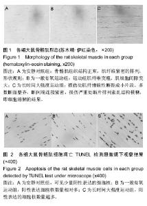
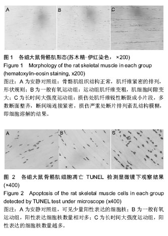
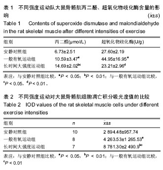
| [1] Kerr JF, Wyllie AH, Currie AR.Apoptosis: a basic biological phenomenon with wide-ranging implications in tissue kinetics. Br J Cancer.1972;26: 239-257.[2] Schoser BG, Wehling S, Blottner D. Cell death and apoptosis-related proteins in muscle biopsies of sporadic amyotrophic lateral sclerosis and polyneuropathy. Muscle Nerve. 2001;24:1083-1089.[3] 郑澜,潘珊珊,张胜年.骨骼肌细胞凋亡研究进展[J].中国运动医学杂志,2000,19(3):301-303.[4] 谢昆,李兴权. 细胞凋亡的信号通路[J]. 山东农业大学学报:自然科学版,2015,46(04):514-518. [5] 张颖, 梁美杨. 8周有氧运动对大鼠骨骼肌细胞凋亡的影响[J].哈尔滨体育学院学报, 2015,33(2):87-91.[6] 张梦颖,吕志跃,吴忠道.细胞凋亡发生机制研究进展[J].热带医学杂志,2016,16(10):1346-1349+1352.[7] 韩奇鹏,罗玲,揭红东,等. 细胞凋亡及其调控因子研究进展[J]. 饲料博览,2016,(4):9-13. [8] 蔡保塔,余斌,赵江萍,等. 不同运动强度与骨骼肌细胞凋亡的时序性实验研究[J]. 南方医科大学学报,2006,26(7):1017-1019.[9] 刘丽萍. 细胞凋亡与运动训练关系的研究进展[J]. 河北体育学院学报, 2000,14(1):75-80.[10] 李莎,缪律,林缘西. 运动训练与细胞自噬、细胞凋亡的分子机制[J]. 南京体育学院学报:自然科学版,2016,(1):27-34.[11] 金其贯. 慢性力竭性训练对大鼠骨骼肌细胞凋亡的影响[J].体育与科学,1999,20(5):23-28+56.[12] 金其贯,冯美云.运动与细胞凋亡的研究进展[J]. 西安体育学院学报, 2001,21(3):30-34+40.[13] Tezini GC, Silveira LC, Maida KD, et al. The effect of ovariectomy on cardiac autonomic control in rats submitted to aerobic physical training. Auton Neurosci. 2008;143(1):5-11.[14] 胡亚哲,王姝玉,王和平,等.不同强度运动大鼠心肌细胞凋亡及一氧化氮合酶变化[J]. 中国运动医学杂志,2007,26(2): 222-225+ 254.[15] 田振军,熊正英,郭进,等.大鼠游泳过度训练模型的建立及心室构型改建研究[J]. 陕西师范大学学报:自然科学版,1996,(3): 108-113. [16] 王爱莲,史晓燕.统计学[M].西安:西安交通大学出版社, 2010: 212.[17] 谢康玲,刘遂心,蔡颖,等. 长期中等强度运动对小鼠骨骼肌HIF-1α mRNA的表达及葡萄糖转运的影响[J].中国康复医学杂志, 2012,27(6):514-518.[18] 刘军. Ebselen预处理对大强度耐力训练大鼠骨骼肌结构和功能的保护及可能机制研究[J].体育科学,2014,34(8):91-98.[19] 陈英杰,郭庆芳.延迟性肌肉损伤与自由基代谢异常[J].中国运动医学杂志, 1993, 12(2): 65-69.[20] 赵甜甜. 紫甘薯花青素对过度训练大鼠骨骼肌氧化损伤保护作用的研究[D].江苏师范大学,2013[21] Gore M, Fiebig R, Hollander J. Endurance training alters antioxidant enzyme gene expression in rat skeletal muscle. Can J Physiol Pharmacol.1998;76(12):1139-1145.[22] 聂园园,仇小强. 心肌细胞凋亡与心血管疾病关系的研究进展[J].实用医学杂志,2012,28(2):324-326.[23] 袁海燕,胡亚哲.不同强度有氧运动对大鼠肝细胞的影响[J].中国组织工程研究,2015,19(51):8311-8316.[24] Munsch D, Watanabe-Fukunaga R, Bourdon JC, et al.Human and mouse Fas (APO-1/CD95) death receptor genes each contain a p53-responsive element that is activated by p53 mutants unable to induce apoptosis. J Biol Chem.2000;275(6): 3867-3872. [25] Volk T, Hensel M, Kox WJ. Transient Ca2+ changes in endothelial cells induced by low doses of reactive oxygen species: role of hydrogen peroxide. Mol Cell Biochem.1997; 171(1-2):11-21. [26] 任小双,张兰,张燕宁,等. 昆虫细胞凋亡的线粒体通路研究进展[J]. 安徽农业科学,2017,45(3):167-170+189.[27] 向军军,赖菁菁,胡跃强. 近3年线粒体介导细胞凋亡的研究进展[J]. 中西医结合心脑血管病杂志,2016,14(13):1497-1499. [28] 李兴太,刘德文,张新,等. 线粒体通透性转换与细胞凋亡[J].中国公共卫生,2015,31(4):529-532.[29] 王佺荃,张文丽,元小冬,等. 线粒体在细胞凋亡中的作用[J]. 中国医学创新,2015,12(06):143-146.[30] Cain K, Bratton SB, Langlais C, et al. Apaf-1 oligomerizes into biologically active approximately 700-kDa and inactive approximately1.4-MDa apoptosome complexes. J Biol Chem. 2000;275(9):6067-6070. [31] Attisano L, Wrana JL. Signal transduction by the TGF-beta superfamily. Science.2002;296(5573):1646-1647. [32] Munsch D, Watanabefukunaga R, Bourdon J, et al. Human and Mouse Fas (APO-1/CD95) Death Receptor Genes Each Contain a p53-responsive Element That Is Activated by p53 Mutants Unable to Induce Apoptosis. J Biol Chem. 2000; 275(6):3867-3872.[33] 王长青,李雷,郑师陵. 游泳训练后大鼠骨骼肌细胞自由基代谢、线粒体膜电位变化与细胞凋亡的关系[J]. 中国运动医学杂志, 2002,21(3): 256-260.[34] 黄文聪,阮利民,王安利.不同负荷游泳训练对大鼠骨骼肌HSP70应答及其脂质过氧化的影响[J]. 体育科学,2008,28(3):63-66+ 78.[35] 李烨,王逸安,张君,等. 丙二醛对体外培养下小鼠骨髓间充质干细胞凋亡的诱导作用[J]. 生命科学研究,2014,18(2):129-133.[36] Munsch D, Watanabefukunaga R, Bourdon J, et al. Human and Mouse Fas (APO-1/CD95) Death Receptor Genes Each Contain a p53-responsive Element That Is Activated by p53 Mutants Unable to Induce Apoptosis. J Biol Chem. 2000; 275(6):3867-3872.[37] 韩宁. 大鼠脑出血模型中神经细胞凋亡与自由基水平的相关性研究[D].第二军医大学,2007. |