Chinese Journal of Tissue Engineering Research ›› 2018, Vol. 22 ›› Issue (4): 593-599.doi: 10.3969/j.issn.2095-4344.0096
Previous Articles Next Articles
Three-dimensional reconstructed human skin model: a powerful tool to study skin homeostasis and epidermal basement membrane morphogenesis
Jiang Dan-dan, Morgan Dos Santos
- JALA R&D Center, Shanghai 200233, China
-
Received:2017-11-23Online:2018-02-08Published:2018-02-08 -
Contact:Jiang Dan-dan, JALA R&D Center, Shanghai 200233, China -
About author:Jiang Dan-dan, Master, JALA R&D Center, Shanghai 200233, China -
Supported by:the Shanghai Special Fund of Capacity Building Project for Enterprise Technology Center, No. J-2013-45
CLC Number:
Cite this article
Jiang Dan-dan, Morgan Dos Santos. Three-dimensional reconstructed human skin model: a powerful tool to study skin homeostasis and epidermal basement membrane morphogenesis[J]. Chinese Journal of Tissue Engineering Research, 2018, 22(4): 593-599.
share this article
Add to citation manager EndNote|Reference Manager|ProCite|BibTeX|RefWorks
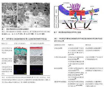
2.1 表皮基底膜的结构和分子组成 表皮基底膜区域可分为4部分结构,图1为正常人皮肤组织中表皮基底膜的透射电镜图片,图中从表皮到真皮的方向依次是:与角质细胞紧紧相邻的胞膜层(plasma membrane)、透明层"
| [1] Ko MS, Marinkovich MP. Role of dermal-epidermal basement membrane zone in skin, cancer, and developmental disorders. Dermatol Clin. 2010;28(1):1-16.[2] Martins VL, Vyas JJ, Chen M, et al. Increased invasive behaviour in cutaneous squamous cell carcinoma with loss of basement-membrane type VII collagen. J Cell Sci. 2009; 122(Pt 11):1788-1799.[3] Hagedorn EJ, Sherwood DR. Cell invasion through basement membrane the anchor cell breaches the barrier. Curr Opin Cell Biol. 2011;23(5):589-596. [4] Whitelock JM, Melrose J, Iozzo RV. Diverse cell signaling events modulated by perlecan. Biochemistry. 2008; 47(43): 11174-11183.[5] Löffek S, Hurskainen T, Jackow J, et al. Transmembrane collagen XVII modulates integrin dependent keratinocyte migration via PI3K/Rac1 signaling. PloS One. 2014; 9(2): e87263.[6] Yurchenco PD. Basement membranes: cell scaffoldings and signaling platforms. Cold Spring Harb Perspect Biol. 2011;3(2). pii: a004911.[7] Kruegel J, Miosge N. Basement membrane components are key players in specialized extracellular matrices. Cell Mol Life Sci. 2010;67(17):2879-2895.[8] Sher I, Zisman-Rozen S, Eliahu L, et al. Targeting perlecan in human keratinocytes reveals novel roles for perlecan in epidermal formation. J Biol Chem. 2006; 281(8):5178-5187.[9] Breitkreutz D, Koxholt I, Thiemann K, et al. Skin Basement Membrane: The Foundation of Epidermal Integrity—BM Functions and Diverse Roles of Bridging Molecules Nidogen and Perlecan. Biomed Res Int. 2013;2013:179784.[10] Chaudhari PR, Vaidya MM. Versatile hemidesmosomal linker proteins: structure and function. Histol Histopathol. 2015; 30(4):425-434.[11] Winograd-Katz SE, Fässler R, Geiger B, et al. The integrin adhesome: from genes and proteins to human disease. Nat Rev Mol Cell Biol; 2014; 15(4):273-288.[12] Rognoni E, Ruppert R, Fässler R. The kindlin family: functions, signaling properties and implications for human disease. J Cell Sci. 2016; 129(1):17-27.[13] Ozawa T, Tsuruta D, Jones JC, et al. Dynamic relationship of focal contacts and hemidesmosome protein complexes in live cells. J Invest Dermatol. 2010; 130(6):1624-1635.[14] Hohenester E, Yurchenco PD. Laminins in basement membrane assembly. Cell Adh Migr. 2013; 7(1):56-63.[15] Vidal EM. The Basement Membrane Zone: Making the Connection. 2013. https://www.aad.org/File%20Library/ Main%20navigation/Education/Basement%20Membrane%20Zone/The-Basement-Membrane-Zone--Textbook--pdf.pdf[16] Turcan I, Jonkman MF. Blistering disease: insight from the hemidesmosome and other components of the dermal-epidermal junction. Cell Tissue Res. 2015; 360(3):545-569.[17] Geary SM, Cowin AJ, Copeland B, et al. The role of the tetraspanin CD151 in primary keratinocyte and fibroblast functions: implications for wound healing. Exp Cell Res. 2008; 314(11-12):2165-2175.[18] Veit G, Zwolanek D, Eckes B, et al. Collagen XXIII, novel ligand for integrin alpha2beta1 in the epidermis. J Biol Chem. 2011; 286(31):27804-27813.[19] Truong T, Shams H, Mofrad MR. Mechanisms of integrin and filamin binding and their interplay withCollagen XXIII, novel ligand for integrin alpha2beta1 in the epidermis talin during early focal adhesion formation. Integr Biol (Camb). 2015; 7(10):1285-1296.[20] Nader GP, Ezratty EJ, Gundersen GG. FAK, talin and PIPKIγ regulate endocytosed integrin activation to polarize focal adhesion assembly. Nat Cell Biol. 2016; 18(5):491-503.[21] Nyström A, Velati D, Mittapalli VR, et al. Collagen VII plays a dual role in wound healing. J Clin Invest. 2013; 123(8): 3498-3509.[22] Bruckner-Tuderman L, Has C. Disorders of the cutaneous basement membrane zone—The paradigm of epidermolysis bullosa. Matrix Biol. 2014;33:29-34. [23] Has C, Nyström A. Epidermal basement membrane in health and disease. Curr Top Membr. 2015; 76:117-170. [24] Kasperkiewicz M, Sadik CD, Bieber K, et al. Epidermolysis Bullosa Acquisita: From Pathophysiology to Novel Therapeutic Options. J Invest Dermatol. 2016; 136(1):24-33.[25] Nishie W. Update on the pathogenesis of bullous pemphigoid: an autoantibody-mediated blistering disease targeting collagen XVII. J Dermatol Sci. 2014; 73(3):179-186.[26] Yamase A, Kono T, Ishii N, et al. An autoimmune bullous dermatosis with clinical, histopathological, and immunological features of bullous pemphigoid and epidermolysis bullosa acquisita in an adult. Br J Dermatol. 2016.[27] Masunaga T. Epidermal basement membrane, its molecular organization and blistering disorders. Connect Tissue Res. 2006;47(2):55-66.[28] Fine JD, Bruckner-Tuderman L, Eady RA, et al. Inherited epidermolysis bullosa: updated recommendations on diagnosis and classification. J Am Acad Dermatol. 2014; 70(6):1103-1126.[29] Has C, Kiritsi D. Therapies for inherited skin fragility disorders. Exp Dermatol. 2015; 24(5):325-331.[30] Hsu CK, Wang SP, Lee JY, et al. Treatment of hereditary epidermolysis bullosa: updates and future prospects. Am J Clin Dermatol. 2014;15(1):1-6.[31] Zouboulis CC, Makrantonaki E. Clinical aspects and molecular diagnostics of skin aging. Clin Dermatol. 2011; 29(1):3-14.[32] Liao YH, Kuo WC, Chou SY, et al. Quantitative analysis of intrinsic skin aging in dermal papillae by in vivo harmonic generation microscopy. Biomed Opt Express. 2014; 5(9): 3266-3279.[33] Mondon P, Hillion M, Peschard O, et al. Evaluation of dermal extracellular matrix and epidermal-dermal junction modifications using matrix-assisted laser desorption/ionization mass spectrometric imaging, in vivo reflectance confocal microscopy, echography, and histology: effect of age and peptide applications. J Cosmet Dermatol. 2015; 14(2): 152-160.[34] Feru J, Delobbe E, Ramont L, et al. Aging decreases collagen IV expression in vivo in the dermo-epidermal junction and in vitro in dermal fibroblasts: possible involvement of TGF-β1. Eur J Dermatol. 2016;26(4):350-360.[35] Fisher G, Rittié L. Restoration of the basement membrane after wounding: a hallmark of young human skin altered with aging. J Cell Commun Signal. 2017.[36] Dos Santos M, Michopoulou A, André-Frei V, et al. Perlecan expression influences the keratin 15-positive cell population fate in the epidermis of aging skin. Aging (Albany NY). 2016; 8(4):751-768.[37] Tsunenaga M. Heparanase inhibitors facilitate the assembly of the basement membrane in artificial skin. Curr Tissue Eng. 2016; 5(2):113-122.[38] Nicholas MN, Jeschke MG, Amini-Nik S. Methodologies in creating skin substitutes. Cell Mol Life Sci. 2016; 73(18): 3453-3472.[39] Nyame TT, Chiang HA, Leavitt T, et al. Tissue-Engineered Skin Substitutes. Plast Reconstr Surg. 2015;136(6): 1379-1388.[40] Schlotmann K, Kaeten M, Black AF, et al. Cosmetic efficacy claims in vitro using a three-dimensional human skin model. Int J Cosmet Sci. 2001;23(5):309-318.[41] Auxenfans C, Fradette J, Lequeux C, et al. Evolution of three dimensional skin equivalent models reconstructed in vitro by tissue engineering. Eur J Dermatol. 2009;19(2):107-113. [42] Nishiyama T, Amano S, Tsunenaga M, et al. The importance of laminin 5 in the dermal-epidermal basement membrane. J Dermatol Sci. 2000;24 Suppl 1:S51-59.[43] Dos Santos M, Metral E, Boher A, et al. In vitro 3-D model based on extending time of culture for studying chronological epidermis aging. Matrix Biol. 2015; 47:85-97.[44] Pageon H, Bakala H, Monnier VM, et al. Collagen glycation triggers the formation of aged skin in vitro. Eur J Dermatol. 2007;17(1):12-20.[45] Pageon H, Técher MP, Asselineau D. Reconstructed skin modified by glycation of the dermal equivalent as a model for skin aging and its potential use to evaluate anti-glycation molecules. Exp Gerontol. 2008;43(6):584-588.[46] Amano S, Akutsu N, Matsunaga Y, et al. Importance of balance between extracellular matrix synthesis and degradation in basement membrane formation. Exp Cell Res. 2001;271(2):249-262.[47] Amano S, Ogura Y, Akutsu N, et al. Protective effect of matrix metalloproteinase inhibitors against epidermal basement membrane damage: skin equivalents partially mimic photoageing process. Br J Dermatol. 2005; 153 Suppl 2: 37-46.[48] Iriyama S, Tsunenaga M, Amano S. Key role of heparan sulfate chains in assembly of anchoring complex at the dermal-epidermal junction. Exp Dermatol. 2011; 20(11): 953-955.[49] Varkey M, Ding J, Tredget EE. Superficial dermal fibroblasts enhance basement membrane and epidermal barrier formation in tissue-engineered skin: implications for treatment of skin basement membrane disorders. Tissue Eng Part A. 2014; 20(3-4):540-552.[50] Wojtowicz AM, Oliveira S, Carlson MW, et al. The importance of both fibroblasts and keratinocytes in a bilayered living cellular construct used in wound healing. Wound Repair Regen. 2014; 22(2):246-255.[51] Phase I/II ex vivo gene therapy clinical trial for recessive dystrophic epidermolysis bullosa using skin equivalent grafts genetically corrected with a COL7A1-encoding SIN retroviral vector (GENEGRAFT). Hum Gene Ther Clin Dev. 2014; 25(2):65-66.[52] Hirsch T, Rothoeft T, Teig N, et al. Regeneration of the entire human epidermis using transgenic stem cells. Nature. 2017; 551(7680):327-332.[53] Bernerd F, Asselineau D. An organotypic model of skin to study photodamage and photoprotection in vitro. J Am Acad Dermatol. 2008; 58(5 Suppl 2):S155-159.[54] Bernerd F. Human skin reconstructed in vitro as a model to study the keratinocyte, the fibroblast and their interactions: photodamage and repair processes. J Soc Biol. 2005; 199(4):313-320.[55] Ponec M, Kempenaar J. Use of human skin recombinants as an in vitro model for testing the irritation potential of cutaneous irritants. Skin Pharmacol. 1995; 8(1-2):49-59.[56] Dickson MA, Hahn WC, Ino Y, et al. Human keratinocytes that express hTERT and also bypass a p16(INK4a)-enforced mechanism that limits life span become immortal yet retain normal growth and differentiation characteristics. Mol Cell Biol. 2000; 20(4):1436-1447.[57] Reijnders CM, van Lier A, Roffel S, et al. Development of a full-thickness human skin equivalent in vitro model derived from TERT-immortalized keratinocytes and fibroblasts. Tissue Eng Part A. 2015; 21(17-18):2448-2459. |
| [1] | Pu Rui, Chen Ziyang, Yuan Lingyan. Characteristics and effects of exosomes from different cell sources in cardioprotection [J]. Chinese Journal of Tissue Engineering Research, 2021, 25(在线): 1-. |
| [2] | Zhang Tongtong, Wang Zhonghua, Wen Jie, Song Yuxin, Liu Lin. Application of three-dimensional printing model in surgical resection and reconstruction of cervical tumor [J]. Chinese Journal of Tissue Engineering Research, 2021, 25(9): 1335-1339. |
| [3] | Zhang Yu, Tian Shaoqi, Zeng Guobo, Hu Chuan. Risk factors for myocardial infarction following primary total joint arthroplasty [J]. Chinese Journal of Tissue Engineering Research, 2021, 25(9): 1340-1345. |
| [4] | Zhang Chao, Lü Xin. Heterotopic ossification after acetabular fracture fixation: risk factors, prevention and treatment progress [J]. Chinese Journal of Tissue Engineering Research, 2021, 25(9): 1434-1439. |
| [5] | Zhou Jihui, Li Xinzhi, Zhou You, Huang Wei, Chen Wenyao. Multiple problems in the selection of implants for patellar fracture [J]. Chinese Journal of Tissue Engineering Research, 2021, 25(9): 1440-1445. |
| [6] | Wang Debin, Bi Zhenggang. Related problems in anatomy mechanics, injury characteristics, fixed repair and three-dimensional technology application for olecranon fracture-dislocations [J]. Chinese Journal of Tissue Engineering Research, 2021, 25(9): 1446-1451. |
| [7] | Chen Junming, Yue Chen, He Peilin, Zhang Juntao, Sun Moyuan, Liu Youwen. Hip arthroplasty versus proximal femoral nail antirotation for intertrochanteric fractures in older adults: a meta-analysis [J]. Chinese Journal of Tissue Engineering Research, 2021, 25(9): 1452-1457. |
| [8] | Li Jing, Xie Jianshan, Cui Huilin, Cao Ximei, Yang Yanping, Li Hairong. Expression and localization of diacylglycerol kinase zeta and protein kinase C beta II in mouse back skin with different coat colors [J]. Chinese Journal of Tissue Engineering Research, 2021, 25(8): 1196-1200. |
| [9] | Ji Zhixiang, Lan Changgong. Polymorphism of urate transporter in gout and its correlation with gout treatment [J]. Chinese Journal of Tissue Engineering Research, 2021, 25(8): 1290-1298. |
| [10] | Yuan Mei, Zhang Xinxin, Guo Yisha, Bi Xia. Diagnostic potential of circulating microRNA in vascular cognitive impairment [J]. Chinese Journal of Tissue Engineering Research, 2021, 25(8): 1299-1304. |
| [11] | Wang Xianyao, Guan Yalin, Liu Zhongshan. Strategies for improving the therapeutic efficacy of mesenchymal stem cells in the treatment of nonhealing wounds [J]. Chinese Journal of Tissue Engineering Research, 2021, 25(7): 1081-1087. |
| [12] | Wan Ran, Shi Xu, Liu Jingsong, Wang Yansong. Research progress in the treatment of spinal cord injury with mesenchymal stem cell secretome [J]. Chinese Journal of Tissue Engineering Research, 2021, 25(7): 1088-1095. |
| [13] | Liao Chengcheng, An Jiaxing, Tan Zhangxue, Wang Qian, Liu Jianguo. Therapeutic target and application prospects of oral squamous cell carcinoma stem cells [J]. Chinese Journal of Tissue Engineering Research, 2021, 25(7): 1096-1103. |
| [14] | Zhao Min, Feng Liuxiang, Chen Yao, Gu Xia, Wang Pingyi, Li Yimei, Li Wenhua. Exosomes as a disease marker under hypoxic conditions [J]. Chinese Journal of Tissue Engineering Research, 2021, 25(7): 1104-1108. |
| [15] | Xie Wenjia, Xia Tianjiao, Zhou Qingyun, Liu Yujia, Gu Xiaoping. Role of microglia-mediated neuronal injury in neurodegenerative diseases [J]. Chinese Journal of Tissue Engineering Research, 2021, 25(7): 1109-1115. |
| Viewed | ||||||
|
Full text |
|
|||||
|
Abstract |
|
|||||