Chinese Journal of Tissue Engineering Research ›› 2024, Vol. 28 ›› Issue (10): 1540-1546.doi: 10.12307/2024.248
Previous Articles Next Articles
In vitro experiment of stem cell engineered two-sided anisotropic electrospun membranes for promoting dural repair
Xu Jingzhi, Wang Wenbo, Sun Huiwen, Gu Yong
- Department of Orthopedics, First Affiliated Hospital of Soochow University, Suzhou 215000, Jiangsu Province, China
-
Received:2022-11-16Accepted:2023-02-08Online:2024-04-08Published:2023-08-19 -
Contact:Gu Yong, Associate chief physician, Associate professor, Master’s supervisor, Department of Orthopedics, First Affiliated Hospital of Soochow University, Suzhou 215000, Jiangsu Province, China -
About author:Xu Jingzhi, Master candidate, Department of Orthopedics, First Affiliated Hospital of Soochow University, Suzhou 215000, Jiangsu Province, China Wang Wenbo, Physician, Department of Orthopedics, First Affiliated Hospital of Soochow University, Suzhou 215000, Jiangsu Province, China -
Supported by:National Natural Science Foundation of China, No. 82072438 (to GY); Jiangsu Provincial Outstanding Youth Fund, No. BK20211504 (to GY)
CLC Number:
Cite this article
Xu Jingzhi, Wang Wenbo, Sun Huiwen, Gu Yong. In vitro experiment of stem cell engineered two-sided anisotropic electrospun membranes for promoting dural repair[J]. Chinese Journal of Tissue Engineering Research, 2024, 28(10): 1540-1546.
share this article
Add to citation manager EndNote|Reference Manager|ProCite|BibTeX|RefWorks

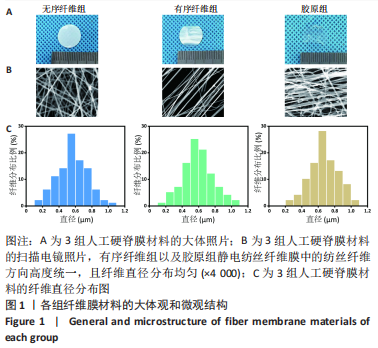
2.1 人工硬脊膜的表面特征 从大体上看,相较于无序纤维组,有序纤维组能明显看到表面纵行的拓扑结构,胶原组纤维膜由于胶原的自组装表面变得湿润透明,见图1A。扫描电镜下可见,有序纤维组以及胶原组静电纺丝纤维膜中的纺丝纤维方向高度统一,且纤维直径分布均匀,无序纤维组、有序纤维组、胶原组的纤维直径分别为(0.55±0.02),(0.57±0.03),(0.64±0.06) μm,见图1B,C,其中胶原组纤维膜可见树枝状的纳米级线性胶原纤维缠绕在聚乳酸纤维表面。"
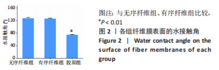
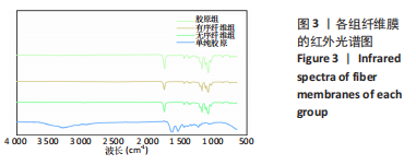
无序纤维组及有序纤维组纤维膜表面水接触角为120°左右,经胶原蛋白组装后,胶原组纤维膜表面水接触角为(54.02±9.83)°,表面亲水性能显著提高,见图2。"
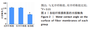

傅里叶变换红外光谱提示,单纯的Ⅰ型胶原有2个主要的特征带,分别在1 650 cm-1处有酰胺Ⅰ(C=O)带,在1 550 cm-1处有酰胺Ⅱ(N-H)带;胶原蛋白与聚乳酸纤维自组装后在胶原组纤维表面也发现了这2个特征峰,见图3,证实了胶原蛋白的成功组装。同时通过能谱仪表面元素测定得出,聚乳酸纤维膜主要含有C、O两种元素,而胶原组纤维膜增加了N元素,见图4,再次证明了胶原蛋白的成功组装。"
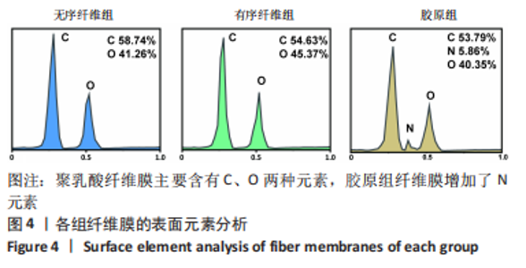
"
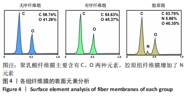

2.2 人工硬脊膜的力学拉伸实验结果 由于胶原蛋白的加入,3组纤维膜的力学强度出现部分差异。从三者的应力-应变曲线可以得出,无序纤维组、有序纤维组及胶原组纤维膜的断裂强度分别为(3.78±0.17),(3.65±0.21),(5.44±0.22) MPa,断裂伸长率分别为(47.42±1.66)%,(47.17±2.48)%和(58.52±1.48)%,进一步计算曲线的斜率可以得到3组材料的弹性模量分别为(0.24±0.02),(0.22±0.01)和(0.27±0.02) MPa,见图5。通过以上结果可以得出,含有胶原蛋白的胶原组纤维膜比单纯聚乳酸纤维膜的力学强度有所提高。"
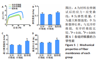
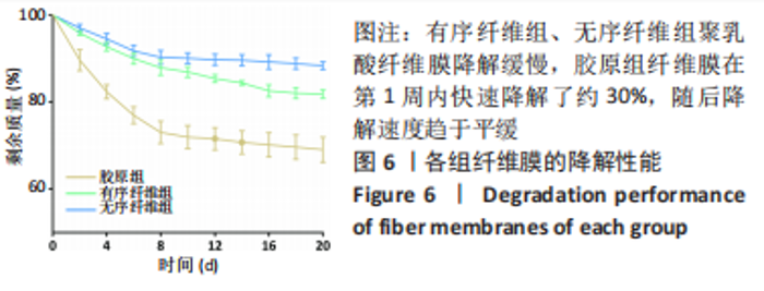
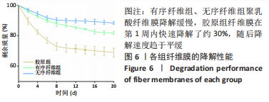
2.3 人工硬脊膜的降解曲线 序纤维组、无序纤维组聚乳酸纤维膜降解缓慢,在3周内降解约20%,而胶原组纤维膜在第1周内快速降解了约30%,随后降解速度趋于平缓(图6),这是由于单纯的胶原蛋白降解性能优于聚乳酸纤维,能在早期快速降解。"
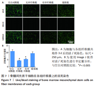
2.4 人工硬脊膜与骨髓间充质干细胞共培养实验结果 2.4.1 活/死染色 由图7可见,3组材料以及空白对照组都可以看见密度均匀的活细胞(绿色荧光),其中胶原组和空白对照组的荧光强度明显高于无序及有序纤维组,且胶原组和空白对照组红色荧光强度(即死亡细胞)均显著弱于无序及有序纤维组,展现出优异的生物相容性。"

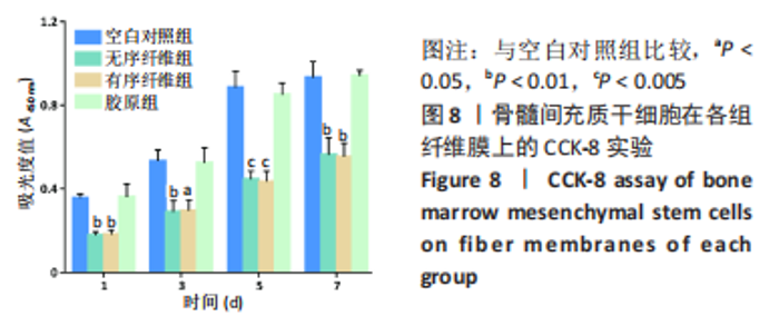
2.4.2 CCK-8实验 各组细胞的吸光度值均随着时间的延长持续增长,综合1,3,5 d的CCK-8结果可以得出:在3个不同时间点,空白对照组的吸光度值均稍高于胶原组,两组比较差异无显著性意义(P > 0.05),无序及有序纤维组的细胞吸光度值明显低于胶原组及空白对照组(P < 0.05,P < 0.01,P < 0.005 ),见图8。"

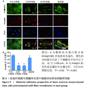
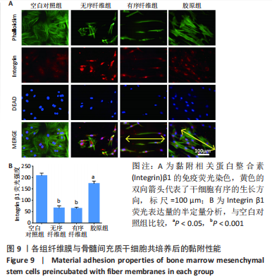
2.4.3 黏附性能评估 通过整合素β1荧光染色图可以看到,空白对照组及无序纤维组的细胞铺展均匀,红色荧光代表的整合素β1表达强烈,但没有统一的生长方向;有序纤维组和胶原组的细胞都沿着纺丝纤维的方向定向生长,这归功于定向聚乳酸纤维的定向拓扑结构,见图9A。空白对照组、无序纤维组、有序纤维组及胶原组的红色荧光强度半定量分别为208.48±10.66,66.59±9.08,64.57±4.92和173.68±9.98,胶原组的整合素β1表达明显高于无序及有序纤维组(P < 0.01),见图9B。"
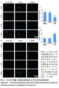
2.5 各组纤维膜与骨髓巨噬细胞共培养实验结果 硬脊膜破损大都是脊柱手术所导致,手术所致的创伤会在硬脊膜破损局部形成一个炎症环境,巨噬细胞在3-7 d内聚集,进一步导致损伤局部瘢痕的形成[33]。如图10所示,通过免疫荧光染色分别标记了促炎型巨噬细胞(M1型)及抗炎型巨噬细胞(M2型),空白对照组、胶原组促炎型巨噬细胞荧光强度明显高于干细胞组,而抗炎型巨噬细胞的荧光强度显著低于干细胞组,同时,骨髓巨噬细胞的形态也从圆形变为M2巨噬细胞典型的长梭形。"


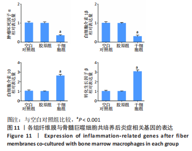
采用qRT-PCR技术检测炎症相关基因的表达,结果显示,相较于空白对照组、胶原组,干细胞组促炎基因白细胞介素1β、肿瘤坏死因子α明显下调,抑炎基因白细胞介素10、转化生长因子β显著上调,见图11。这进一步验证了骨髓间充质干细胞通过对炎症相关基因表达的调整来改变巨噬细胞极化的方向,从而达到免疫调节的目的。"
| [1] GUERIN P, EL FEGOUN AB, OBEID I, et al. Incidental durotomy during spine surgery: incidence, management and complications. A retrospective review. Injury. 2012;43(4):397-401. [2] LUSZCZYK MJ, BLAISDELL GY, WIATER BP, et al. Traumatic dural tears: what do we know and are they a problem? Spine J. 2014;14(1):49-56. [3] WEBER C, PIEK J, GUNAWAN D. Health care costs of incidental durotomies and postoperative cerebrospinal fluid leaks after elective spinal surgery. Eur Spine J. 2015;24(9):2065-2068. [4] HEO DH, HA JS, LEE DC, et al. Repair of Incidental Durotomy Using Sutureless Nonpenetrating Clips via Biportal Endoscopic Surgery. Global Spine J. 2022;12(3): 452-457. [5] SKOVSTED YDE S, BRUNBJERG ME, GUDMUNDSDOTTIR G, et al. Dural repair using porcine ADM: two cases and a literature review. Case Reports Plast Surg Hand Surg. 2017;4(1):5-8. [6] STENDEL R, DANNE M, FISS I, et al. Efficacy and safety of a collagen matrix for cranial and spinal dural reconstruction using different fixation techniques. J Neurosurg. 2008;109(2):215-521. [7] BOHOUN C A, GOTO T, MORISAKO H, et al. Skull Base Dural Repair Using Autologous Fat as a Dural Substitute: An Efficient Technique. World Neurosurg. 2019;127:e896-e900. [8] FAN B, WEI Z, YAO X, et al. Microenvironment Imbalance of Spinal Cord Injury. Cell Transplant. 2018;27(6):853-866. [9] VENKATALAXMI A, PADMAVATHI BS, AMARANATH T. A general solution of unsteady Stokes equations. Fluid Dyn Res. 2004;35(3):229-236. [10] LI Q, MA L, GAO C. Biomaterials for in situ tissue regeneration: development and perspectives. J Mater Chem B. 2015;3(46):8921-8938. [11] SCHMALZ P, GRIESSENAUER C, OGILVY CS, et al. Use of an Absorbable Synthetic Polymer Dural Substitute for Repair of Dural Defects: A Technical Note. Cureus. 2018;10(1):e2127. [12] LI J, TIAN J, LI C, et al. A hydrogel spinal dural patch with potential anti-inflammatory, pain relieving and antibacterial effects. Bioact Mater. 2022;14:389-401. [13] LI Q, MU L, ZHANG F, et al. A novel fish collagen scaffold as dural substitute. Mater Sci Eng C Mater Biol Appl. 2017;80:346-351. [14] WANG Z, CUI W. Two Sides of Electrospun Fiber in Promoting and Inhibiting Biomedical Processes. Advanced Therapeutics. 2020;4(1): 2000096. [15] VIGANI B, ROSSI S, SANDRI G, et al. Design and criteria of electrospun fibrous scaffolds for the treatment of spinal cord injury. Neural Regen Res. 2017;12(11): 1786-1790. [16] XU Y, CUI W, ZHANG Y, et al. Hierarchical Micro/Nanofibrous Bioscaffolds for Structural Tissue Regeneration. Adv Healthc Mater. 2017;6(13). doi: 10.1002/adhm.201601457. [17] CHEN AY, ZHONG C, LU TK. Engineering living functional materials. ACS Synth Biol. 2015;4(1):8-11. [18] TANG TC, AN B, HUANG Y, et al. Materials design by synthetic biology. Na Rev Mater. 2020;6(4):332-350. [19] SHANG L, SHAO C, CHI J, et al. Living Materials for Life Healthcare. Acc Mater Res. 2020;2(1):59-70. [20] WANG S, QU X, ZHAO RC. Clinical applications of mesenchymal stem cells. J Hematol Oncol. 2012;5:19. [21] BRUNELLE AR, HORNER CB, LOW K, et al. Electrospun thermosensitive hydrogel scaffold for enhanced chondrogenesis of human mesenchymal stem cells. Acta Biomater. 2018;66:166-176. [22] LV B, ZHANG X, YUAN J, et al. Biomaterial-supported MSC transplantation enhances cell-cell communication for spinal cord injury. Stem Cell Res Ther. 2021;12(1):36. [23] MOTHE AJ, TATOR CH. Advances in stem cell therapy for spinal cord injury . J Clin Invest. 2012;122(11): 824-3834. [24] MARDPOUR S, HAMIDIEH AA, TALEAHMAD S, et al. Interaction between mesenchymal stromal cell-derived extracellular vesicles and immune cells by distinct protein content. J Cell Physiol. 2019;234(6):8249-8258. [25] ANTON K, BANERJEE D, GLOD J. Macrophage-associated mesenchymal stem cells assume an activated, migratory, pro-inflammatory phenotype with increased IL-6 and CXCL10 secretion. PLoS One. 2012;7(4):e35036. [26] CARTY F, MAHON BP, ENGLISH K. The influence of macrophages on mesenchymal stromal cell therapy: passive or aggressive agents? Clin Exp Immunol. 2017; 188(1):1-11. [27] CHUNG E, SON Y. Crosstalk between mesenchymal stem cells and macrophages in tissue repair. Tissue Eng Regen Med. 2014;11(6):431-438. [28] DAFFORD EE, ANDERSON PA. Comparison of dural repair techniques. Spine J. 2015;15(5):1099-1105. [29] XI K, GU Y, TANG J, et al. Microenvironment-responsive immunoregulatory electrospun fibers for promoting nerve function recovery. Nat Commun. 2020; 11(1):4504. [30] WU L, GU Y, LIU L, et al. Hierarchical micro/nanofibrous membranes of sustained releasing VEGF for periosteal regeneration. Biomaterials. 2020;227:119555. [31] TANG J, XI K, CHEN H, et al. Flexible Osteogenic Glue as an All‐In‐One Solution to Assist Fracture Fixation and Healing. Adv Funct Mater. 2021;31(38):2102465.1-2102465.16. [32] BAI J, WANG H, CHEN H, et al. Biomimetic osteogenic peptide with mussel adhesion and osteoimmunomodulatory functions to ameliorate interfacial osseointegration under chronic inflammation. Biomaterials. 2020;255:120197. [33] LEE HG, KANG MS, KIM SY, et al. Dural Injury in Unilateral Biportal Endoscopic Spinal Surgery. Global Spine J. 2021;11(6):845-851. [34] NAGEL SJ, REDDY CG, FRIZON LA, et al. Spinal dura mater: biophysical characteristics relevant to medical device development. J Med Eng Technol. 2018;42(2):128-139. [35] SHIN JK, YOUN MS, SEONG YJ, et al. Iatrogenic dural tear in endoscopic lumbar spinal surgery: full endoscopic dural suture repair (Youn’s technique). Eur Spine J. 2018;27(Suppl 3):544-548. [36] SHIMADA Y, HONGO M, MIYAKOSHI N, et al. Dural substitute with polyglycolic acid mesh and fibrin glue for dural repair: technical note and preliminary results. J Orthop Sci. 2006;11(5):454-458. [37] IWATA A, TAKAHATA M, KADOYA K, et al. Effective Repair of Dural Tear Using Bioabsorbable Sheet With Fibrin Glue. Spine (Phila Pa 1976). 2017;42(18):1362-1366. [38] PENG Z, GAO W, YUE B, et al. Promotion of neurological recovery in rat spinal cord injury by mesenchymal stem cells loaded on nerve-guided collagen scaffold through increasing alternatively activated macrophage polarization. J Tissue Eng Regen Med. 2018;12(3):e1725-e736. [39] LUZ-CRAWFORD P, JORGENSEN C, DJOUAD F. Mesenchymal Stem Cells Direct the Immunological Fate of Macrophages. Results Probl Cell Differ. 2017;62:61-72. [40] ORR MB, GENSEL JC. Spinal Cord Injury Scarring and Inflammation: Therapies Targeting Glial and Inflammatory Responses. Neurotherapeutics. 2018;15(3):541-553. |
| [1] | Shen Jiangyong, He Xi, Tang Yuting, Wang Jianjun, Liu Jinyi, Chen Yuanyuan, Wang Xinyi, Liu Tong, Sun Haoyuan. RAS-selective lethal small molecule 3 inhibits the fibrosis of pathological scar fibroblasts [J]. Chinese Journal of Tissue Engineering Research, 2024, 28(8): 1168-1173. |
| [2] | Wang Wu, Fan Xiaolei, Xie Jie, Hu Yihe, Zeng Min. Hydroxyapatite-polyvinyl alcohol/collagen-chitosan-gelatin composite hydrogel for repairing rabbit osteochondral defect [J]. Chinese Journal of Tissue Engineering Research, 2024, 28(5): 682-689. |
| [3] | Xu Rong, Wang Haojie, Geng Mengxiang, Meng Kai, Wang Hui, Zhang Keqin, Zhao Huijing. Research advance in preparation and functional modification of porous polytetrafluoroethylene artificial blood vessels [J]. Chinese Journal of Tissue Engineering Research, 2024, 28(5): 759-765. |
| [4] | Yin Tong, Yang Jilei, Li Yourui, Liu Zhuoran, Jiang Ming. Application of core-shell structured nanofibers in oral tissue regeneration [J]. Chinese Journal of Tissue Engineering Research, 2024, 28(5): 766-770. |
| [5] | Lin Feng, Cheng Ling, Gao Yong, Zhou Jianye, Shang Qingqing. Hyaluronic acid hydrogel-encapsulated bone marrow mesenchymal stem cells promote cardiac function in myocardial infarction rats (III) [J]. Chinese Journal of Tissue Engineering Research, 2024, 28(3): 355-359. |
| [6] | Bi Yujie, Ma Dujun, Peng Liping, Zhou Ziqiong, Zhao Jing, Zhu Houjun, Zhong Qiuhui, Yang Yuxin. Strategy and significance of Chinese medicine combined with medical hydrogel for disease treatment [J]. Chinese Journal of Tissue Engineering Research, 2024, 28(3): 419-425. |
| [7] | Ran Lei, Han Haihui, Xu Bo, Wang Jianye, Shen Jun, Xiao Lianbo, Shi Qi. Molecular docking analysis of the anti-inflammatory mechanism of Cibotium barometz and Epimedium for rheumatoid arthritis: animal experiment validation [J]. Chinese Journal of Tissue Engineering Research, 2024, 28(2): 208-215. |
| [8] | Chen Ligang, He Xiaoming, Tan Yu, Xiao Yuzhi, Ma Chuntao, Guo Liang. Bone metabolism in patients with osteonecrosis of the femoral head based on etiology and Association Research Circulation Osseous staging [J]. Chinese Journal of Tissue Engineering Research, 2024, 28(16): 2461-2466. |
| [9] | Zhao Shasha, He Qing, Li Jia, Wu Ying. Effects of recombinant human collagen supplementation on extracellular matrix remodeling in mouse skeletal muscle after eccentric exercise [J]. Chinese Journal of Tissue Engineering Research, 2024, 28(16): 2542-2549. |
| [10] | Yao Siqi, Li Wenzheng, Wang Hong. Transforming growth factor beta/Smad signaling pathway and targeted therapy of keloid scars [J]. Chinese Journal of Tissue Engineering Research, 2024, 28(16): 2619-2624. |
| [11] | Zhang Hongxia, Li Xingchao, Gao Xixin, Zhang Xiao, Mei Shuang, Ma Hanxi, Zhang Tian. The histological and thickness changes of attached gingiva following grafting with different soft tissue substitutes in the labial region of the cuspids in Beagles [J]. Chinese Journal of Tissue Engineering Research, 2024, 28(11): 1660-1665. |
| [12] | Lyu Shangyi, He Huiyu, Wufanbieke · Baheti, Yang Quan, Ma Lisha, Han Xiangzhen. Physicochemical properties and biocompatibility of graphene oxide-hydroxyapatite composite coating materials [J]. Chinese Journal of Tissue Engineering Research, 2024, 28(10): 1477-1483. |
| [13] | Su Kunyang, Chen Bineng, Chen Yiliang, Jin Shaofeng. Strontium ranelate-loaded sodium alginate/collagen hydrogel promotes bone defect repair in osteoarthritis [J]. Chinese Journal of Tissue Engineering Research, 2024, 28(10): 1568-1574. |
| [14] | Jiang Yu, He Feng, Liu Huan, Wu Ruixin. Application of melt electrowriting technology in tissue engineering [J]. Chinese Journal of Tissue Engineering Research, 2024, 28(10): 1606-1612. |
| [15] | Fan Yongjing, Wang Shu, Jin Wulong. Characteristics, advantages and application of osteogenic differentiation of jaw bone marrow mesenchymal stem cells [J]. Chinese Journal of Tissue Engineering Research, 2024, 28(1): 100-106. |
| Viewed | ||||||
|
Full text |
|
|||||
|
Abstract |
|
|||||