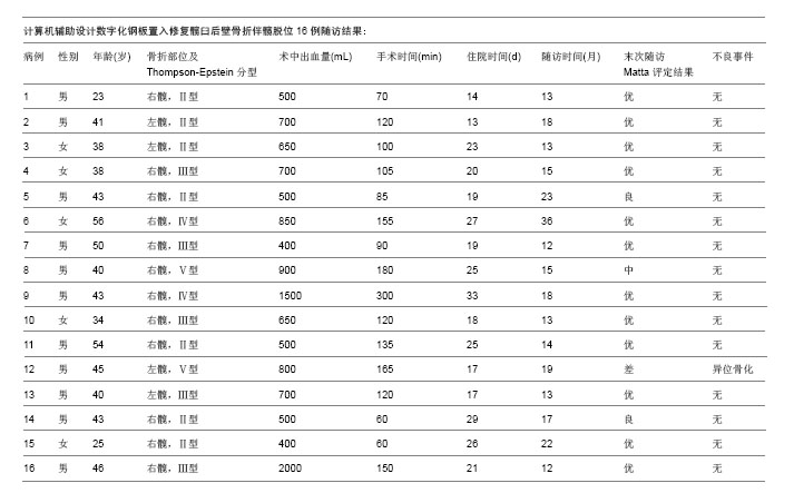
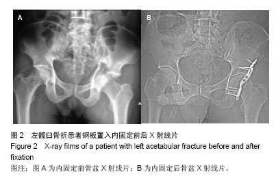
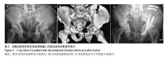
| [1]Gänsslen A, Hildebrand F, Kretek C. Transverse posterior wall fractures of the acetabulum: epidemiology, operative management and long-term results. Acta Chir Orthop Traumatol Cech. 2013; 80(1):27-33.
[2]马玉鹏,周东生,李连欣,等.ISO-C3D计算机导航技术经皮微创内固定治疗髋臼骨折[J].中国组织工程研究杂志,2013,17(52):9023-9028.
[3]孟波,孙海钰,李明,等.计算机辅助在骨盆骨折诊断与治疗方面的临床价值[J].中国组织工程研究杂志,2014,18(17):2673-2678.
[4]Thompson VP, Epstein HC. Traumatic dislocation of the hip: a surgery of two hundred and four cases covering a period of twenty-one years. J Bone Joint Surg (Am). 1951;33(3): 746-748.
[5]Matta JM. Fractures of the acetabulum: accuracy of reduction and clinical result s in patients managed operatively within three weeks after the injury. J Bone Joint Surg (Am). 1996; 78(11): 1632-1645.
[6]Judet R, Judet J, Letournel E. Fractures of the acetabulum: classification and surgical approaches for open reduction. J Bone Joint Surg(Am). 1964;46:1615-1638.
[7]d’Aubigné RM, Postel M. The classic: functional results of hip arthroplasty with acrylic prosthesis. 1954. Clin Orthop Relat Res. 2009;467(1): 7-27.
[8]Brooker AF, Bowerman JW, Robinson RA, et al. Ectopic ossification following total hip replacement. Incidence and a method of classification. J Bone Joint Surg (Am). 1973;55: 1629-1632.
[9]周方.脊柱与四肢骨折的治疗策略[M].北京:北京大学医学出版社, 2010:82-99.
[10]张发平,李玉桥,罗仕武,等.手术治疗髋臼骨折的临床疗效[J].临床骨科杂志,2014,17(4):422-425.
[11]胡杰挺,陈建生,钱钟.手术与非手术复位法治疗髋臼后壁骨折的临床研究[J].中华全科医学杂志,2014,12(8):1239-1241.
[12]贾献荣,赵育威,吴苏琪,等.髋臼后壁骨折临床疗效的相关因素分析[J].中华全科医学杂志,2014,12(8):1245-1247.
[13]黄凯,丁亮华,胡新宇,等.计算机辅助设计数字化个体钢板内固定治疗复杂胫骨平台骨折短期临床疗效分析[J].中国骨与关节损伤杂志,2013,4(28):324-326.
[14]路博,孔繁林,丁亮华,等.数字化钢板在尺桡桡骨干骨折中的临床临床应用[J].中国骨与关节损伤杂志,2014,5(29):457-459.
[15]郭晓山.骨盆与髋臼骨折的治疗展望[J].中医正骨,2013,6(25):3-5.
[16]任龙韬,张云鹏,郭志坚,等.计算机辅助设计股骨远端个性化解剖型接骨板[J].中国组织工程研究与临床康复, 2011, 15(13): 2309-2312.
[17]刘瑞平,孙荣彬,黄用,等.计算机辅助设计胫骨平台后柱骨折个性化解剖型接骨板[J].江苏医药,2012,38(23):2823-2825.
[18]章莹,夏远军,万磊,等.计算机三维仿真技术与常规手术治疗胫骨平台骨折的疗效对比分析[J].中国骨与关节损伤杂志, 2010, 8(25):686-688.
[19]刘寿堂,柳青峰,李连,等.计算机辅助设计个体化塑形钛网修补大面积额颞顶部颅骨缺损[J].中国美容整形外科杂志,2008,19(3): 210-211.
[20]龚振宇,刘彦普,周树夏,等.基于反求工程和快速原型的下颌骨缺损的修复[J].中华口腔医学杂志, 2004,39(1): 9-11.
[21]归来,左锋,张智勇,等.颅骨缺损的个性化修复[J].中华整形外科杂志, 2004, 20(2): 98-100.
[22]宋涛,归来,张智勇,等.个性化钛植入体修复大面积颅骨缺损[J].创伤外科杂志, 2007, 9(6): 509-511.
[23]Sunil G. RP inmedicine: a case study in cranial reconstructive surgery. Rapid Prot J.2004;3: 207-211.
[24]姜涛,孙大军,刘建国.个体化股骨假体的CAD实践及模拟力学实验[J].生物医学工程学杂志, 2007, 24(6): 1286-1290.
[25]Martini F, Sell S, Kremling E, et al.Determination of periprosthetie bone density with the DEXA method after implantation of cuatom - made uncemented femoral stems.Int Orthop. 1996;4: 218-221.
[26]Zadeh AG, Hua J, Walker PS, et al.Uncemented total hip arthroplasty with sub trochanteric derotational osteotomy for severe femoral anteversion. J Arthroplasty. 1999;6: 682.
[27]王臻,滕勇,李涤尘,等.基于快速成型的个体化人工半膝关节的研制[J].中国修复重建外科杂志,2004,18(5): 347-351.
[28]王臻,滕勇,李涤尘,等.基于快速成型技术的个体化人工股骨髁关节面的设计与应用[J].中华外科杂志, 2004, 42(12): 746-749.
[29]邱贵兴主译.骨盆与髋臼骨折[M].北京:人民卫生出版社, 2006: 666-667.
[30]周程鹏,高仕长,刘杰,等.髋臼后壁钢板螺钉固定安全区域的应用解剖学研究[J].中国临床解剖学杂志, 2013,31(1): 13-18.
[31]王愉思,何畔,王家让,等.髋臼后柱安全置钉的临床解剖学研究[J].中国骨与关节损伤杂志,2009,24(7):584-586.
[32]牛云飞,许硕贵,张春才.髋臼后壁厚度的解剖学测量及其意义[J].中国临床解剖学杂志,2007,25(4):400-402.
[33]Wang XQ, Cai JF, Cao XC, et al. A quantitative anatomic study of plate-screw fixation of the acetabular anterior column through an anterior approach. Arch Orthop Trauma Surg. 2010; 130: 257-262.
[34]蓝旭,梁军,文益民,等.髋臼骨折手术并发症分析[J].中国现代手术学杂志,2007,11(5):374-377.
[35]周炎,瞿新丛,易成腊,等.髋臼后壁骨折伴髋关节后脱位诊疗失误及并发症分析[J].创伤外科杂志,2013,15(3):216-219.
[36]危伟浪.影响髋臼后壁骨折手术疗效的相关因素[J].中国伤残医学杂志,2013,21(4):83-84.
[37]Magu NK, Gogna P, Singh A, et al. Long term results after surgical management of posterior wall acetabular fractures.J Orthop Traumatol. 2014. [Epub ahead of print]
[38]Shen F, Chen B, Guo Q, et al. Augmented reality patient-specific reconstruction plate design for pelvic and acetabular fracture surgery.Int J Comput Assist Radiol Surg. 2013;8(2):169-179.
[39]Reddix RN Jr, Webb LX. Computer-assisted preoperative planning in the surgical treatment of acetabular fractures.J Surg Orthop Adv. 2007;16(3):138-143.
[40]Hu Y, Li H, Qiao G, et al. Computer-assisted virtual surgical procedure for acetabular fractures based on real CT data. Injury. 2011;42(10):1121-1124. |
.jpg)