中国组织工程研究 ›› 2026, Vol. 30 ›› Issue (30): 8000-8007.doi: 10.12307/2026.928
• 组织构建综述 tissue construction review • 上一篇
细胞外基质力学特性与骨再生:从分子机制到仿生材料
晋嘉璐1,2,喻 鹏1,余钒源1,2,吴佳益1,3,叶 玲1,2
- 1口腔疾病防治全国重点实验室,国家口腔医学中心,国家口腔疾病临床医学研究中心,四川省成都市 610041;四川大学华西口腔医院,2牙体牙髓科,3锦江门诊部,四川省成都市 610041
-
收稿日期:2025-12-17修回日期:2026-05-15出版日期:2026-10-28发布日期:2026-09-15 -
通讯作者:叶玲,博士,教授,口腔疾病防治全国重点实验室,国家口腔医学中心,国家口腔疾病临床医学研究中心,四川省成都市 610041;四川大学华西口腔医院牙体牙髓科,四川省成都市 610041 共同通讯作者:吴佳益,博士,主治医师,口腔疾病防治全国重点实验室,国家口腔医学中心,国家口腔疾病临床医学研究中心,四川省成都市610041;四川大学华西口腔医院锦江门诊部,四川省成都市 610041 -
作者简介:晋嘉璐,女,2001年生,重庆市人,汉族,四川大学华西口腔医学院在读硕士,主要从事生物材料及骨组织工程方面的研究。 -
基金资助:四川省自然科学基金重大项目(2026NSFSCZY0003),项目负责人:叶玲;四川省自然科学基金青年科学基金项目(2026NSFSC1586),项目负责人:吴佳益;国家自然科学基金青年科学基金项目(82501119),项目负责人:吴佳益
Extracellular matrix mechanical properties in bone regeneration: from molecular mechanisms to biomimetic materials
Jin Jialu1, 2, Yu Peng1, Yu Fanyuan1, 2, Wu Jiayi1, 3, Ye Ling1, 2
- 1State Key Laboratory of Oral Diseases & National Center for Stomatology & National Clinical Research Center for Oral Diseases, Chengdu 610041, Sichuan Province, China; 2Department of Endodontics, 3Department of Jinjiang Outpatient, West China Hospital of Stomatology, Sichuan University, Chengdu 610041, Sichuan Province, China
-
Received:2025-12-17Revised:2026-05-15Online:2026-10-28Published:2026-09-15 -
Contact:Ye Ling, MD, Professor, State Key Laboratory of Oral Diseases & National Center for Stomatology & National Clinical Research Center for Oral Diseases, Chengdu 610041, Sichuan Province, China; 2Department of Endodontics, West China Hospital of Stomatology, Sichuan University, Chengdu 610041, Sichuan Province, China Co-corresponding author: Wu Jiayi, MD, Attending doctor, State Key Laboratory of Oral Diseases & National Center for Stomatology & National Clinical Research Center for Oral Diseases, Chengdu 610041, Sichuan Province, China; Department of Jinjiang Outpatient, West China Hospital of Stomatology, Sichuan University, Chengdu 610041, Sichuan Province, China -
About author:Jin Jialu, MS candidate, State Key Laboratory of Oral Diseases & National Center for Stomatology & National Clinical Research Center for Oral Diseases, Chengdu 610041, Sichuan Province, China; Department of Endodontics, West China Hospital of Stomatology, Sichuan University, Chengdu 610041, Sichuan Province, China -
Supported by:Major Program of the Natural Science Foundation of Sichuan Province, No. 2026NSFSCZY0003 (to YL); Youth Scientific Program of the Natural Science Foundation of Sichuan Province, No. 2026NSFSC1586 (to WJY); National Natural Science Foundation of China (Youth Program), No. 82501119 (to WJY)

摘要:

文题释义:
细胞外基质:是由细胞分泌的多种结构蛋白和糖胺聚糖等组成的三维网络结构,为细胞提供机械支撑,并参与组织发育、损伤修复及骨重建等生理过程。
仿生材料:是指在结构与功能上模拟天然细胞外基质力学特性的生物材料,通过调节材料的力学参数,可实现对成骨相关细胞行为的精准调控,从而促进骨组织再生。
背景:细胞外基质不仅可作为细胞黏附和迁移的物理支架,还通过特有的力学特性调控细胞行为,在骨再生中发挥重要作用。
目的:系统总结细胞外基质的主要力学特性及分子基础,阐述这些力学特性如何通过机械信号通路调控成骨相关细胞的行为,深入理解细胞外基质的力学特性及信号通路,为构建具有仿生力学微环境的骨修复材料提供新思路。
方法:在PubMed、Web of Science及中国知网数据库中,以英文检索词“Extracellular matrix,Mechanical properties,Osteogenic differentiation,Bone regeneration,Bone tissue engineering”,中文检索词“细胞外基质,力学特性,成骨分化,骨再生,骨组织工程”进行文献检索,筛选并分析与细胞外基质力学特性及骨修复相关的研究,排除原创性不足、与主题关系不大以及陈旧的文献,检索时限为1995年6月至2025年9月,纳入135篇文献进行综述。
结果与结论:①细胞外基质的刚度、黏弹性、塑性、纤维取向及孔隙-渗透特征共同决定力学微环境,从而调控成骨基因表达;②细胞外基质主要由纤维蛋白、糖蛋白、蛋白聚糖及无机成分协同构成,细胞外基质的交联密度、水合作用及矿物分布决定力学特性;③细胞外基质力学信号通过整合素-黏着斑激酶、Yes相关蛋白/PDZ结合域转录共激活因子-Hippo、Ras同源家族成员A-Rho相关卷曲螺旋蛋白激酶、Wnt/β-catenin及机械敏感离子通道-钙信号通路传导,参与细胞增殖、分化与矿化,维持骨形成与吸收的动态平衡;④仿生材料通过交联、矿物相引入与复合结构设计等方式优化支架的刚度、孔隙结构与力学稳定性,能够为细胞提供更合适的支撑与微环境,促进细胞浸润、血管长入与成骨分化,从而提升骨缺损修复效果。
https://orcid.org/0009-0006-9962-974X(晋嘉璐);https://orcid.org/0000-0002-9710-5882(吴佳益)
中国组织工程研究杂志出版内容重点:干细胞;骨髓干细胞;造血干细胞;脂肪干细胞;肿瘤干细胞;胚胎干细胞;脐带脐血干细胞;干细胞诱导;干细胞分化;组织工程
中图分类号:
引用本文
晋嘉璐, 喻 鹏, 余钒源, 吴佳益, 叶 玲. 细胞外基质力学特性与骨再生:从分子机制到仿生材料[J]. 中国组织工程研究, 2026, 30(30): 8000-8007.
Jin Jialu, Yu Peng, Yu Fanyuan, Wu Jiayi, Ye Ling. Extracellular matrix mechanical properties in bone regeneration: from molecular mechanisms to biomimetic materials[J]. Chinese Journal of Tissue Engineering Research, 2026, 30(30): 8000-8007.
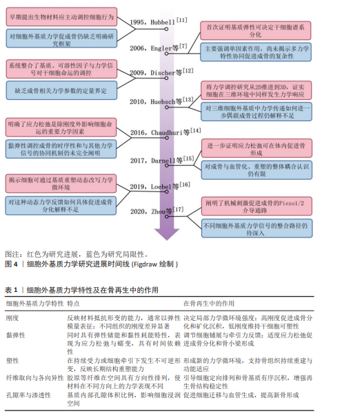
2.2 细胞外基质的力学特性 细胞外基质的力学特性包括多个方面,主要有刚度、黏弹性、塑性、纤维取向与各向异性以及孔隙率与渗透性(表1),这些特征共同塑造骨组织的微环境,调控成骨相关细胞的黏附与分化,决定骨形成的速度与质量,在骨修复与再生过程中发挥重要作用。
2.2.1 刚度 刚度通常指细胞外基质在外力作用下抵抗形变的能力,是细胞外基质最重要的力学特性之一。细胞外基质的刚度常通过弹性模量来量化,即材料在拉伸时的应力与应变之比。不同组织的刚度差异极大,例如脑组织为0.1-1 kPa,横纹肌为8-17 kPa,而关节软骨的弹性模量多为0.5-1.7 MPa,成熟皮质骨则通常为17-20 GPa[18-20]。这种显著的差异决定了不同组织独特的生物力学微环境,影响细胞的形态、功能及再生潜能[21-23]。
研究显示,骨髓间充质干细胞的分化命运与基质刚度密切相关:当基质刚度处于较低水平时(如0.1-1 kPa),细胞更倾向维持较高可塑性;随着基质刚度升高至20 kPa以上,细胞铺展、黏附及成骨相关活性逐渐增强,在40-80 kPa范围内可观察到更为明显的成骨表型[24-25]。
脂肪间充质干细胞在刚度约3 kPa的基质上观察到成脂分化,在刚度约35 kPa的基质上更有利于成骨分化[26]。基质刚度还影响免疫细胞的行为,部分研究发现刚度变化会影响巨噬细胞黏附、铺展、炎症因子分泌及M1/M2极化平衡,间接调控成骨和骨重塑过程[27]。
2.2.2 黏弹性 黏弹性是指细胞外基质在受力过程中同时表现出弹性固体的能量储存特性和黏性流体的能量消耗特性,具有显著的时间依赖性[28–30]。在恒定应变条件下,细胞外基质的应力会随时间逐渐衰减,即应力松弛;恒定应力条件下应变会随着时间逐渐增加,即蠕变[31]。
细胞外基质的黏弹性通常通过储能模量、损耗模量以及应力松弛半衰期等参数进行表征[32]。
骨组织具有一定的黏弹性,在长期机械负荷作用下通过缓冲应力、消耗部分能量和减少疲劳断裂来维持整体的力学强度与韧性[31]。在骨质疏松等病理状态下,骨组织黏弹性下降与应力松弛加快导致组织脆性增加;在骨再生过程中,新生细胞外基质的黏弹性直接影响骨小梁形成速度与结构质量[33]。
细胞外基质的黏弹性通过力学信号传导调控成骨相关细胞的黏附、迁移与分化行为。研究表明,应力松弛快速的基质显著促进间充质干细胞及成骨细胞的黏附与铺展,低松弛的基质影响形态扩展与骨样结构形成[31,34]。黏弹性基质能够在细胞牵引作用下发生局部变形与结构重排,为细胞迁移创造有利空间,增强在骨修复环境中的定向迁移能力[28,32]。在细胞分化方面,快速应力松弛可促使细胞体积变大、核张力增强,促进成骨分化及矿化形成[34-35]。具有一定应力松弛能力的微环境,还可刺激成骨细胞与软骨细胞分泌新的细胞外基质,形成正反馈,进一步优化骨再生的局部微环境[32]。
2.2.3 塑性 塑性指细胞外基质在细胞长期牵引或外界持续受力时会发生不可逆的形变。基质中的胶原纤维被细胞不断拉动、重排或重新交联,外力解除也无法完全恢复原状,这类力学环境常出现在骨缺损修复、慢性炎症、纤维化、骨质疏松、长期应力过载(如咬合创伤)等情况中,此时细胞会持续施加牵引力以重建受损组织,细胞外基质逐渐变成不同于原本的状态,这种永久性的结构改变相当于在微观层面保留了细胞施加过的力学刺激信息,持续影响后续细胞的黏附、迁移与分化,在重复受力或长期修复过程中维持骨组织的韧性与结构稳定性[36-38]。
塑性特性对长期力学稳定性及骨修复过程具有重要意义:塑性通过逐步形成新的力学微环境,决定基质是否能被细胞持续改造并生成新的骨样组织[36];在生理条件下,适度的塑性有助于骨组织在应力负荷下实现结构适应,从而维持良好的力学性能和抗疲劳能力。但是在代谢性疾病或衰老状态下,胶原纤维的重塑能力减弱,会导致骨组织韧性下降、脆性增加,易发生骨折等结构性损伤[39-40]。
在骨修复过程中,骨细胞外基质的“形成-降解-重塑”循环决定其力学特性随时间的动态变化。当损伤修复不完全或细胞外基质过度沉积时,局部可形成存在差异的力学微环境,不仅改变了细胞在组织中的分布方式和停留位置,也使细胞与周围基质的接触和附着状态发生改变,进而影响新骨的结构重建与再生进程[41–42]。最新研究发现,基质降解性与应力松弛之间存在功能耦合关系:在整体刚度不变的情况下,适度的降解即可显著增强应力松弛能力,从而重塑细胞所感知的时间依赖性力学环境,最终调控细胞的铺展、黏附与力传导模式[42]。
2.2.4 纤维取向与各向异性 纤维取向指的是细胞外基质中胶原等纤维在空间上的特定排列方向。细胞外基质在不同方向上表现出不同的力学和传导特性,如抗拉强度、抗弯性能以及渗透性等[43]。在天然骨组织中,各向异性是一种普遍存在的结构特征,尤其在皮质骨中表现最为典型,是同时兼具高强度与良好韧性的重要基础之一[43-45]。
从细胞行为角度来看,有取向的纤维结构能够为细胞提供明确的接触导向信号,使骨髓间充质干细胞和成骨细胞沿纤维方向发生极化与定向排列,这一过程常伴随应力纤维与细胞核取向的同步重排,从而激活成骨相关信号通路,促进矿化及成骨表型的形成[43,46]。
具有方向性孔道或纤维排列的材料能够在骨缺损早期引导细胞和血管沿固定方向生长,加速细胞浸润与血管化过程,提高新生骨量并加快力学功能恢复[47-48]。沿受力方向设计的结构在“受力-传导-重塑”的动态过程中提供方向性支撑与负载转移,能够促进骨小梁按功能方向组织化生长,增强再生骨的结构完整性与耐力[49-50]。近期研究还表明,纤维取向不仅调控成骨细胞行为,也可间接影响免疫微环境。特定取向的基质能够调节巨噬细胞的极化状态,促进其向抗炎型表型转化,加速炎症消退,构建有利于成骨与组织重建的局部环境[48]。
2.2.5 孔隙率与渗透性 孔隙率是指细胞外基质内部孔隙体积占总体积的比例;渗透性则反映流体在细胞外基质孔隙网络中流动与扩散的能力,二者的大小主要受孔径、孔隙连通性及空间分布等因素共同影响[51-52]。
在骨修复过程中,较高的孔隙率与良好的孔隙连通性有助于骨髓间充质干细胞在基质内部黏附、迁移与均匀分布,为血管生成提供充足的空间支撑[53-55]。既往研究认为,孔径至少大于100 μm才能满足细胞迁移和物质运输,300-500 μm的孔径更有利于血管化和新骨长入;总孔隙率则常设计在50%-90%范围内,以在成骨效率与力学支撑之间取得平衡[54];渗透性直接影响局部环境中氧气与营养物质的扩散速率,调控细胞代谢与成骨活性。实验研究表明,过低的渗透性会导致细胞缺氧和营养不足,抑制成骨分化;而过高的渗透性则使信号分子难以在局部富集,降低成骨诱导效率[56-57]。
适度的孔隙-渗透平衡有利于维持骨组织稳态与促进新骨形成。骨质疏松骨细胞外基质的孔隙率过高且渗透性过度增加,导致骨量下降与骨组织脆性增强[58]。合理的孔隙率与渗透性不仅影响骨小梁的形成速度与空间分布,还决定最终的重塑质量[51]。结构上相互连通的孔隙促进细胞深层浸润与血管长入,在骨形成过程中有效分散局部应力,减少坏死区域的出现,从而提升新生骨的连续性与力学整合[53]。
2.2.6 细胞外基质力学特性的相互作用 除各单一力学特性外,多种力学特性之间的相互作用同样值得关注。骨再生过程中细胞所感知的并不是独立的力学参数,而是由刚度、黏弹性、塑性、纤维取向及孔隙-渗透特征共同构成并相互耦合的综合力学微环境。各因素之间存在明显的联动关系:刚度决定细胞受到牵引时基质提供的支撑强度,但这种支撑并非静态不变,还会受到黏弹性的影响,即同样的刚度条件下,基质应力释放和形变恢复的速度不同,细胞实际经历的受力过程也会不同;塑性则进一步决定基质在持续牵引下是否发生不可逆重排,从而改变后续细胞再次感受到的局部力学状态。纤维取向不仅影响基质在不同方向上的力学表现,还会通过改变细胞排列方向、黏着斑分布和细胞骨架张力,使力的传递呈现明显方向性;而孔隙率与渗透性除影响细胞浸润和营养交换外,也会影响局部应力扩散、液体流动及力学信号在组织内部的传导效率。因此刚度决定“支撑有多强”, 黏弹性和塑性决定“这种支撑如何随时间变化”,纤维取向决定“力沿什么方向传递”,孔隙-渗透特征则决定“细胞和组织内部是否能够有效承接和分散这些力学刺激”。细胞外基质力学特性对骨再生的影响并不是各单一因素的简单叠加,而是多种力学因素在空间和时间上相互作用的结果,最终综合调控细胞黏附、铺展、迁移、分化及骨组织重塑过程。
2.3 细胞外基质力学特性的决定因素 细胞外基质的力学特性由组成成分、分子结构及动态重塑过程共同决定[59]。在骨再生过程中,纤维蛋白、黏附性糖蛋白、蛋白聚糖与糖胺聚糖以及矿物相之间的比例与相互作用状态对骨细胞外基质的力学特性起到了至关重要的作用,而这些力学特性又直接关系着成骨相关干细胞的行为[60-61](表2)。由此可见,揭示细胞外基质力学特性的决定因素,是理解骨再生中力学生物学机制并指导仿生材料设计的关键点。
2.3.1 纤维蛋白 纤维蛋白的主要组成成分是胶原蛋白,包括Ⅰ、Ⅱ、Ⅲ和Ⅳ型等,还有含量较少的弹性蛋白[62]。胶原纤维通过分子排列和多级结构构建细胞外基质的主要力学支撑框架,提供抗拉强度和刚度稳定结构[63]。近年来研究发现,胶原纤维在组织受力时重新排列,它的柔韧性与交联特征决定了细胞外基质的弹性与塑性。酶促反应形成的交联在增强纤维稳定性的同时保持适度的柔韧性,但是在衰老或高糖环境下,葡萄糖代谢产物与胶原发生非酶促糖基化反应,形成异常交联,导致基质僵硬且脆性增加[64-65]。弹性蛋白虽然含量相对较低,但它较强的可伸展性可提升细胞外基质的回弹性能,帮助组织在反复应力环境下维持形态与功能稳定[66]。因此,纤维蛋白提供兼具刚性、弹性与耐疲劳性的多层次力学结构,构建细胞外基质在生物力学环境中的稳固支撑[62-66]。
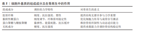
2.3.2 黏附性糖蛋白 黏附性糖蛋白,如纤连蛋白和层粘连蛋白,在细胞外基质中不仅承担黏附功能,还参与力学特性的调控[67]。黏附性糖蛋白通过调节纤维之间的连接密度与排列方式影响基质的整体致密性与结构稳定性[68]。当细胞外基质中纤连蛋白与胶原比例合适,细胞牵引力有效拉紧纤维网络促进纤维重排并增强整体刚度[69],但是纤连蛋白比例失衡,网络结构松散或弹性不足就会导致细胞外基质变软、加剧形变,降低组织韧性[70]。而且糖蛋白还能与整合素等受体结合,形成细胞-基质黏着连接,将细胞牵引力传导至细胞外基质并诱导纤维重新取向,从而动态调节基质的刚度与黏弹性[59]。不同类型和分布的糖蛋白还会改变细胞外基质的力学响应模式,使其在受力后的恢复性和抗变形能力不同,进而影响细胞对外界硬度信号的感知与反应[71-72]。黏附性糖蛋白通过调控细胞外基质网络的结构形成与力学稳定性以及结合细胞力学反馈,双向调节细胞外基质的刚度与黏弹行为,为成骨细胞提供合适的力学微环境。
2.3.3 蛋白聚糖与糖胺聚糖 蛋白聚糖和糖胺聚糖是细胞外基质中富含负电荷的长链多糖成分,它们结合大量水分形成高水合状态的凝胶相,让基质在受压或拉伸时表现出很好的黏弹响应能力[73]。蛋白聚糖和糖胺聚糖填充在胶原纤维网络中,调节纤维间的相互作用与应变传递,让细胞外基质具备更高的形变适应性和能量耗散能力[71]。在骨再生过程中,特殊类型的蛋白聚糖,如骨桥蛋白和小纤维聚蛋白,与水分子形成稳定结合在矿化区域中,水分让骨组织更具韧性,在反复受力情况下保持结构稳定[74]。蛋白聚糖和糖胺聚糖为细胞提供了稳定可调的微环境,提高骨组织的延展性和抗疲劳性能,这些有利于骨组织再生与重塑。
2.3.4 无机成分 无机与有机组分间的相互作用是维系细胞外基质协调与稳定的重要机制[75]。无机成分中羟基磷灰石等矿物晶体是决定细胞外基质硬度和抗压能力的重要因素,这些矿物沉积在胶原纤维上形成的矿化复合结构提高了基质的抗变形能力。矿物与胶原的协同作用让细胞外基质在刚度与韧性之间实现力学平衡,从而兼具高强度与能量耗散能力[76-79]。研究表明,纤维内矿化有效地传递应力并维持结构稳定,而过度或不均匀的矿化则可能导致组织过硬、脆性增加[80]。随着年龄增长,矿物晶体平均尺寸变大的同时晶体取向一致性下降,导致细胞外基质表现出更高的硬度但韧性降低,这也是老龄骨组织易碎的重要原因之一[81]。无机矿物提供刚性支撑增强细胞外基质的承载能力,其晶体特征与分布方式调节整体的韧性与塑性。
2.4 细胞外基质力学介导的成骨信号机制 虽然细胞外基质存在多种不同的力学特性,但细胞对这些特性的响应往往依赖于一系列信号通路(表3),这些通路既是细胞将外界力学刺激“翻译”成分子信号的关键环节,也是成骨细胞命运决定和骨组织修复的核心调控点(图5)。
2.4.1 整合素-黏着斑激酶-丝裂原活化蛋白激酶/细胞外信号调节激酶通路 整合素是由α和β亚基组成的跨膜受体蛋白,连接细胞外基
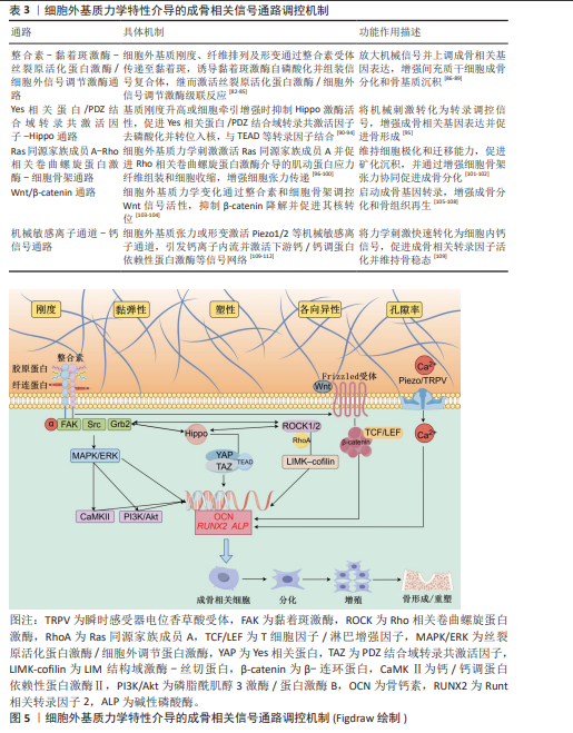
质与细胞骨架,具有双向传导信号的能力,是细胞感知细胞外基质力学环境的核心受体[82]。在“整合素-黏着斑激酶-丝裂原活化蛋白激酶/细胞外信号调节激酶”轴上,成骨相关细胞能够将刚度、纤维取向等物理刺激翻译成分子信号,激活该信号轴可增强骨髓间充质干细胞的成骨潜能,而阻断整合素或抑制黏着斑激酶会削弱成骨分化与骨形成信号[83-85]。当整合素与胶原或纤连蛋白结合后,会促使黏着斑激酶在黏着斑处自磷酸化并作为信号“中继站”,与Src(一种非受体型酪氨酸激酶)、Grb2等组装成为信号复合体,继而放大并将机械信号耦合到丝裂原活化蛋白激酶/细胞外信号调节激酶通路,最终驱动成骨相关基因的表达,从而增强成骨分化与矿化[86-87]。同时,在骨代谢相关细胞(包括成骨细胞、骨细胞、破骨细胞及骨髓间充质干细胞)中,整合素触发的机械应答通过黏着斑激酶-细胞外信号调节激酶通路放大后,可与钙/钙调蛋白依赖性蛋白激酶、磷脂酰肌醇3激酶/蛋白激酶B等信号网络交叉整合,共同上调成骨相关基因的表达,从而促进骨形成与组织再生[88-89]。
2.4.2 Yes相关蛋白/PDZ结合域转录共激活因子–Hippo通路 Yes相关蛋白与PDZ结合域转录共激活因子是转录调节蛋白,平时在抑制性Hippo激酶级联通路作用下被磷酸化并留在胞质;而当细胞处于更高基质刚度、形变或牵引力环境时,细胞骨架张力上升、Hippo抑制减弱,Yes相关蛋白/PDZ结合域转录共激活因子转位入核,在机械信号与转录反应之间发挥桥梁作用,通过与TEAD等转录因子协同激活成骨相关基因的表达[90]。Yes相关蛋白/PDZ结合域转录共激活因子对基质刚度、细胞形状和细胞铺展度、应力纤维张力、基质黏弹性及周期性应变高度敏感,其入核程度与成骨基因表达呈正相关;相反,软基质或张力不足时,Yes相关蛋白/PDZ结合域转录共激活因子被排除出核,分化倾向转弱或转向其他谱系[91-92]。Hippo负责“刹车”,Yes相关蛋白/PDZ结合域转录共激活因子则是“油门”,当细胞外基质给予足够的力学刺激时,油门开启,成骨被推进[93]。同时,在骨修复过程中,Hippo-Yes相关蛋白/PDZ结合域转录共激活因子还可与整合素-黏着斑激酶、Ras同源家族成员A/Rho相关卷曲螺旋蛋白激酶、Wnt/β-catenin及免疫微环境发生交叉调控,既能响应细胞外基质力学属性的变化,又可以反过来调节成骨细胞基因与基质重塑,因此被视为连接“力-基因-骨形成”的关键通路[94-95]。
2.4.3 Ras同源家族成员A-Rho相关卷曲螺旋蛋白激酶-细胞骨架通路 Ras同源家族成员A-Rho相关卷曲螺旋蛋白激酶-细胞骨架通路由作为分子开关的小GTP酶 Ras同源家族成员A、主要效应激酶Rho相关卷曲螺旋蛋白激酶1/2,以及磷酸化的下游肌球蛋白轻链与LIMK-cofilin轴共同构成[96]。它能够感知基质刚度与纤维排列方向,并调控肌动蛋白-肌球蛋白应力纤维的组装与收缩,同时维持应力纤维与黏着斑之间的稳定性[96-97]。在骨修复过程中Ras同源家族成员A/Rho相关卷曲螺旋蛋白激酶通路维持细胞张力与极化,促进骨髓间充质干细胞与成骨细胞迁移,有利于骨小梁的有序排列与骨基质沉积,并可在机械牵拉或形变增强时驱动Yes相关蛋白/PDZ结合域转录共激活因子入核,同时还能与细胞外信号调节激酶等通路协同,上调成骨基因表达[98-100],而且它也参与骨吸收侧的调控,过度激活的Ras同源家族成员A/Rho相关卷曲螺旋蛋白激酶会促进蛋白激酶B–哺乳动物雷帕霉素靶蛋白-活化T细胞核因子1介导的破骨分化与骨吸收[101]。因此Ras同源家族成员A/Rho相关卷曲螺旋蛋白激酶通路在成骨与破骨过程发挥双向调节作用,是维持骨修复动态平衡的关键枢纽[96,98,102]。
2.4.4 Wnt/β-catenin通路 Wnt/β-catenin通路的核心组成包括外源Wnt配体(Wnt3a、Wnt1等)、细胞膜上的Frizzled受体和共受体LRP5/6[103]。在细胞质中,当无Wnt信号时,β-catenin被糖原合成酶激酶3β/Axin/APC复合体持续磷酸化并降解,而细胞外基质的刚度以及孔隙率等机械属性的变化能通过整合素-黏着斑激酶通路间接调控Wnt信号的表达,糖原合成酶激酶3β/Axin/APC复合体被抑制,
β-catenin积累并转入细胞核与TCF/LEF转录因子结合,启动成骨基因表达,促进成骨分化与骨组织再生[103-104]。早期研究和近年的实验证据一致表明,该通路在维持骨量、促进成骨细胞分化与活性、提升骨密度以及促进新骨形成和矿化方面发挥着关键作用[105-108]。因此,无论是遗传水平的信号激活,还是通过材料和外部刺激手段的调控,Wnt/β-catenin活化都应被视为推动骨组织结构重建与功能恢复的关键点。
2.4.5 机械敏感离子通道-钙信号通路 以Piezo/瞬时感受器电位香草酸受体为代表的机械敏感离子通道在细胞外基质的刚度、张力或黏弹性等机械属性变化时被激活,从而引发钙离子流入触发钙信号通路,以此调控成骨相关转录因子和下游基因的表达,促进骨髓间充质干细胞向成骨方向分化、增强成骨细胞的矿化活性,并在骨重塑过程中协调成骨与破骨的平衡[109-110]。已有研究证实,失去Piezo1/2的骨祖细胞或成骨细胞会削弱对细胞外基质刚度的感知、降低钙离子内部流动,导致成骨分化受阻;而机械负载时Piezo1激活则能增强成骨和抑制骨吸收[111]。近年更多研究进一步表明,该通路在维持骨量稳态和加速骨修复中发挥着重要作用[111-112]。因此Piezo/瞬时感受器电位香草酸受体-钙信号通路是细胞外基质力学属性与骨再生过程之间的关键桥梁,是成骨过程中力学信号转化为生物学效应的重要节点。
2.5 基于细胞外基质力学特性的仿生材料研究进展
2.5.1 天然来源材料 纯天然来源材料主要包括胶原、透明质酸、海藻酸盐以及脱细胞基质等,它们的优势在于本身就具备接近天然细胞外基质的理化性质与结构,所以更容易还原骨修复所需的力学微环境,被广泛用于骨修复与组织工程领域。天然胶原形成的束状纤维结构与多尺度孔隙网络直接模拟骨组织中的结构支撑框架[113],而明胶是胶原的天然降解产物,在结构上有细胞黏附的结合位点,具备更高的含水量与更适宜的柔韧性[114],这两类材料增强细胞与基质之间的黏附作用,促进黏着斑形成及细胞骨架重组,激活整合素-黏着斑激酶-丝裂原活化蛋白激酶/细胞外信号调节激酶通路以及Ras同源家族成员A-Rho相关卷曲螺旋蛋白激酶-细胞骨架通路,上调成骨相关基因表达。透明质酸作为细胞外基质中重要的高分子多糖,具备天然的高亲水性、黏弹性和应力松弛特性,使它在细胞牵引和基质变形过程中形成类软骨和骨髓腔的缓冲层[115],调节细胞机械信号及Yes相关蛋白/PDZ结合域转录共激活因子活性,影响细胞铺展、黏附和成骨分化。脱细胞基质最大限度保留细胞外基质的纤维蛋白、蛋白聚糖、糖蛋白以及三维纤维网络、孔隙结构与层级力学特征,可能协同影响多条机械转导相关通路,从而促进细胞浸润、基质重塑和新骨形成[116]。
OLIVEIRA等[117]研究指出Ⅰ型胶原海绵的三维孔隙结构、连通性以及接近骨与软骨界面的弹性模量为成骨细胞提供支撑和机械线索,从而促进新骨形成,这提示胶原类材料的主要优势在于其成分与天然细胞外基质高度接近、生物相容性好,并具备天然细胞黏附位点,但不足之处在于力学强度有限、降解较快,单独用于大体积骨缺损时空间维持能力不足。在之后的研究中,W?HNERT等[118]利用明胶海绵填充大鼠颅骨缺损,结果显示这种天然多孔材料能稳定支撑初期血凝块形成、促进细胞浸润并提高新骨形成速度和骨小梁结构的重建程度。明胶作为胶原的变性产物,具有来源广泛、成本较低、易于加工成型等优点,且保留了一定的生物活性,但力学稳定性不足、降解速度较快,不适合单独承担长期骨支撑功能。DE BRITO BEZERRA等[119]的早期实验表明高分子质量透明质酸凝胶通过固有的高亲水性和黏弹行为模拟软骨/骨髓周围的机械环境,有助于细胞响应应力传递和形变调节。与胶原和明胶相比,透明质酸的优势体现在优异的水化能力和微环境调节功能,而不足之处在于机械强度偏低、骨传导支撑能力有限。LEE等[120]在动物颅骨缺损模型中使用脱细胞骨基质作为填充材料,这种保留了原生纤维网络和孔隙层级结构的天然细胞外基质替代物支持新骨深度长入和稳定整合。脱细胞骨基质的最大优势在于高度仿生,较完整地保留天然骨组织的结构和生化信号,但是来源、批次一致性、脱细胞彻底性及标准化制备仍面临一定限制。因此未经任何化学改性或机械强化的天然来源材料,通过原生的三维孔隙、力学可变形性和黏弹行为,可模拟细胞外基质的关键信号环境促进骨再生与组织重构,为进一步的材料改性和力学调控设计提供了基础。
2.5.2 基于天然基质的改性材料 研究者对天然材料进行结构优化和改性研发了更接近骨细胞外基质力学特性的改性材料,这类材料的核心设计思路是在保留胶原、透明质酸等天然成分的同时通过结构重建使材料的力学表现更接近骨细胞外基质,为成骨过程提供合适的力学微环境[121],常见方法包括调节交联密度、引入矿物相以及构成复合网络结构,改善材料的刚度、弹性恢复能力和力学稳定性[122]。例如,交联增强材料结构稳定性使材料在细胞牵引力作用下保持形变而不塌陷;矿物化处理提高材料刚度;而复合结构设计具有力学支撑和应力缓冲功能,更有效传递机械信号并促进成骨[123-125]。
早期研究中,UEDA等[126]利用交联胶原海绵构建多孔支架,发现它结构稳定性增强,能促进骨组织长入;HANDA等[127]在明胶中引入钙磷矿物,使材料的弹性模量接近天然骨组织,促进了骨缺损修复;ZHOU等[128]利用矿化胶原支架模拟天然骨的矿物-胶原复合结构,增强细胞牵引力传递,通过Yes相关蛋白/PDZ结合域转录共激活因子介导的机械转导及经典Wnt/β-catenin信号促进成骨相关基因表达。近年来,CHAUDHURI等[14]构建具有可控应力松弛行为的透明质酸基质调节细胞牵引反馈和机械信号,从而增强干细胞的成骨分化能力;ZHU等[129]构建更接近天然骨细胞外基质黏弹行为的双网络透明质酸复合水凝胶材料,有效促进骨缺损区域的骨组织再生。这些研究表明调节天然基质的力学特性有效模拟了细胞外基质环境并促进骨再生过程。
2.5.3 人工聚合物与复合材料 相比天然材料,人工合成材料具有更好的可控性和可设计性,研究者更精确地模拟细胞外基质的力学特征,主要包括可降解聚酯、聚合物-陶瓷复合材料等。研究者通过调节材料的组成和结构,控制材料的刚度、孔隙率和力学稳定性,更接近天然骨细胞外基质的力学范围[130]。例如,增加矿物相含量提高材料刚度并增强应力传递能力,调节孔隙结构有助于细胞迁移和血管长入,促进骨组织形成[131]。
随着材料技术的发展,研究者开发具有可调力学行为的复合材料,材料在骨修复过程中逐渐改变力学特性,使它更接近天然细胞外基质的动态环境。早期研究中,WILLIAMS等[132]构建多孔聚合物支架证明了材料的结构和力学特性可以影响骨组织再生。随后,PETRETTA 等[133]在聚合物中引入生物活性玻璃,提高材料刚度并增强骨整合能力。近年来,ZHANG等[134]通过聚乳酸-羟基乙酸共聚物-羟基磷灰石复合支架调控材料弹性模量和孔隙结构,发现材料可促进干细胞成骨分化并加速骨修复;BAI
等[135]开发具有可调力学特性的3D打印聚合物复合支架,材料刚度逐渐匹配骨愈合过程中的力学变化,提高了骨再生质量。这些研究表明人工合成材料精确调控力学特性,有效模拟细胞外基质环境并促进骨再生。

| [1] XUE N, DING X, HUANG R, et al. Bone Tissue Engineering in the Treatment of Bone Defects. Pharmaceuticals. 2022;15(7):879. [2] ALONZO M, PRIMO FA, KUMAR SA, et al. Bone tissue engineering techniques, advances and scaffolds for treatment of bone defects. Curr Opin Biomed Eng. 2021;17:100248. [3] SCHULZE F, LANG A, SCHOON J, et al. Scaffold Guided Bone Regeneration for the Treatment of Large Segmental Defects in Long Bones. Biomedicines. 2023;11(2):325. [4] LIANG H, CHEN S, SHI M, et al. Global epidemiology and burden of osteoporosis among postmenopausal women: insights from the Global Burden of Disease Study 2021. NPJ Aging. 2025;11(1):78. [5] 杨宁,韩泽奎,王心彧,等.自体骨与混合骨在引导骨再生过程中血管再生及成骨效应的差异[J]. 中国组织工程研究,2026,30(24):6207-6213. [6] ZHU G, ZHANG T, CHEN M, et al. Bone physiological microenvironment and healing mechanism: Basis for future bone-tissue engineering scaffolds. Bioact Mater. 2021;6(11):4110-4140. [7] ENGLER AJ, SEN S, SWEENEY HL, et al. Matrix elasticity directs stem cell lineage specification. Cell. 2006;126(4):677-689. [8] DUPONT S, MORSUT L, ARAGONA M, et al. Role of YAP/TAZ in mechanotransduction. Nature. 2011; 474(7350):179-183. [9] CHAUDHURI O. Viscoelastic hydrogels for 3D cell culture. Biomater Sci. 2017;5(8):1480-1490. [10] 高建朋,朱正国,李明,等.基于天然骨仿生的骨组织工程设计综述[J].解放军医学院学报, 2024,45(7):789-793. [11] HUBBELL J. Biomaterials in Tissue Engineering. Nat Biotechnol. 1995;13:565-576. [12] DISCHER DE, MOONEY DJ, ZANDSTRA PW. Growth factors, matrices, and forces combine and control stem cells. Science. 2009;324(5935):1673-1677. [13] HUEBSCH N, ARANY PR, MAO AS, et al. Harnessing traction-mediated manipulation of the cell/matrix interface to control stem-cell fate. Nat Mater. 2010; 9(6):518-526. [14] CHAUDHURI O, GU L, KLUMPERS D, et al. Hydrogels with tunable stress relaxation regulate stem cell fate and activity. Nat Mater. 2016;15(3):326-334. [15] DARNELL M, YOUNG S, GU L, et al. Substrate Stress-Relaxation Regulates Scaffold Remodeling and Bone Formation In Vivo. Adv Healthc Mater. 2017;6(1):10. [16] LOEBEL C, MAUCK RL, BURDICK JA. Local nascent protein deposition and remodelling guide mesenchymal stromal cell mechanosensing and fate in three-dimensional hydrogels. Nat Mater. 2019; 18(8):883-891. [17] ZHOU T, GAO B, FAN Y, et al. Piezo1/2 mediate mechanotransduction essential for bone formation through concerted activation of NFAT-YAP1-β-catenin. Elife. 2020;9:e52779. [18] JANMEY PA, FLETCHER DA, REINHART-KING CA. Stiffness Sensing by Cells. Physiol Rev. 2020;100(2):695-724. [19] GUIMARÃES CF, GASPERINI L, MARQUES AP, et al. The stiffness of living tissues and its implications for tissue engineering. Nat Rev Mater. 2020;5:351-370. [20] URBANSKA M, WINZI M, NEUMANN K, et al. Single-cell mechanical phenotype is an intrinsic marker of reprogramming and differentiation along the mouse neural lineage. Development. 2017;144(23):4313-4321. [21] JIANG Y, ZHANG H, WANG J, et al. Targeting extracellular matrix stiffness and mechanotransducers to improve cancer therapy. J Hematol Oncol. 2022; 15(1):34. [22] YUAN Z, LI Y, ZHANG S, et al. Extracellular matrix remodeling in tumor progression and immune escape: from mechanisms to treatments. Mol Cancer. 2023;22(1):48. [23] SU Z, CHEN Z, MA K, et al. Molecular determinants of intrinsic cellular stiffness in health and disease. Biophys Rev. 2022;14(5):1197-1209. [24] 刘洋,韩东,华闻达,等.基底硬度与形貌协同对大鼠骨髓间充质干细胞成骨分化的影响[J].医用生物力学,2016;31(3):218-226. [25] GOSSETT DR, TSE HT, LEE SA, et al. Hydrodynamic stretching of single cells for large population mechanical phenotyping. Proc Natl Acad Sci U S A. 2012;109(20):7630-7635. [26] GONZÁLEZ-CRUZ RD, FONSECA VC, DARLING EM. Cellular mechanical properties reflect the differentiation potential of adipose-derived mesenchymal stem cells. Proc Natl Acad Sci U S A. 2012;109(24):E1523-E1529. [27] WANG Y, WANG J, ZHANG J, et al. Stiffness sensing via Piezo1 enhances macrophage efferocytosis and promotes the resolution of liver fibrosis. Sci Adv. 2024;10(23):eadj3289. [28] CHAUDHURI O, COOPER-WHITE J, JANMEY PA, et al. Effects of extracellular matrix viscoelasticity on cellular behaviour. Nature. 2020;584(7822):535-546. [29] LI ZY, LI TY, YANG HC, et al. Design and Fabrication of Viscoelastic Hydrogels as Extracellular Matrix Mimicry for Cell Engineering. Chem Bio Eng. 2024;1(11):916-933. [30] MARTINEZ-GARCIA FD, DE HILSTER RHJ, SHARMA PK, et al. Architecture and Composition Dictate Viscoelastic Properties of Organ-Derived Extracellular Matrix Hydrogels. Polymers. 2021; 13(18):3113. [31] SARASWATHIBHATLA A, INDANA D, CHAUDHURI O. Cell-extracellular matrix mechanotransduction in 3D. Nat Rev Mol Cell Biol. 2023;24(7):495-516. [32] COURBOT O, ELOSEGUI-ARTOLA A. The role of extracellular matrix viscoelasticity in development and disease. NPJ Biol Phys Mech. 2025;2(1):10. [33] HUANG D, HUANG Y, XIAO Y, et al. Viscoelasticity in natural tissues and engineered scaffolds for tissue reconstruction. Acta Biomater. 2019;97:74-92. [34] ESLAMI H, DARVISHI A. Extracellular Matrix Viscoelasticity: A Dynamic Regulator of Cellular Behavior. Ann Biomed Eng. 2025;53(9):2029-2046. [35] LUO P, CHENG Y, LUO Y, et al. Hydrogel Enhanced Organoid Multidirectional Differentiation via Yap/Tead4 Mechanotransduction for Accelerated Tissue Regeneration. ACS Appl Mater Interfaces. 2025; 17(26):37601-37616. [36] WOOLLEY W, CHIN N, YU SM, et al. Fluorescent collagen hybridizing peptide for quantifying collagen denaturation in cortical bone. Bone Rep. 2025;26:101855. [37] ZITNAY JL, JUNG GS, LIN AH, et al. Accumulation of collagen molecular unfolding is the mechanism of cyclic fatigue damage and failure in collagenous tissues. Sci Adv. 2020;6(35):eaba2795. [38] ZITNAY JL, LI Y, QIN Z, et al. Molecular level detection and localization of mechanical damage in collagen enabled by collagen hybridizing peptides. Nat Commun. 2017;8:14913. [39] ROSENBERG J, SUNDARESH V, GILILLAND J, et al. Glycemic Marker Correlation with Collagen Denaturation and Non-Enzymatic Collagen Cross-Linking in Age-Associated Bone Resistance. JOM. 2024;76:5684-5691. [40] ROSENBERG JL, WOOLLEY W, ELNUNU I, et al. Effect of non-enzymatic glycation on collagen nanoscale mechanisms in diabetic and age-related bone fragility. Biocell. 2023;47(7):1651-1659. [41] MAYORCA-GUILIANI AE, LEEMING DJ, HENRIKSEN K, et al. ECM formation and degradation during fibrosis, repair, and regeneration. NPJ Metab Health Dis. 2025;3(1):25. [42] NARASIMHAN BN, FRALEY SI. Matrix degradation enhances stress relaxation, regulating cell adhesion and spreading. Proc Natl Acad Sci U S A. 2025;122(13) :e2416771122. [43] CHEN J. Current advances in anisotropic structures for enhanced osteogenesis. Colloids Surf B Biointerfaces. 2023;231:113566. [44] WANG X, BANK RA, TEKOPPELE JM, et al. The role of collagen in determining bone mechanical properties. J Orthop Res. 2001;19(6):1021-1026. [45] 董翔宇.细胞外基质纤维结构调控细胞力学感应的研究[D].成都:四川大学,2023. [46] TANG S, YAN Y, LU X, et al. Nanocomposite magnetic hydrogel with dual anisotropic properties induces osteogenesis through the NOTCH-dependent pathways. NPG Asia Mater. 2024;16(1):1-15. [47] WINGENDER B, BRADLEY P, SAXENA N, et al. Biomimetic organization of collagen matrices to template bone-like microstructures. Matrix Biol. 2016;52-54:384-396. [48] XU R, ZHANG H, LUO Y, et al. Magnetically bioprinted anisotropic hydrogels promote BMSC osteogenic differentiation for bone defect repair. Mater Today Bio. 2025;32:101885. [49] CHEN S, WANG H, MCCARTHY A, et al. Three-Dimensional Objects Consisting of Hierarchically Assembled Nanofibers with Controlled Alignments for Regenerative Medicine. Nano Lett. 2019;19(3):2059-2065. [50] WANG L, CHEN X, GUO R, et al. Wood-inspired highly anisotropic scaffold activated with in situ mineralized collagen for advanced bone regeneration. Chem Eng J. 2024;494:153297. [51] SEBASTIANI S, BUCCINO F, QIN Z, et al. Structural influences on bone tissue engineering: A review and perspective. Matter. 2025;8:102252. [52] GABRIELI R, SCHIAVI A, BAINO F. Determining the Permeability of Porous Bioceramic Scaffolds: Significance, Overview of Current Methods and Challenges Ahead. Materials. 2024;17(22):5522. [53] SUN Y, WANG Y, DONG B, et al. Understanding the relationship between pore structure and properties of triply periodic minimal surface bone scaffolds. J Mater Sci Mater Med. 2025;36(1):6. [54] JIAO J, HONG Q, ZHANG D, et al. Influence of porosity on osteogenesis, bone growth and osteointegration in trabecular tantalum scaffolds fabricated by additive manufacturing. Front Bioeng Biotechnol. 2023;11:1117954. [55] ZAHRA M, JOSTEIN H H, DAGNIJA L, et al. Review on the strategies to improve the mechanical strength of highly porous bone bioceramic scaffolds. J Eur Ceram Soc. 2024;44:23-42. [56] LU Y, CHENG L, YANG Z, et al. Relationship between the morphological, mechanical and permeability properties of porous bone scaffolds and the underlying microstructure. PLoS One. 2020;15(9):e0238471. [57] ZHANG B, HUANG J, NARAYAN RJ. Gradient scaffolds for osteochondral tissue engineering and regeneration. J Mater Chem B. 2020;8(36):8149-8170. [58] PORRELLI D, ABRAMI M, PELIZZO P, et al. Trabecular bone porosity and pore size distribution in osteoporotic patients - A low field nuclear magnetic resonance and microcomputed tomography investigation. J Mech Behav Biomed Mater. 2022; 125:104933. [59] FRANCHI M, PIPERIGKOU Z, MASTRONIKOLIS NS, et al. Extracellular matrix biomechanical roles and adaptation in health and disease. FEBS J. 2024; 291(3):430-440. [60] ALCORTA-SEVILLANO N, MACÍAS I, INFANTE A, et al. Deciphering the Relevance of Bone ECM Signaling. Cells. 2020;9(12):2630. [61] DZOBO K, DANDARA C. The Extracellular Matrix: Its Composition, Function, Remodeling, and Role in Tumorigenesis. Biomimetics. 2023;8(2):146. [62] SELVARAJ V, SEKARAN S, DHANASEKARAN A, et al. Type 1 collagen: Synthesis, structure and key functions in bone mineralization. Differentiation. 2024;136:100757. [63] HE Q, FENG T, XIE Y, et al. Recent Advances in the Development and Application of Cell-Loaded Collagen Scaffolds. Int J Mol Sci. 2025;26(9):4009. [64] KAMML J, ACEVEDO C, KAMMER DS. Advanced-Glycation Endproducts: How cross-linking properties affect the collagen fibril behavior. J Mech Behav Biomed Mater. 2023;148:106198. [65] FOMOVSKY GM, THOMOPOULOS S, HOLMES JW. Contribution of extracellular matrix to the mechanical properties of the heart. J Mol Cell Cardiol. 2010;48(3):490-496. [66] HARTLEY A, WILLIAMS PM, MATA A. A Comparison of the Mechanical Properties of ECM Components and Synthetic Self-Assembling Peptides. Adv Healthc Mater. 2025;14(11):e2402385. [67] TÖPFER U. Basement membrane dynamics and mechanics in tissue morphogenesis. Biol Open. 2023;12(8):bio059980. [68] BROWN BN, BADYLAK SF. Extracellular matrix as an inductive scaffold for functional tissue reconstruction. Transl Res. 2014;163(4):268-285. [69] SUBRAMANIAN BALACHANDAR VA, STEWARD RL JR. Extracellular matrix composition alters endothelial force transmission. Am J Physiol Cell Physiol. 2023;325(1):C314-C323. [70] AHN S, JAIN A, KASUBA KC, et al. Engineering fibronectin-templated multi-component fibrillar extracellular matrices to modulate tissue-specific cell response. Biomaterials. 2024;308:122560. [71] BERDIAKI A, NEAGU M, TZANAKAKIS P, et al. Extracellular Matrix Components and Mechanosensing Pathways in Health and Disease. Biomolecules. 2024;14(9):1186. [72] 樊廷俊,田梦,赵君.细胞外基质对细胞行为调控作用的研究进展[J].生命科学,2021,33(7): 844-852. [73] FARACH-CARSON MC, WU D, FRANÇA CM. Proteoglycans in Mechanobiology of Tissues and Organs: Normal Functions and Mechanopathology. Proteoglycan Res. 2024;2(2):e21. [74] HUA R, HAN Y, NI Q, et al. Pivotal roles of biglycan and decorin in regulating bone mass, water retention, and bone toughness. Bone Res. 2025;13(1):2. [75] NYMAN JS, ROY A, SHEN X, et al. The influence of water removal on the strength and toughness of cortical bone. J Biomech. 2006;39(5):931-938. [76] GARCÍA-ORTIZ D, MARTÍNEZ-SANMIGUEL JJ, ZÁRATE TRIVIÑO DG, et al. Unveiling the role of hydroxyapatite and hydroxyapatite/silver composite in osteoblast-like cell mineralization: An exploration through their viscoelastic properties. Bone. 2024;184:117090. [77] 王欢.仿生细胞外基质的凝胶材料的构建及其用于骨组织工程的研究[D].成都:西南交通大学,2024. [78] LI R, XU S, GUO Y, et al. Application of collagen in bone regeneration. J Orthop Translat. 2025;50:129-143. [79] WU S, CHEN Y, GUO X, et al. Collagen mineralization and its applications in hard tissue repair. Mater Chem Front. 2021;5:7071-7087. [80] DOYLE ME, DALGARNO K, MASOERO E, et al. Advances in biomimetic collagen mineralisation and future approaches to bone tissue engineering. Biopolymers. 2023;114(1):e23527. [81] RAVAZZANO L, COLAIANNI G, TARAKANOVA A, et al. Multiscale and multidisciplinary analysis of aging processes in bone. NPJ Aging. 2024;10(1):28. [82] PANG X, HE X, QIU Z, et al. Targeting integrin pathways: mechanisms and advances in therapy. Signal Transduct Target Ther. 2023;8(1):1. [83] SALASZNYK RM, KLEES RF, WESTCOTT AM, et al. Focal adhesion kinase signaling pathways regulate the osteogenic differentiation of human mesenchymal stem cells. Experimental Cell Research. 2006;313(1):22-37. [84] YANG L, CHEN H, YANG C, et al. Research progress on the regulatory mechanism of integrin-mediated mechanical stress in cells involved in bone metabolism. J Cell Mol Med. 2024;28(7):e18183. [85] MAO L, WANG L, XU J, et al. The role of integrin family in bone metabolism and tumor bone metastasis. Cell Death Discov. 2023;9(1):119. [86] 王鹏,魏强.细胞感应基质机械力学信号的过程与机制[J].生命的化学,2023,43(7):947-958. [87] 彭彦.循环牵张应力通过整合素αVβ3-微丝轴调控人成纤维细胞及间充质干细胞成骨分化[D].广州:南方医科大学,2022. [88] TOLLABI M, POURSALEHI Z, MEHRAFSHAR P, et al. Insight into the role of integrins and integrins-targeting biomaterials in bone regeneration. Connect Tissue Res. 2024;65(5):343-363. [89] LI X, HU G, GUO J, et al. Role and mechanism of Integrin α5β1 in bone formation and disease. Front Cell Dev Biol. 2025;13:1632710. [90] FU M, HU Y, LAN T, et al. The Hippo signalling pathway and its implications in human health and diseases. Signal Transduct Target Ther. 2022;7(1):376. [91] JAFARINIA H, KHALILIMEYBODI A, BARRASA-FANO J, et al. Insights gained from computational modeling of YAP/TAZ signaling for cellular mechanotransduction. NPJ Syst Biol Appl. 2024;10(1):90. [92] PANCIERA T, AZZOLIN L, CORDENONSI M, et al. Mechanobiology of YAP and TAZ in physiology and disease. Nat Rev Mol Cell Biol. 2017;18(12):758-770. [93] HANSEN CG, MOROISHI T, GUAN KL. YAP and TAZ: a nexus for Hippo signaling and beyond. Trends Cell Biol. 2015;25(9):499-513. [94] WANG H, YU H, HUANG T, et al. Hippo-YAP/TAZ signaling in osteogenesis and macrophage polarization: Therapeutic implications in bone defect repair. Genes Dis. 2023;10(6):2528-2539. [95] SHAO C, CHEN H, LIU T, et al. The Hippo pathway in bone and cartilage: implications for development and disease. PeerJ. 2025;13:e19334. [96] AMANO M, NAKAYAMA M, KAIBUCHI K. Rho-kinase/ROCK: A key regulator of the cytoskeleton and cell polarity. Cytoskeleton. 2010;67(9):545-554. [97] TOJKANDER S, GATEVA G, LAPPALAINEN P. Actin stress fibers--assembly, dynamics and biological roles. J Cell Sci. 2012;125(Pt 8):1855-1864. [98] YE J, WANG J, ZHAO J, et al. RhoA/ROCK-TAZ Axis regulates bone formation within calvarial trans-sutural distraction osteogenesis. Cell Signal. 2024;121:111300. [99] KIM E, RIEHL BD, BOUZID T, et al. YAP mechanotransduction under cyclic mechanical stretch loading for mesenchymal stem cell osteogenesis is regulated by ROCK. Front Bioeng Biotechnol. 2024;11:1306002. [100] PARDO PS, DANILA D, MISRA RDK, et al. Sensing the Stiffness: Cellular Mechano-Sensing at the Implant Interface. Cells. 2025;14(14):1101. [101] WANG J, XU C, ZHANG J, et al. RhoA promotes osteoclastogenesis and regulates bone remodeling through mTOR-NFATc1 signaling. Mol Med. 2023; 29(1):49. [102] MELZER M, BURK J, GUEST DJ, et al. Influence of Rho/ROCK inhibitor Y-27632 on proliferation of equine mesenchymal stromal cells. Front Vet Sci. 2023;10:1154987. [103] HU L, CHEN W, QIAN A, et al. Wnt/β-catenin signaling components and mechanisms in bone formation, homeostasis, and disease. Bone Res. 2024;12(1):39. [104] DU J, ZU Y, LI J, et al. Extracellular matrix stiffness dictates Wnt expression through integrin pathway.Sci Rep. 2016;6:20395. [105] HOLMEN SL, ZYLSTRA CR, MUKHERJEE A, et al. Essential role of beta-catenin in postnatal bone acquisition. J Biol Chem. 2005;280(22):21162-21168. [106] YU F, ZHANG G, WENG J, et al. Repair of Osteoporotic Bone Defects in Rats via the Sirtuin 1-Wnt/β-catenin Signaling Pathway by Novel Icariin/Porous Magnesium Alloy Scaffolds. Biomater Res. 2024;28:0090. [107] HAN Y, GAO H, GAO J, et al. Low-intensity pulsed ultrasound regulates bone marrow mesenchymal stromal cells differentiation and inhibits bone loss by activating the IL-11-Wnt/β-catenin signaling pathway. Int Immunopharmacol. 2024;143(Pt 1):113380. [108] AGNES CJ, WILLIE BM, BOCCACCINI AR, et al. Advances in bone tissue engineering using biomaterial based scaffolds, purine crosslinking and Wnt signaling. Explor Biomat X. 2025;2:101327. [109] CHEN C, YANG F, CHEN R, et al. TRPV Channels in Osteoarthritis: A Comprehensive Review. Biomolecules. 2024;14(3):292. [110] HAO Y, YANG N, SUN M, et al. The role of calcium channels in osteoporosis and their therapeutic potential. Front Endocrinol. 2024;15:1450328. [111] LEI L, WEN Z, CAO M, et al. The emerging role of Piezo1 in the musculoskeletal system and disease. Theranostics. 2024;14(10):3963-3983. [112] HUANG Z, HUANG Y, NING X, et al. The functional effects of Piezo channels in mesenchymal stem cells. Stem Cell Res Ther. 2023;14(1):222. [113] SHOULDERS MD, RAINES RT. Collagen structure and stability. Annu Rev Biochem. 2009;78:929-958. [114] ANDREAZZA R, MORALES A, PIENIZ S, et al. Gelatin-Based Hydrogels: Potential Biomaterials for Remediation. Polymers. 2023;15(4):1026. [115] AN C, LI H, ZHAO Y, et al. Hyaluronic acid-based multifunctional carriers for applications in regenerative medicine: A review. Int J Biol Macromol. 2023;231:123307. [116] XU P, KANKALA RK, WANG S, et al. Decellularized extracellular matrix-based composite scaffolds for tissue engineering and regenerative medicine. Regen Biomater. 2023;11:rbad107. [117] OLIVEIRA SM, RINGSHIA RA, LEGEROS RZ, et al. An improved collagen scaffold for skeletal regeneration. J Biomed Mater Res A. 2010;94(2):371-379. [118] WÄHNERT D, KOETTNNITZ J, MERTEN M, et al. Spongostan™ leads to increased regeneration of a rat calvarial critical size defect compared to NanoBone® and Actifuse. Materials. 2021;14(8):1961. [119] DE BRITO BEZERRA B, MENDES BRAZÃO MA, DE CAMPOS ML, et al. Association of hyaluronic acid with a collagen scaffold may improve bone healing in critical-size bone defects. Clin Oral Implants Res. 2012;23(8):938-942. [120] LEE DJ, DIACHINA S, LEE YT, et al. Decellularized bone matrix grafts for calvaria regeneration. J Tissue Eng. 2016;7:2041731416680306. [121] SHOU Y, TEO XY, WU KZ, et al. Dynamic stimulations with bioengineered extracellular matrix-mimicking hydrogels for mechano cell reprogramming and therapy. Adv Sci (Weinh). 2023;10(21):e2300670. [122] BAI X, GAO M, SYED S, et al. Bioactive hydrogels for bone regeneration. Bioact Mater. 2018;3(4):401-417. [123] KLOXIN AM, KLOXIN CJ, BOWMAN CN, et al. Mechanical properties of cellularly responsive hydrogels and their experimental determination. Adv Mater. 2010;22(31):3484-3494. [124] YI B, XU Q, LIU W. An overview of substrate stiffness guided cellular response and its applications in tissue regeneration. Bioact Mater. 2021;15:82-102. [125] SUN J, XIAO W, TANG Y, et al. Biomimetic interpenetrating polymer network hydrogels based on methacrylated alginate and collagen for 3D pre-osteoblast spreading and osteogenic differentiation. Soft Matter. 2012;8(8):2398-2404. [126] UEDA H, NAKAMURA T, YAMAMOTO M, et al. Repairing of rabbit skull defect by dehydrothermally crosslinked collagen sponges incorporating transforming growth factor beta1. J Control Release. 2003;88(1):55-64. [127] HANDA T, ANADA T, HONDA Y, et al. The effect of an octacalcium phosphate co-precipitated gelatin composite on the repair of critical-sized rat calvarial defects. Acta Biomaterialia. 2012;8(3):1190-1200. [128] ZHOU Q, LYU S, BERTRAND AA, et al. Stiffness of Nanoparticulate Mineralized Collagen Scaffolds Triggers Osteogenesis via Mechanotransduction and Canonical Wnt Signaling. Macromol Biosci. 2021;21(3):e2000370. [129] ZHU M, ZHANG H, ZHOU Q, et al. Dynamic GelMA/DNA Dual-Network Hydrogels Promote Woven Bone Organoid Formation and Enhance Bone Regeneration. Adv Mater. 2025;37(24):e2501254. [130] LEE SS, DU X, KIM I, et al. Scaffolds for bone-tissue engineering: design criteria and recent advances. Matter. 2022;5(9):2722-2759. [131] ZHU L, LUO D, LIU Y. Effect of the nano/microscale structure of biomaterial scaffolds on bone regeneration. Bone Res. 2020;8:6. [132] WILLIAMS JM, ADEWUNMI A, SCHEK RM, et al. Bone tissue engineering using polycaprolactone scaffolds fabricated via selective laser sintering. Biomaterials. 2005;26(23):4817-4827. [133] PETRETTA M, GAMBARDELLA A, BOI M, et al. Composite Scaffolds for Bone Tissue Regeneration Based on PCL and Mg-Containing Bioactive Glasses. Biology. 2021; 10(5):398. [134] ZHANG N, WANG Y, XU W, et al. Poly(lactide-co-glycolide)/Hydroxyapatite Porous Scaffold with Microchannels for Bone Regeneration. Polymers. 2016;8:218. [135] BAI J, HAN C, GAO Y, et al. UV-assisted direct ink writing of near-infrared light-responsive shape memory plant-based resin bone scaffold. Biomater Adv. 2025;177:214381. |
| [1] | 赖 渝, 陈跃平, 章晓云. 生物活性材料治疗骨感染的研究热点与前沿趋势[J]. 中国组织工程研究, 2026, 30(8): 2132-2144. |
| [2] | 吴妍廷, 李 宇, 廖金凤. 氧化镁纳米粒调控成骨与血管生成相关基因表达促进骨缺损愈合[J]. 中国组织工程研究, 2026, 30(8): 1885-1895. |
| [3] | 李镇宇, 张思明, 柏家祥, 朱 晨. 蛇床子素改善高糖环境下骨髓间充质干细胞的成骨分化功能[J]. 中国组织工程研究, 2026, 30(7): 1641-1648. |
| [4] | 金东升, 赵张红, 朱子银, 张 森, 孙祖延, 邓 江. 淫羊藿苷缓释微球三维支架对兔骨髓间充质干细胞成骨分化的影响[J]. 中国组织工程研究, 2026, 30(7): 1658-1668. |
| [5] | 邹玉莲, 陈朝沛, 黄海霞, 兰玉燕, 刘 敏, 黄 婷. 白藜芦醇在炎症微环境下促进骨髓间充质干细胞的成骨分化[J]. 中国组织工程研究, 2026, 30(7): 1669-1678. |
| [6] | 刘新月, 李春年, 李一卓, 徐世芳. 口腔牙槽骨缺损的再生修复[J]. 中国组织工程研究, 2026, 30(5): 1247-1259. |
| [7] | 王厚元, 肖嘉聪, 邵首嘉, 何拍岸, 杨 楠, 姜自伟, . 杜仲苷促进小鼠颅顶前成骨细胞MC3T3-E1的成骨分化[J]. 中国组织工程研究, 2026, 30(36): 9381-9392. |
| [8] | 徐洪涛, 王建平, 徐月红, 李 琴, 漆启华, 夏琪鹏. 积雪草苷促进骨质疏松大鼠的成骨分化[J]. 中国组织工程研究, 2026, 30(35): 9113-9119. |
| [9] | 吴 凡, 杨文龙, 刘江源, 夏汉庭, 曾志奎, 杨凤云. 加味阳和汤含药血清抑制过氧化氢诱导成骨细胞的氧化损伤[J]. 中国组织工程研究, 2026, 30(33): 8583-8590. |
| [10] | 闫小龙, 赵灿斌, 邵 将, 孙宏章, 管东辉, 秦 英, 李晓阳, 陈东峰. 姜黄素促进成骨分化治疗骨质疏松的机制[J]. 中国组织工程研究, 2026, 30(33): 8591-8606. |
| [11] | 金 媛, 周加兵. 水凝胶在运动损伤相关骨组织工程修复中的应用与进展[J]. 中国组织工程研究, 2026, 30(32): 8512-8520. |
| [12] | 朱骥生, 滕建祥, 邹梓豪, 潘家曌, 周天骐, 舒小林, 何 成, 袁代柱, 田晓滨. 负载脱细胞软骨基质可光交联纳米纤维支架在软骨组织工程中的应用[J]. 中国组织工程研究, 2026, 30(32): 8402-8412. |
| [13] | 余启明, 狄静怿, 张 昊, 陈纪龙, 肖 辉, 胡图强. 锰掺杂羟基磷灰石/聚多巴胺复合材料的表征及生物学性能[J]. 中国组织工程研究, 2026, 30(32): 8443-8453. |
| [14] | 杨彦军, 朱 林, 顾永春, 言湛军. 镁植入物激活整合素α10β1促进骨膜干细胞成骨分化的作用机制[J]. 中国组织工程研究, 2026, 30(32): 8319-8326. |
| [15] | 杨 平, 戚晓阳, 雷智杰, 陈一心, 邱旭升. 新型胶原膜修复大鼠颅骨缺损[J]. 中国组织工程研究, 2026, 30(32): 8364-8371. |
目前,临床骨缺损修复常用的材料主要包括自体骨、同种异体骨、异种骨和异质骨。自体骨成骨效果较好,但存在来源有限、供区损伤和手术负担增加等问题;同种异体骨和异种骨有免疫排斥、疾病传播风险及修复效果不稳定等局限[5]。近年来,骨组织工程的发展为骨缺损治疗提供了新的思路,通过重建损伤区域细胞外基质微环境调控细胞行为、促进组织精准再生,逐渐成为骨再生领域的研究热点。作为天然的三维支架,细胞外基质为细胞黏附、迁移、排列及组织重塑提供结构基础,也是细胞进行机械感知与信号传递的重要界面[6]。不仅如此,骨细胞外基质的多种力学特性,包括弹性刚度、黏弹行为、应力松弛、纤维取向和孔隙结构,决定细胞所能感受到的外界机械特征,还会通过整合素-黏着斑激酶、Ras同源家族成员A-Rho相关卷曲螺旋蛋白激酶、Yes相关蛋白/PDZ结合域转录共激活因子、Wnt/β-catenin等机械转导信号通路影响干细胞的谱系分化、细胞骨架的张力调控以及成骨相关基因的表达[7-8],这些力学特性使细胞外基质传递瞬时机械信号,在持续变形中释放或重构力学应力,从而调控细胞的张力感应、骨架重组和核转录反应,促进成骨相关干细胞的定向分化,维持骨组织稳态,推动骨缺损再生[9]。
综上,模拟或重建细胞外基质的多维力学特性已成为骨再生仿生材料设计的重要方向[10]。
为深化对这一领域的理解,与以往侧重单一力学特性或单类材料的综述不同,该文章系统梳理细胞外基质的关键力学特性及其结构与分子基础,分析这些力学特性如何通过力学传导途径影响成骨相关细胞行为,并结合近年来的研究进展,总结基于细胞外基质力学特性设计的骨修复材料(图1),以期为材料科学、组织工程与临床骨科的交叉研究提供参考。
中国组织工程研究杂志出版内容重点:干细胞;骨髓干细胞;造血干细胞;脂肪干细胞;肿瘤干细胞;胚胎干细胞;脐带脐血干细胞;干细胞诱导;干细胞分化;组织工程
1.1 资料来源
1.1.1 检索人及检索时间 第一作者于2025年9月进行文献检索。
1.1.2 检索文献时限 1995年6月至2025年9月。
1.1.3 检索数据库 中国知网、PubMed、Web of Science。
1.1.4 检索词 中文检索词为“细胞外基质,力学特性,成骨分化,骨再生,骨组织工程”,英文检索词为“Extracellular matrix,Mechanical properties,Osteogenic differentiation,Bone regeneration,Bone tissue engineering”。
1.1.5 检索文件类型 综述、病例报告、学位论文、研究原著等。
1.1.6 手工检索情况 无。
1.1.7 检索策略 见图2。
1.2 入选标准
1.2.1 纳入标准 ①与细胞外基质、力学特性、成骨分化及骨修复相关的研究;②近期发表或发表于权威期刊的文献;③语言为中文或英文。
1.2.2 排除标准 ①与研究内容不相关的文献;②内容陈旧、逻辑不完整、结论不客观或重复性高的文献;③会议摘要、简报或非研究性论文。
1.3 文献质量评估及数据提取 通过计算机初步检索共获得文献1 856篇,其中 PubMed数据库742篇,Web of Science数据库936篇,中国知网294篇,阅读标题与摘要后进行筛选,排除与研究主题关联性较弱、原创性不足、数据或方法描述不完整及重复发表的文献;同时结合研究设计规范性、结果完整性及文献代表性对纳入文献进行综合评估,最终筛选出135篇符合标准的文献进行综述,见图3。

3.2 作者综述区别于他人他篇的特点 该综述从细胞外基质的多种力学特征出发,归纳总结这些特性在成骨过程中的影响,又深入整理了基于这些独特力学特性的成骨相关分子机制,最后系统梳理相关仿生材料的研究进展,形成“机制探究向材料转化”的完整框架。与以往侧重某一单一方面的综述相比,该文章更注重整体框架的构建,按照“力学属性-决定因素-作用机制-材料进展”的逻辑展开,力求更全面地反映细胞外基质力学调控骨再生领域的研究现状。
3.3 综述的局限性 文中研究结果多来源于小动物模型,大动物研究及相关临床试验数据有限,因而结论的临床可推广性受限;不同研究在材料制备与力学测量方法上存在差异,因而无法总结出细胞外基质力学特性衍生材料的最佳力学参数范围;该文章侧重探讨力学途径直接对成骨生物学过程的影响,未深入讨论力学信号间接调控免疫、代谢、神经等途径对成骨过程的交叉影响。
3.4 综述的重要意义 该综述系统整理了细胞外基质力学特性及影响因素,归纳梳理了力学信号在骨修复中的作用机制。由此可见,骨组织的形成与改建除依赖生长因子等化学信号外,还与力学微环境传递的物理信号密切相关,对相关作用机制的深入挖掘可为优化骨缺损修复材料设计提供新的思路与方向。
3.5 课题组专家对未来的建议 未来研究应在现有基础上寻找成骨过程中力学环境变化规律,结合不同检测技术明确不同阶段的力学特性。在此基础上,加强基础研究与临床应用之间的衔接,推动基于细胞外基质力学调控理念的骨修复材料向临床转化。此外,力学信号除直接作用于细胞发挥成骨作用之外,与免疫、代谢等相关通路可能会有交叉作用,从而间接参与骨形成。例如,力学微环境变化可影响巨噬细胞极化、炎症反应及细胞能量代谢状态间接参与骨形成与重塑过程。未来,这些“力学-免疫-代谢”相关的深化研究将有助于全面理解骨再生的调控机制,为临床骨修复方法提供新的方向。
中国组织工程研究杂志出版内容重点:干细胞;骨髓干细胞;造血干细胞;脂肪干细胞;肿瘤干细胞;胚胎干细胞;脐带脐血干细胞;干细胞诱导;干细胞分化;组织工程
近年来,细胞外基质力学特性对骨再生的调控作用已成为骨组织工程领域的研究热点。相关研究由早期单一关注基质刚度,扩展到黏弹性、塑性、纤维取向及孔隙-渗透等多维力学属性,并进一步深入解析整合素–黏着斑激酶、Yes相关蛋白/PDZ结合域转录共激活因子、Ras同源家族成员A–Rho相关卷曲螺旋蛋白激酶、Wnt/β-catenin及机械敏感离子通道等信号转导机制。骨修复材料的设计理念也由单纯强调生物相容性,转向更加重视力学仿生、结构调控及动态微环境匹配。该领域正处于由基础力学生物学研究向仿生材料构建和转化应用发展阶段。该文章从力学特性类型、决定因素、分子机制到材料应用进行了较系统梳理,对把握领域研究现状和发展趋势具有重要参考价值。
中国组织工程研究杂志出版内容重点:干细胞;骨髓干细胞;造血干细胞;脂肪干细胞;肿瘤干细胞;胚胎干细胞;脐带脐血干细胞;干细胞诱导;干细胞分化;组织工程
| 阅读次数 | ||||||
|
全文 |
|
|||||
|
摘要 |
|
|||||