中国组织工程研究 ›› 2026, Vol. 30 ›› Issue (32): 8364-8371.doi: 10.12307/2026.208

• 膜生物材料 membrane biomaterials • 上一篇    下一篇

新型胶原膜修复大鼠颅骨缺损

杨  平1,戚晓阳2,雷智杰3,陈一心1,2,邱旭升1,2,3   

  1. 1南京中医药大学鼓楼临床医学院骨科,江苏省南京市   210008;2南京大学医学院附属鼓楼医院骨科,江苏省南京市   210008;3江苏大学鼓楼临床医学院骨科,江苏省南京市   210008
  • 接受日期:2025-09-07 出版日期:2026-11-18 发布日期:2026-04-23
  • 通讯作者: 邱旭升,博士,主任医师,南京中医药大学鼓楼临床医学院骨科,江苏省南京市 210008;南京大学医学院附属鼓楼医院骨科,江苏省南京市 210008;江苏大学鼓楼临床医学院骨科,江苏省南京市 210008
  • 作者简介:杨平,男,1996年生,安徽省阜阳市人,汉族,硕士在读,主要从事骨缺损方面的研究。
  • 基金资助:
    南京市卫生科技发展专项项目(ZKX21029),课题名称:骨引导膜的构建及其在长骨感染性大段骨缺损中的应用和相关机理研究,项目负责人:邱旭升

Novel collagen membrane in repairing skull bone defects in rats

Yang Ping1, Qi Xiaoyang2, Lei Zhijie3, Chen Yixin1, 2, Qiu Xusheng1, 2, 3   

  1. 1Department of Orthopedics, Nanjing Drum Tower Hospital Clinical College of Nanjing University of Chinese Medicine, Nanjing 210008, Jiangsu Province, China; 2Department of Orthopedics, Nanjing Drum Tower Hospital Affiliated to Nanjing University Medical School, Nanjing 210008, Jiangsu Province, China; 3Department of Orthopedics, Nanjing Drum Tower Hospital Clinical College of Jiangsu University, Nanjing 210008, Jiangsu Province, China 
  • Accepted:2025-09-07 Online:2026-11-18 Published:2026-04-23
  • Contact: Qiu Xusheng, MD, Chief physician, Department of Orthopedics, Nanjing Drum Tower Hospital Clinical College of Nanjing University of Chinese Medicine, Nanjing 210008, Jiangsu Province, China; Department of Orthopedics, Nanjing Drum Tower Hospital Affiliated to Nanjing University Medical School, Nanjing 210008, Jiangsu Province, China; Department of Orthopedics, Nanjing Drum Tower Hospital Clinical College of Jiangsu University, Nanjing 210008, Jiangsu Province, China
  • About author:Yang Ping, Master candidate, Department of Orthopedics, Nanjing Drum Tower Hospital Clinical College of Nanjing University of Chinese Medicine, Nanjing 210008, Jiangsu Province, China
  • Supported by:
    Nanjing Municipal Health Science and Technology Development Special Project, No. ZKX21029 (to QXS) 

摘要:

文题释义:
引导骨再生技术:是口腔颌面外科常用的一种骨缺损修复技术,该技术的基本原理:根据各类组织细胞迁移速度不同的特点,将屏障膜置于软组织和骨缺损之间建立生物屏障,创造一个相对封闭的组织环境,阻止结缔组织细胞和上皮细胞进入骨缺损区,允许有潜在生长能力、迁移速度较慢的前体成骨细胞优先进入骨缺损区优势生长,同时保护血凝块,减缓压力,实现缺损区的骨修复性再生。
屏障膜:是引导骨再生技术中所使用的一种膜,用于隔离骨缺损区与周围软组织。屏障膜分为不可吸收屏障膜(如钛膜、膨体聚四氟乙烯膜)和可吸收屏障膜(如胶原膜),不可吸收屏障膜有需要二次手术取出的缺点,而现有商用胶原膜存在机械强度差的缺点。

背景:引导骨再生技术常用的可吸收屏障膜——Bio-Gide胶原膜(猪皮来源)存在机械强度差、降解速度快的缺点,限制了它的临床应用。因此,研制理化性质更优的胶原膜具有重要意义。
目的:制备猪膀胱来源新型胶原膜,表征该膜的理化特性以及修复大鼠颅骨缺损的效果。
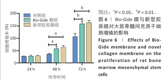
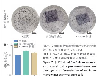
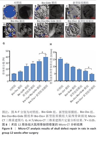
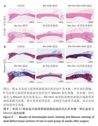
方法:①去除猪膀胱的浆膜层和部分肌肉层,制备新型胶原膜。表征新型胶原膜与Bio-Gide胶原膜的表面形貌、吸水率、孔隙率、降解率与拉伸弹性模量、极限载荷。②将大鼠骨髓间充质干细胞分别与新型胶原膜与Bio-Gide胶原膜共培养,以单独培养的细胞为对照,CCK-8法检测细胞增殖;成骨诱导后7 d,通过碱性磷酸酶染色评估成骨分化情况。③在18只SD大鼠颅骨矢状缝左右两侧各制作1个直径5 mm的圆形全层骨缺损,36个缺损部位随机分6组干预:对照组(n=6)缺损处不植入任何材料,Bio-Oss组(n=6)缺损处填充Bio-Oss骨粉,Bio-Gide组(n=6)缺损处覆盖Bio-Gide膜,新型胶原膜组(n=6)缺损处覆盖新型胶原膜,Bio-Oss+Bio-Gide组(n=6)缺损处填充Bio-Oss骨粉后覆盖Bio-Gide膜,Bio-Oss+新型胶原膜组(n=6)缺损处填入Bio-Oss骨粉后再覆盖新型胶原膜。术后12周取材,分别进行Micro-CT扫描与组织学观察。
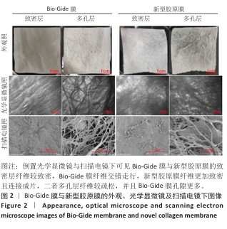
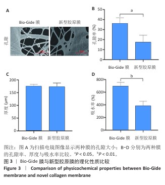
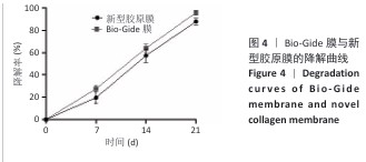
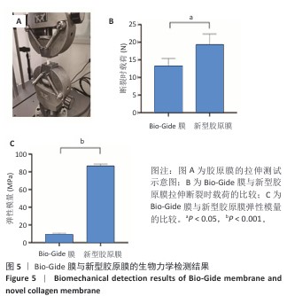
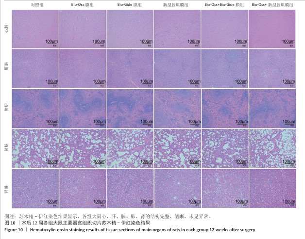
结果与结论:①扫描电镜下可见Bio-Gide膜与新型胶原膜的致密层纤维较致密,Bio-Gide膜纤维交错走行,新型胶原膜纤维更加致密且连接成片,二者多孔层纤维较疏松,并且Bio-Gide膜孔隙更多。新型胶原膜的孔隙率、吸水率均低于Bio-Gide膜(P < 0.05),拉伸弹性模量与极限载荷均高于Bio-Gide膜(P < 0.05)。②相较于Bio-Gide膜,新型胶原膜的降解更稳定。CCK-8检测显示,Bio-Gide膜与新型胶原膜均可促进大鼠骨髓间充质干细胞的增殖。碱性磷酸酶染色显示,Bio-Gide膜与新型胶原膜均不影响大鼠骨髓间充质干细胞的成骨分化。③Micro-CT扫描结果显示,对照组骨缺损处有少量新骨形成,其他5组骨缺损处有大量新骨形成,其中Bio-Oss+新型胶原膜组新骨形成量与骨结构成熟度最佳。苏木精-伊红染色与Masson染色显示,对照组缺损区新生骨形成较少,其余5组缺损区新生骨形成较多,其中Bio-Oss+新型胶原膜组新生骨质密度更高、结构更成熟。④结果表明,新型胶原膜的理化特性优于Bio-Gide膜,在配合使用Bio-Oss骨粉时,新型胶原膜表现出比Bio-Gide膜更优的骨修复效果。
https://orcid.org/0009-0004-6290-4213 (杨平) 

中国组织工程研究杂志出版内容重点:生物材料;骨生物材料;口腔生物材料;纳米材料;缓释材料;材料相容性;组织工程

关键词: 引导骨再生">, 新型胶原膜">, Bio-Gide膜">, 大鼠">, 颅骨缺损">, 骨髓间充质干细胞">, 疗效">, 微型计算机断层扫描

Abstract: BACKGROUND: The commonly used absorbable barrier membrane in guided bone regeneration technology is Bio-Gide collagen membrane (source of pig skin), which has the disadvantages of poor mechanical strength and fast degradation rate, thus limiting its clinical application. Therefore, it is of great significance to develop collagen membranes with better physicochemical properties.
OBJECTIVE: To prepare a novel collagen membrane derived from porcine bladder, characterize its physicochemical properties and its effect on repairing rat skull bone defects. 
METHODS: (1) The serosal layer and partial muscle layer of porcine bladders were removed to prepare a novel collagen membrane. The surface morphology, water absorption, porosity, degradation rate, tensile modulus, and ultimate load of the novel and Bio-Gide collagen membranes were characterized. (2) Rat bone marrow mesenchymal stem cells were co-cultured with the novel and Bio-Gide collagen membranes, respectively. Cells cultured alone were used as controls. Cell proliferation was assessed by CCK-8 assay. Osteogenic differentiation was assessed by alkaline phosphatase staining 7 days after osteogenic induction. (3) A 5-mm-diameter circular, full-thickness bone defect was created on each side of the sagittal suture of the skull in 18 SD rats. Thirty-six defect sites were randomly divided into six intervention groups: the control group (n=6) received no implantation at the defect site; the Bio-Oss group (n=6) had the defect filled with Bio-Oss bone powder; the Bio-Gide group (n=6) had the defect covered with a Bio-Gide membrane; the novel collagen membrane group (n=6) had the defect covered with a novel collagen membrane; the Bio-Oss+Bio-Gide group (n=6) had the defect filled with Bio-Oss bone powder and then covered with a Bio-Gide membrane; and the Bio-Oss+novel collagen membrane group (n=6) had the defect filled with Bio-Oss bone powder and then covered with a novel collagen membrane. Twelve weeks after surgery, the tissue samples were harvested for micro-computed tomography and histological observation.
RESULTS AND CONCLUSION: (1) Scanning electron microscopy revealed that the fibers in the compact layers of the Bio-Gide and novel collagen membranes were denser, with the fibers in the Bio-Gide membrane interlaced. The fibers in the novel collagen membrane were denser and connected into sheets. The fibers in the porous layers of both membranes were looser, with the Bio-Gide membrane having more pores. The porosity and water absorption of the novel collagen membrane were lower than those of the Bio-Gide membrane (P < 0.05), while the tensile elastic modulus and ultimate load were higher than those of the Bio-Gide membrane (P < 0.05). (2) Compared with the Bio-Gide membrane, the novel collagen membrane was more stable to degradation. CCK-8 assay showed that both the Bio-Gide and novel collagen membranes promoted the proliferation of rat bone marrow mesenchymal stem cells. Alkaline phosphatase staining revealed that neither the Bio-Gide nor the novel collagen membranes affected the osteogenic differentiation of rat bone marrow mesenchymal stem cells. (3) Micro-computed tomography revealed a small amount of new bone formation at the bone defect site in the control group, while substantial new bone formation was observed in the other five groups. The Bio-Oss+novel collagen membrane group demonstrated the greatest amount of new bone formation and structural maturity. Hematoxylin-eosin and Masson staining revealed less new bone formation at the defect site in the control group, while more was observed in the other five groups. The Bio-Oss+novel collagen membrane group demonstrated higher new bone density and more mature structure. (4) The results demonstrated that the novel collagen membrane exhibited superior physical and chemical properties to the Bio-Gide membrane. When combined with Bio-Oss bone powder, the novel collagen membrane demonstrated superior bone repair efficacy compared with the Bio-Gide membrane.

Key words: guided bone regeneration">, novel collagen membrane">, Bio-Gide membrane">, rat">, skull bone defect">, bone marrow mesenchymal stem cell">, efficacy">, micro-computed tomography

中图分类号: