中国组织工程研究 ›› 2026, Vol. 30 ›› Issue (31): 8237-8246.doi: 10.12307/2026.336
• 干细胞综述 stem cell review • 上一篇 下一篇
单细胞RNA测序技术在膝关节各组织相关疾病中的研究与应用
杨文戟1,2,戴 祝2
- 1南华大学,湖南省衡阳市 421000;2南华大学附属第一医院,湖南省衡阳市 421000
-
收稿日期:2025-06-06接受日期:2025-08-15出版日期:2026-11-08发布日期:2026-05-25 -
通讯作者:戴祝,博士,教授,博士生导师,南华大学附属第一医院创伤骨科,湖南省衡阳市 421000 -
作者简介:杨文戟,男,1998年生,湖南省株洲市人,汉族,南华大学在读硕士,主要从事关节疾病与运动损伤的研究。 -
基金资助:湖南省卫生健康委临床重点课题项目(20201907),项目负责人:戴祝;湖南省自然科学基金项目(2023JJ60050),项目负责人:戴祝
Applications and research advances of single-cell RNA sequencing in diseases of knee joint tissues
Yang Wenji1, 2, Dai Zhu2
- 1University of South China, Hengyang 421000, Hunan Province, China; 2The First Affiliated Hospital of University of South China, Hengyang 421000, Hunan Province, China
-
Received:2025-06-06Accepted:2025-08-15Online:2026-11-08Published:2026-05-25 -
Contact:Dai Zhu, PhD, Professor, Doctoral supervisor, The First Affiliated Hospital of University of South China, Hengyang 421000, Hunan Province, China -
About author:Yang Wenji, MS candidate, University of South China, Hengyang 421000, Hunan Province, China; The First Affiliated Hospital of University of South China, Hengyang 421000, Hunan Province, China -
Supported by:Clinical Key Research Project of Hunan Provincial Health Commission, No. 20201907 (to DZ); Hunan Province Natural Science Foundation, No. 2023JJ60050 (to DZ)

摘要:

文题释义:
单细胞RNA测序技术:是一种在单细胞分辨率下解析组织转录组异质性的高通量技术,其核心优势在于能够精确识别膝关节组织中不同细胞亚群的基因表达特征。该技术可同时检测单个细胞中数千至数万个基因的表达量(检测灵敏度达0.1-1个转录本/细胞),并能鉴定传统测序方法无法区分的稀有细胞类型(如占比<1%的前软骨干细胞)。在膝关节研究中,单细胞RNA测序技术可定量分析软骨、滑膜等组织中各细胞亚群的差异表达基因(如骨关节炎中软骨细胞的Ⅱ型胶原α1表达下调>50%,基质金属蛋白酶13上调3-5倍),并通过拟时序分析揭示细胞状态转变的分子机制。该技术为膝关节疾病的细胞特异性靶点发现提供了单细胞精度的研究手段。膝关节组织:是由多种异质性细胞群构成的复杂功能单元,主要包括软骨(占关节体积15%-20%)、滑膜(厚度50-100 μm)、半月板(胶原纤维占比>75%)及韧带(Ⅰ型胶原含量>90%)等。在单细胞RNA测序研究中,这些组织可解析出10-30种不同的细胞亚群,如软骨中的肥大软骨细胞、滑膜中的成纤维细胞亚型以及免疫细胞。膝关节组织的细胞组成和基因表达谱直接影响其生理功能及病理进程。单细胞分辨率的研究可精准量化这些细胞群体的分子特征,为膝关节疾病的机制解析提供组织学基础。
摘要
背景:单细胞RNA测序技术是从组织样本中分离出单个细胞为单位进行测序并构建出分子图谱,它能够分析各细胞群体中的细胞异质性,从而为研究组织和疾病发生发展过程提供新的视角。近年来单细胞RNA测序技术不断发展与进步,使得越来越多的研究者将单细胞RNA测序技术应用到各个领域获得新的发现,膝关节的研究就是其中之一。
目的:介绍单细胞RNA测序技术的背景,对其在膝关节的应用及取得的进展进行综述,并对单细胞RNA测序技术所面临的挑战及未来发展的前景进行讨论。
方法:应用计算机系统检索PubMed、Web of Science和中国知网数据库的相关文献,英文检索词为“single-cell RNA sequencing,sequencing technology,articular cartilage,synovium,bone,meniscus,lagement”;中文检索词为“单细胞RNA测序,测序技术,关节软骨,滑膜,骨骼,半月板,韧带”。排除质量较差、内容重复及不相关的文献,最终纳入79篇文献进行综述分析。
结果与结论:①单细胞RNA测序作为一种高通量测序技术,通过解析单个细胞的转录组信息突破了传统批量测序的局限性。与传统测序技术相比,其核心优势包括揭示细胞异质性、追踪细胞分化轨迹、构建细胞间调控网络、识别疾病特异性细胞亚群,这些特性使单细胞RNA测序成为研究复杂组织(如膝关节)细胞组成与功能的关键工具。②单细胞RNA测序技术已被应用于膝关节软骨、滑膜、骨骼、半月板和韧带等组织的研究,探讨不同组织的细胞亚型及独特功能,让研究者更好地了解膝关节各组织细胞的异质性以及启动病理的“驱动”细胞群体。③通过单细胞RNA测序分析,提高了研究者对膝关节各组织相关疾病的了解,揭示了复杂组织中细胞的多样性和相关基因表达水平变化,找到发病机制生物学行为、通路和潜在治疗作用的分子靶点。④尽管单细胞RNA测序技术成果显著,仍面临着技术瓶颈、数据分析复杂性和临床转化障碍的挑战。对于未来发展的方向,则包括多组学整合、精准医学应用、技术优化等。
中图分类号:
引用本文
杨文戟, 戴 祝. 单细胞RNA测序技术在膝关节各组织相关疾病中的研究与应用[J]. 中国组织工程研究, 2026, 30(31): 8237-8246.
Yang Wenji, Dai Zhu. Applications and research advances of single-cell RNA sequencing in diseases of knee joint tissues[J]. Chinese Journal of Tissue Engineering Research, 2026, 30(31): 8237-8246.
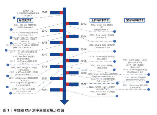
2.1.1 单细胞RNA测序技术的背景 2009年,TANG等[5]率先采用高通量测序技术对单个分离的胚胎细胞进行mRNA转录组研究,这一突破性工作标志着单细胞测序(single cell sequencing,SCS)技术在科研领域的广泛应用正式拉开序幕。随着单细胞测序技术的普及,细胞生物学和分子生物学领域近年来取得了显著进展[5]。2018年,研究人员利用单细胞测序技术构建了斑马鱼和青蛙早期胚胎发育过程中基因表达的动态图谱,通过整合分钟至小时级别的时间尺度上的测序数据,逐个解析细胞,追踪胚胎的最终形成,成功构建了完整的发育过程图谱,揭示了从单个细胞到整个器官的发育轨迹。单细胞测序的广泛应用使得研究人员在多个方面对细胞功能有了更深入和全面的理解[6],包括肿瘤形成[7]、神经退行性变[8]、免疫反应[9]、细胞分化以及基因表达调控等[10-12]。近年来单细胞测序技术得到了快速、全面的发展,许多更优质的技术得到开发,主要发展历程见图3。
2.1.2 单细胞RNA测序技术的原理和流程 单细胞测序的原理是从组织、液体样本里分离出单个细胞,继而对这些单个细胞里的遗传物质进行测序剖析,跟传统的二代测序技术不一样,后者主要对样本里整个细胞群体的“平均基因组”进行检测,因而掩盖了不同细胞亚群在基因表达以及功能上的多样性,单细胞测序可以精准区分样本中的不同细胞亚群,且在单细胞水平探究这些亚群之间基因表达的差异,进而揭示样本中细胞亚群的异质性,该方法为探索组织和疾病的发生机制提供了全新的研究切入点。
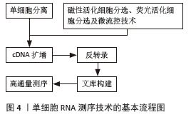
单细胞RNA测序技术是单细胞测序中应用最为广泛的技术之一,以下简要介绍单细胞RNA测序技术的基本流程:单细胞分离,反转录,cDNA扩增,文库构建,高通量测序。其中,单细胞分离是单细胞RNA测序技术的初始步骤,也是最为关键的环节,目前,常用的单细胞分离方法包括磁性活化细胞分选、荧光活化细胞分选以及微流控技术。磁性活化细胞分选利用抗体介导的超顺磁纳米粒子标记目标细胞表面的特定蛋白质,随后通过外部磁场分离标记细胞,而未标记的细胞则被洗脱。流式细胞分选技术是一种常用的高纯度单细胞分离方法,其通过荧光单克隆抗体识别特定的细胞表面标记,从而对不同细胞群进行分类[13]。微流控技术则通过微米级流道在微升或毫升级别的样本中分离单细胞,具有高灵敏度、高特异性以及高通量的优势。单细胞高通量液滴测序是微流控技术中获取单细胞的代表性方法,该方法使用条形码珠,珠上的寡核苷酸包含用于PCR扩增的引物、识别单细胞的唯一分子标识符以及捕获单细胞mRNA的寡核苷酸。基于单细胞高通量液滴测序的原理,10X Genomics开发了一个广泛应用的单细胞测序文库制备平台,其通过油滴微流体装置高效捕获更多细胞,具有更高的灵敏度[14]。此外,BD Rhapsody是另一个广泛使用的单细胞测序平台。与单细胞高通量液滴测序不同,BD Rhapsody采用CytoSeq独特的细胞面板,而非通过微流控通道发射细胞并与磁珠碰撞[15]。具体而言,细胞悬浮液通过注射孔注入后自然沉降到反应孔中,随后通过同样的方式将珠子插入反应孔,从而将细胞捕获在单个反应孔中,实现细胞的物理隔离,见图4。
2.1.3 单细胞RNA测序技术的优势 相较于传统的批量RNA测序,单细胞RNA测序技术在生物学研究中展现出四大优势。其一,该技术能够解析细胞间的异质性或识别特定的细胞亚群;其二,它可以刻画生理和病理过程中不同细胞状态、转变及分化的动态路径;其三,单细胞RNA测序技术能够通过富集分析构建异质性细胞信号模型;其四,它能够揭示细胞群体间的调控网络[16]。单细胞RNA测序技术的研究进展深化了对这些疾病的理解,从而为预防、干预及治愈这些疾病提供了潜在的策略。
2.2 单细胞RNA测序技术在膝关节各组织中的应用与研究 膝关节结构复杂,由关节囊内的骨骼、软骨、韧带、半月板和滑膜组成[17]。最常见的关节疾病是骨关节炎和类风湿关节炎[4],两者的病因有明显区别,骨关节炎主要是在软骨组织中,而类风湿关节炎主要是在滑膜组织中。类风湿关节炎的特征是伴有大量浸润的免疫细胞,如常驻的滑膜成纤维细胞[18],炎症滑膜分泌的细胞因子信号促使破骨细胞成熟,导致骨破坏以及软骨细胞外基质降解[19]。骨关节炎早期表现为关节软骨表面纤维化以及细胞外基质逐渐侵蚀,伴随软骨下骨重塑,最终到晚期骨关节炎会出现滑膜炎症,但其滑膜免疫细胞与类风湿关节炎不同[20-21]。目前尚不清楚两种疾病中软骨与滑膜组织的致病细胞群是否重叠,且这种潜在重叠性不仅存在于上述两种疾病,更存在于整个膝关节的不同组织之间,因此了解膝关节各组织细胞的异质性以及启动病理的“驱动”细胞群体至关重要。膝关节的复杂组织结构包含多种细胞类型,除了识别致病细胞群体外,单细胞数据还能够揭示相关基因特征、生物学行为、寻找潜在治疗靶点,从而为理解疾病进展过程中细胞和分子环境的变化提供了更高分辨率的视角。许多研究已将单细胞RNA测序技术应用于膝关节各组织,如软骨[22]、骨[23]、滑膜[24]、半月板等[25]。
2.2.1 关节软骨 膝关节软骨朝向关节腔的面光滑,便于骨与骨之间的运动,软骨本身具有弹性,所以能缓冲相连骨在走、跳及其他运动时的振动和冲击,增加关节活动性,是膝关节的重要组成部分之一[26]。
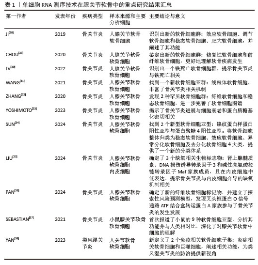
关节软骨破坏在骨关节炎疾病中最常见,其中软骨细胞为关节软骨的主要细胞,因此单细胞RNA测序技术在膝关节软骨中的研究主要集中于软骨细胞[27]。JI等[28]通过对膝关节置换术后骨关节炎患者的关节软骨进行单细胞RNA测序研究,发现了3种新的软骨细胞亚群:效应软骨细胞、调节软骨细胞和稳态软骨细胞,均在骨关节炎发展过程中发挥保护作用。同时该研究还识别出2种不同的肥大软骨细胞亚群:亚型肥大软骨细胞A与软骨发育和结缔组织生成相关的基因表达相关,亚型肥大软骨细胞B则与细胞外基质重塑、骨化及矿化相关的基因表达密切相关,这进一步深化理解了肥大软骨细胞在骨关节炎发展中的作用。CHOU等[29]识别出2种新的软骨细胞群:修复性软骨细胞和前纤维软骨细胞,前者高表达与细胞外基质信号和胶原纤维组织相关的基因,表现出较强的修复能力;后者高表达成纤维细胞相关基因和白细胞介素11,可能在细胞外调节蛋白激酶信号通路中起重要作用。LV等[30]首次在人类骨关节炎样本中识别出一个铁死亡软骨细胞群,激活瞬时感受器电位香草酸受体1可以通过上调谷胱甘肽过氧化酶4来防止软骨细胞发生铁死亡。WANG等[31]对健康软骨细胞的细胞群及其遗传特征进行了鉴定,成功识别出10个细胞簇,并通过细胞标记物进行了详细注释;此外,他们还发现了一个新的亚群——线粒体软骨细胞。为了探究骨关节炎不同阶段关节软骨细胞的表型变化,ZHANG等[32]采用单细胞RNA测序技术识别出2种较为罕见的软骨细胞亚群:纤维软骨细胞和稳态软骨细胞,纤维软骨细胞主要存在于晚期骨关节炎中,与胶原蛋白的形成紧密相关,通过调控纤维软骨细胞的功能,可以影响胶原蛋白的生成;稳态软骨细胞是近年新发现的细胞亚群,有助于调节软骨细胞代谢活动的稳态平衡,以适应软骨微环境的变化。YOSHIMOTO等[33]利用单细胞RNA测序技术研究了骨关节炎患者软骨细胞衰老与蛋白质糖基化之间的联系,研究结果发现在骨关节炎进展过程中,细胞衰老与蛋白质糖基化密切相关,尤其是O-连接糖基化;此外,该家族的表达水平与软骨细胞的衰老进程呈相关性。SUN等[34]对正常与骨关节炎患者的膝关节软骨进行单细胞分析,鉴定出11种软骨细胞亚型,其中包括2个新型软骨细胞亚型:镍纹蛋白样蛋白阳性亚型与蛋白聚糖4阳性亚型。整体归类为稳态软骨细胞、效应软骨细胞、异常分化软骨细胞及去分化软骨细胞4大类。随着细胞分化,细胞亚型逐渐转变为细胞外基质黏附与胶原重塑特征,信号通路也从缺氧诱导因子1转变为Hippo通路。LIU等[35]找到12个缺氧相关差异表达基因,并确定了3个缺氧相关生物标志物(肾上腺素髓质素、DNA损伤诱导转录因子3和碱性亮氨酸拉链转录因子Maf家族成员),同时发现15种具有显著差异的免疫细胞,并证实骨关节炎组内皮细胞中这3个缺氧相关生物标志物的表达水平均低于正常组,为骨关节炎的诊疗提供了理论基础。PAN等[36]鉴定出10种软骨细胞类型,并筛选出6个纤维软骨细胞相关骨关节炎生物标志物(B细胞淋巴瘤因子6、ATP结合盒转运蛋白A5、ATP结合盒转运蛋白A6、Cbp/p300 结合转化激活因子、核受体亚家族1D组成员1和溶质载体家族7成员8),建立了探索性风险预测模型,同时发现叉头框蛋白O信号通路可能与这些标志物相关,其中ATP结合盒转运蛋白A5和ATP结合盒转运蛋白A6涉及软骨细胞胆固醇稳态调控,揭示了ATP结合盒转运蛋白A家族成员作为骨关节炎发病机制的新参与者,为治疗靶点开发提供了新方向。SEBASTIAN等[37]首次报道了健康小鼠膝关节软骨中的9种软骨细胞亚型(Ucmahigh、Cytl1high、Chil1high、Mef2chigh、Krt16high、Tnfaip6high、S100a4high、Neat1high和divC软骨细胞群),其中Ucmahigh和Krt16high群高表达与蛋白质合成和mRNA代谢相关的基因,还发现小鼠中的部分细胞群分别与人类对应细胞群相似。这些研究成果不仅深化了对膝关节软骨中细胞功能的理解,还为骨关节炎的治疗策略开辟了新的可能性。
类风湿关节炎也会导致关节软骨破坏,引起关节畸形、僵硬和功能障碍。YAN等[38]研究了类风湿关节炎患者股骨负重和非负重区域的软骨,鉴定了6个细胞群,其中新定义了2个免疫相关软骨细胞子集(炎症相关软骨细胞和巨噬细胞)。位于整个软骨层和中层的炎症相关软骨细胞亚群表现出很高的免疫活性,主要参与免疫过程,如抗原处理和呈现、组织相容性复合体Ⅱ类蛋白质复合物和免疫受体活性;巨噬细胞亚群分布在整个软骨层,被认为是具有免疫细胞黏附、细胞因子活性等炎症特征的软骨细胞,在白细胞-细胞黏附、免疫突触、细胞因子活性和细胞因子-细胞因子受体相互作用方面起到重要调节作用。
单细胞RNA测序技术在膝关节软骨中的重点研究结果汇总,见表1。
2.2.2 滑膜 滑膜是关节疾病中发生显著变化的关节组织之一,也是通过与其他组织的相互作用在病理过程中起主要作用的组织。在类风湿关节炎和骨关节炎中,一旦稳态屏障被破坏,滑膜中就会出现大量浸润细胞[39]。
类风湿关节炎作为一种主要累及周围关节的系统性自身免疫疾病,以滑膜慢性炎症为典型特征。ZHANG等[40]对类风湿关节炎患者的滑膜炎症进行了单细胞RNA测序分析和流式细胞术分析,通过整合单细胞水平的数据,精确定义了18种独特的细胞亚群,全面展示了类风湿关节炎患者滑膜组织的细胞组成。研究进一步采用细胞因子-细胞亚群关联分析,将白细胞介素6的表达特征精准定位于THY1+HLA-DRhi表型的成纤维细胞亚群,同时将白细胞介素1β的合成来源确认为具有促炎表型的单核细胞群体。
在关于滑膜组织中成纤维细胞的研究中,CROFT等[41]发现,删除FAPα+表型的成纤维细胞可以抑制炎症和骨侵蚀,FAPα+THY1+表型的成纤维细胞亚群主要与严重持续性关节炎的病理过程密切相关;FAPα+THY1-表型的成纤维细胞亚群则特异性参与骨和软骨组织的损伤过程,在炎症反应中的作用较小。WEI等[42]揭示了Notch同源物3信号在CD90(THY1)+成纤维细胞分化和病理性生长中的关键作用,导致炎症性关节炎的发展,抑制Notch同源物3信号通路能够有效减轻炎症反应。ZHENG等[43]鉴定出一种CD45-CD31-PDPN+ITGA5+滑膜成纤维细胞亚群,其具有独特的骨膜蛋白、Ⅲ型胶原A1、CC类趋化因子配体5和转化生长因子β1转录组特征,并在免疫调节通路中显著富集,这群细胞可能通过诱导CXCL13hiPD-1hi TPHs细胞的分化及重塑促炎微环境来调控类风湿关节炎的进展。
在关于类风湿关节炎巨噬细胞的研究中,KUO等[44]发现了一种HBEGF+的炎症巨噬细胞亚群,通过表皮生长因子受体促进成纤维细胞的侵袭,导致关节破坏,同时这些巨噬细胞高表达与炎症反应相关的基因(核受体亚家族4A组成员3、尿激酶型纤溶酶原激活因子受体、CXC 趋化因子配体 2及生长因子肝素结合性表皮生长因子、表皮调节素);阻断这些巨噬细胞与成纤维细胞之间的相互作用,可能为类风湿关节炎的治疗提供新的途径。ALIVERNI等[45]发现,通过激活MerTK受体或相关转录因子诱导MerTKposCD206pos巨噬细胞具有治疗类风湿关节炎的效果,有助于恢复滑膜稳态。CULEMANN等[46]研究揭示,CX3CR1+巨噬细胞亚群能够在滑膜内层形成一种动态的膜状结构,这种结构有助于维持关节内部的紧密连接,从而保护关节免受炎症反应的损害。
对于其他免疫细胞,如T细胞、自然杀伤细胞、嗜酸性粒细胞、单核细胞和B细胞等也有相关研究。KELKKA等[47]在具有侵袭性抗瓜氨酸肽抗体阳性的类风湿关节炎患者的CD8+细胞毒性T细胞中检测到特定的T细胞受体特征,并且这些细胞高表达与骨破坏相关的细胞因子肿瘤坏死因子配体超家族成员14。MEEDNU等[48]识别出在类风湿关节炎滑膜中富集的独特B细胞,可能是滑膜中慢性自身抗原刺激的生物标志物和新的治疗靶点。ANDREEV等[49]发现一种新型的调节性嗜酸性粒细胞亚群存在于关节中,这些细胞能够通过Ⅱ型固有淋巴细胞-白细胞介素5轴在类风湿关节炎缓解期改善慢性关节炎症,调节性嗜酸性粒细胞在关节中的积累也增强了关节结构的保护,表明这些细胞可能促进组织再生。HAN等[50]观察到类风湿关节炎患者中CD56bright和CD56dim NK细胞的活跃干扰素信号通路与疾病进展之间存在联系,干扰素信号通路激活可能影响类风湿关节炎浆细胞中的抗体类别转换和自身抗体的合成。DUNLAP等[51]也对类风湿关节炎患者滑膜中B细胞和T细胞的功能进行分析,系统揭示了类风湿关节炎患者滑膜浸润淋巴细胞中功能异质性群体间的克隆关联。综上这些研究有助于更好地理解类风湿关节炎滑膜的组织学、异质性、进展和治疗。
有研究者进行了骨关节炎中滑膜组织的单细胞RNA测序分析。NANUS等[52]在早期和晚期骨关节炎患者中识别出了7种不同的滑膜成纤维细胞亚群(Cluster 0-6):Cluster 0在骨关节炎晚期更为常见,其高表达与疼痛相关的基因;Cluster 2-5则在早期更常见,主要存在于疼痛区,高表达与纤维化、炎症和神经元生长相关的基因;Cluster 1和6也在早期更常见,但主要存在于无痛区域。CAI等[53]进一步揭示骨关节炎患者的滑膜成纤维细胞在功能状态转变过程中与类风湿关节炎表现出相似性,提示这两种疾病可能存在共同的致病机制,同时探讨了调控滑膜成纤维细胞功能状态的核心信号通路。TANG等[54]发现滑膜成纤维细胞的祖细胞是DPP4+间充质细胞,此外骨关节炎会诱导间充质谱系细胞促纤维化与炎症表型。YU等[55]通过对骨关节炎患者滑膜组织进行单细胞RNA测序分析,确定了4个新的早期骨关节炎的诊断和预测标志物,鉴定的细胞群中滑膜成纤维细胞在每个样本中比例最高。当滑膜成纤维细胞被激活时,细胞分泌细胞因子、趋化因子和降解基质的酶,最终导致关节破坏。LIU等[56]在骨关节炎患者单细胞数据集中发现,CILP+成纤维细胞主要在非损伤软骨细胞区域,而POSTN+成纤维细胞则主要在损伤软骨细胞区域。CILP+成纤维细胞可能对软骨细胞发挥修复保护作用,而POSTN+成纤维细胞加剧软骨退变。这些结果为探索潜在的治疗靶点提供了新的思路,同时也为延缓骨关节炎的疾病进展指明了潜在的研究方向。
单细胞RNA测序技术在膝关节滑膜中的重点研究结果汇总,见表2。

2.2.3 骨骼 骨骼比大多数组织更为复杂,类似于一个器官,因为它包含不同的区域,如骨髓、骨膜和骨内膜,这些区域具有不同的功能,并容纳多种细胞类型,包括多种间充质细胞、成骨细胞、骨细胞及破骨细胞等。
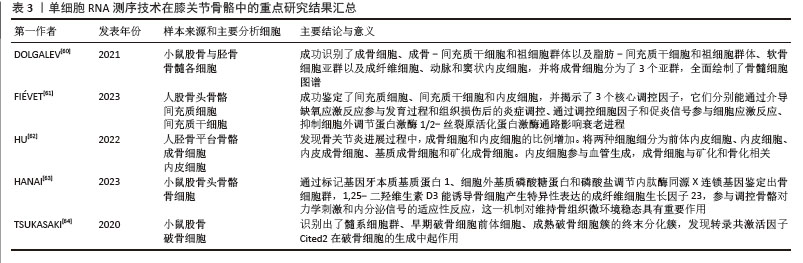
骨髓中含有造血干细胞及其衍生的细胞系,能够分化为骨、软骨、脂肪和基质细胞的多能间充质干细胞,以及内皮细胞和血管细胞。多项小鼠单细胞研究描述了产生不同骨骼细胞的间充质干细胞和祖细胞群体[57-59]。DOLGALEV等[60]通过结合3项不同的小鼠股骨与胫骨骨髓单细胞RNA测序数据,成功识别了成骨-间充质干细胞和祖细胞群体以及脂肪-间充质干细胞和祖细胞群体,此外还识别了软骨细胞亚群以及成纤维细胞、动脉和窦状内皮细胞。FIéVET等[61]取人股骨头进行单细胞RNA测序分析,成功鉴定了间充质细胞、间充质干细胞和内皮细胞,还揭示了3个核心调控模块(早期生长应答因子1、锌指蛋白36和侧支发芽因子同源物1),这些因子可能位于关键通路的核心节点,其中早期生长应答因子1能介导缺氧应激反应、参与发育过程或组织损伤后的炎症调控;锌指蛋白36通过调控细胞因子和生长因子信号(尤其是促炎信号)参与细胞应激反应;侧支发芽因子同源物1则能抑制细胞外调节蛋白激酶1/2-丝裂原活化蛋白激酶通路在脂肪细胞早期分化阶段表达来影响衰老进程。
对于成骨细胞的研究,DOLGALEV等[60]最初将成骨细胞分为3个主要簇:Col16a1high Tnnhigh成骨细胞;Fbn1high Igf1high转分化成骨细胞;Bglaphigh Car3high成熟成骨细胞。此外,整合数据分析全面绘制了成骨细胞上游的基质细胞亚群,还识别了早期定向的成骨祖细胞。HU等[62]为研究成骨细胞和其他细胞群体的差异,从2例接受关节置换手术患者的胫骨平台和受损的内侧平台中分离细胞进行分析,发现在骨关节炎进展过程中,成骨细胞和内皮细胞的比例增加。将两种细胞分为前体内皮细胞、内皮细胞、内皮成骨细胞、基质成骨细胞和矿化成骨细胞,前体内皮细胞在核糖体和外泌体合成方面富集;而内皮细胞在血管生成相关通路中富集;内皮成骨细胞在血管内皮生长因子结合和血小板衍生生长因子受体β信号通路中富集,表明它们可能参与血管生成;基质成骨细胞在细胞外基质沉积相关通路中富集;而矿化成骨细胞在与成熟成骨细胞相关的矿化和骨化通路中富集。
骨细胞作为终末分化的骨组织细胞,在骨中占比较高,其发育过程具有独特的生物学特征。这类细胞由成熟的成骨细胞经过一系列复杂的生物学转变而形成。从细胞学特征来看,成熟的骨细胞网络通过特殊的细胞间连接结构实现细胞间的通讯联系,同时依靠骨小管系统中的组织液流动建立功能性的机械信号传导系统。HANAI等[63]取小鼠股骨头进行单细胞RNA测序分析,通过特异性标志物牙本质基质蛋白1、细胞外基质磷酸糖蛋白和磷酸盐调节内肽酶同源X连锁基因对骨细胞群体进行鉴定,经骨化三醇处理后,部分骨细胞亚群可检测到成纤维细胞生长因子23的特异性表达。1,25-二羟维生素D3能诱导骨细胞产生成纤维细胞生长因子23,还能参与调控骨骼对力学刺激和内分泌信号的适应性反应,这一机制对维持骨组织微环境稳态具有重要作用。
骨重塑需要通过破骨细胞进行骨吸收来实现,TSUKASAKI等[64]从小鼠中分离出骨髓细胞,并通过逐步添加巨噬细胞集落刺激因子和核因子κB受体活化因子配体来诱导破骨细胞生成,对破骨细胞培养第0,1,3天的细胞进行单细胞RNA测序分析,识别出7个细胞簇,包括起始的髓系细胞群、表达树突状细胞标记CD11c的早期破骨细胞前体细胞,以及产生最终成熟破骨细胞簇的终末分化簇。此外,发现删除Cited2(编码p300/CBP介导的转录复合物的转录共激活因子)将导致破骨细胞的生成受到抑制,表明其在破骨细胞命运决定中起关键作用。
单细胞RNA测序技术在膝关节骨骼中的重点研究结果汇总,见表3。
2.2.4 半月板 半月板也是膝关节中的重要组成部分,主要负责吸收振动、分散压力、缓冲冲击以及增强关节的稳定性。然而,由于半月板的血液供应不足,其自我修复能力较弱,容易发生退化,退变的半月板不仅无法有效保护膝关节,还容易发生撕裂,进而损伤膝关节软骨,导致膝关节疼痛和活动受限等症状[65]。
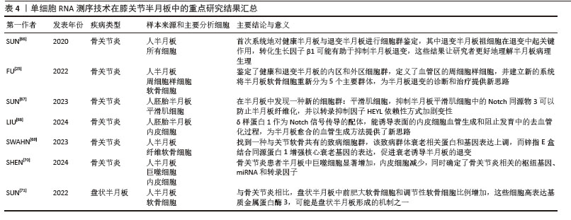
对于半月板组织的单细胞RNA测序研究主要集中于退变因素的分析。SUN等[66]使用单细胞RNA测序技术首次对健康和骨关节炎患者的半月板进行了分析,将健康半月板中的细胞确定为7个不同的簇:纤维软骨细胞、纤维软骨前体细胞、增殖性纤维软骨细胞、软骨祖细胞、调节性软骨细胞、前肥大软骨细胞和内皮细胞。在退变半月板中识别出3个细胞簇:单核细胞衍生的树突状细胞、肥大软骨细胞和退变半月板祖细胞,其中退变半月板祖细胞在半月板退变中起着关键作用,并且可用作评估半月板退变的标志物或作为治疗半月板退变的靶点。转化生长因子β1在健康半月板细胞中高表达,可能有助于抑制半月板退变。FU等[25]分析了健康和退变半月板的内区和外区,内区软骨细胞簇包括软骨细胞、淋巴细胞、髓样细胞、内皮细胞和一组ACTA2+细胞;外区组织除了这些细胞外,还观察到5种类型的神经细胞,并发现一组施万细胞,表明外区可能参与感觉信号传递,甚至可能与疼痛处理有关。定义了主要分布在血管区的ACTA2+周细胞样细胞,主要参与维持血管稳定与促进血管生成;此外将半月板软骨细胞分为5个主要群体(Ch.1-Ch.5),并分析了每种软骨细胞的功能,为退行性疾病的诊断和治疗提供新思路。为研究人类半月板的纤维化过程,SUN等[67]利用人胚胎半月板进行单细胞RNA测序探索纤维化的细胞机制,将胚胎半月板细胞分为3个簇:纤维粒细胞/软骨细胞、内皮细胞和平滑肌细胞/周细胞;将平滑肌细胞与成熟半月板细胞一起分析发现Notch同源物3参与胚胎发育中的细胞命运决定、血管发育、平滑肌生物学和组织纤维化。对Notch通路纤维化相关的下游因子进行分析,找到HEYL转录抑制因子,抑制半月板平滑肌细胞中的Notch同源物3可以防止半月板纤维化,并以HEYL依赖性方式加剧变性。LIU等[68]也利用胚胎半月板与成熟半月板对内皮细胞进行单细胞分析,观察到胚胎半月板12周开始出现内皮细胞无血管形成,14周血管位于半月板表面;δ样蛋白1作为Notch信号传导的配体解释了靠近表面的内皮细胞血管生成较低以及发育中的去血管化过程,为半月板愈合的血管生成方法提供了证据。SWAHN等[69]对健康和骨关节炎患者膝关节软骨和半月板组织进行单细胞RNA测序分析,探索在两种组织中的共同致病因素,结果找到一种致病细胞群,该致病群体的许多衰老相关核心蛋白基因和衰老分泌表型基因表达上调,锌指E盒结合同源蛋白1能够增强核心衰老基因的表达,在促进衰老诱导退变中发挥直接作用。SHEN等[70]利用公共单细胞数据库进行分析,对比健康和骨关节炎半月板发现骨关节炎组巨噬细胞显著增加,半月板中细胞毒性淋巴细胞和内皮细胞减少,确定了10个骨关节炎枢纽基因,同时预测了与枢纽基因密切相关的3个关键的miRNA和13个共享转录因子。
还有研究对盘状半月板进行了单细胞RNA测序分析,盘状外侧半月板是一种无症状的病症,也被称为弹响膝综合征,其特征是半月板增厚且呈椭圆形。SUN等[71]比较了人盘状外侧半月板与骨关节炎半月板,发现盘状外侧半月板中前肥大软骨细胞和调节性软骨细胞簇比例增加,这些细胞以高表达基质金属蛋白酶3为特征,表明异常的基质金属蛋白酶3表达可能是盘状外侧半月板形成的机制之一。
单细胞RNA测序技术在膝关节半月板中的重点研究结果汇总,见表4。
2.2.5 韧带 韧带是纤维结缔组织,膝关节的主要韧带包括前后交叉韧带(前后十字韧带)、内外侧副韧带、髌韧带,围绕膝关节的韧带主要作用是在整个关节活动范围内为关节提供稳定性[72]。
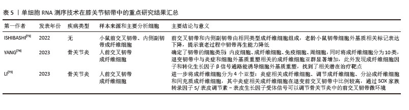
膝关节韧带损伤常见于前交叉韧带和内侧副韧带,细胞成分主要为成纤维细胞类型。ISHIBASHI等[73]对小鼠前交叉韧带与内侧副韧带的细胞成分进行了单细胞RNA测序,前交叉韧带和内侧副韧带由相同类型的成纤维细胞(结区成纤维细胞、“附件”细胞和腱细胞)组成。在前交叉韧带中,免疫细胞(B细胞、T细胞和中性粒细胞)在老年样本中的比例增加,表明与年龄相关的炎症导致前交叉韧带退化,Col14a1-2型细胞的比例随着年龄的增长而增加,而Col14a1-1型细胞的比例相对稳定。在内侧副韧带中,年轻和老年样本之间的免疫细胞比例几乎没有变化,而成纤维细胞的比例不同于Col14a1-1型和2型细胞的比例,随着年龄的增长而减少。老化韧带中细胞外基质相关标记的表达下降,解释了衰老过程中韧带再生能力的功能变化。YANG等[74]对骨关节炎患者膝前交叉韧带和健康膝前交叉韧带进行了单细胞分析,确定了4个细胞类别,包括内皮细胞、成纤维细胞、免疫细胞和周细胞。将成纤维细胞亚聚类,分为10个子集,并对退变组与健康组各细胞比例进行了分析,同时确定了7个血管衍生细胞群,包括5个内皮细胞群和2个周细胞群。退变韧带组织中与炎症和细胞外基质重塑相关的成纤维细胞亚群显著增加,且细胞外基质相关基因在退变组中上调,表明细胞外基质模块在韧带退化过程中发挥着重要作用。此外发现成纤维细胞因子和转化生长因子β信号通路可能会在韧带退变过程中诱导细胞外基质重塑,成纤维细胞因子7-成纤维细胞因子受体1和转化生长因子β1-转化生长因子β受体2可能是治疗这种疾病的潜在靶点。LI等[75]也对骨关节炎患者膝前交叉韧带和健康膝前交叉韧带进行了单细胞RNA测序分析,鉴定了6个细胞群:肌腱干细胞/祖细胞、增殖的成纤维细胞、成纤维细胞、血管平滑肌细胞、内皮细胞和巨噬细胞,成纤维细胞在退变前交叉韧带中比例很高,并且在炎症相关通路中明显富集。进一步将成纤维细胞鉴定为4个亚型:炎症相关成纤维细胞、调节成纤维细胞、分泌成纤维细胞和间充质成纤维细胞,其中炎症相关成纤维细胞比例在退变前交叉韧带中较高。炎症相关成纤维细胞亚群主要富集在炎症和成骨相关途径中,通过SOX家族转录因子5/表皮调节素-表皮生长因子受体信号传导轴途径调节骨关节炎中的前交叉韧带微环境和软骨退变,可以在前交叉韧带和软骨细胞代谢中发挥重要的调节作用。
单细胞RNA测序技术在膝关节韧带中的重点研究结果汇总,见表5。
2.2.6 多组织互作与整合 单细胞RNA测序技术为探究膝关节复杂组织结构提供了前所未有的精细分辨率,展现了骨关节炎和类风湿关节炎等膝关节疾病中跨组织互作网络的动态改变。膝关节作为一个功能化的整体,其关节软骨、滑膜、骨骼、半月板、韧带等组织利用共享微环境、细胞通讯及信号通路,协同构成复杂的相互作用网络,对现有研究的整合分析,提炼出多组织互作的核心特性。首先体现为代谢微环境共享,滑膜脂肪细胞释放出游离脂肪酸,被软骨细胞以及半月板细胞摄取后引起线粒体功能障碍,引起跨组织的代谢紊乱现象,软骨下骨中的异常血管生成,进一步对局部氧环境作出改变,影响软骨代谢[76]。然后是免疫炎症网络扩展,滑膜中的巨噬细胞、T细胞等免疫细胞,借助分泌白细胞介素1β、肿瘤坏死因子α等促炎因子,激活软骨细胞和韧带的炎症反应,软骨细胞退变释放的损伤相关分子还可召集更多免疫细胞,形成自我增益的循环[77]。再者是机械应力协同传导现象,半月板退变引起机械缓冲功能丧失,异常应力以软骨-软骨下骨单元为途径传导,引发骨细胞机械敏感通路,推进病理性骨的重塑。最后所提及的是基因表观协同调控,从上述各组织的研究进展可以看出,SOX转录因子家族中的SOX5、SOX9等在多个组织中协调细胞分化与基质代谢,其失调造成多组织的稳态呈现失衡现象。进一步探究跨组织间的相互协作,软骨下骨中的骨细胞经成纤维细胞生长因子23-无机磷酸盐通路来调节磷代谢,在骨关节炎进程中,骨重塑加速引起钙磷稳态被破坏,直接影响软骨基质的矿化进程,骨细胞网络借助骨小管系统察觉机械应力的变化,协调成骨细胞、破骨细胞的活性水平,异常的骨重塑引起软骨下骨板硬度的改变,影响到所覆盖软骨的生物力学环境,可见骨骼与软骨的钙磷代谢有着耦合的关系。滑膜成纤维细胞为炎症扩散的核心中介,在骨关节炎和类风湿关节炎相关疾病中,滑膜成纤维细胞被激活为类似POSTN+成纤维细胞的炎症表型,释放CC趋化因子5、CXC趋化因子12等趋化因子,募集单核细胞和T细胞,这些细胞呈现出对基质金属蛋白酶13、血小板反应蛋白解整合素金属肽酶5的高表达,直接引起软骨基质的降解;DPP4+间充质祖细胞受骨关节炎刺激后分化成促纤维化和炎症性滑膜成纤维细胞,此过程的调控依赖于转化生长因子β和Wnt信号,与类风湿关节炎的致病机制相近,体现两种疾病存在共同的炎性转分化渠道;出现铁死亡的软骨细胞释放出脂质过氧化物丙二醛,作为强大的炎症递质可激活滑膜以及韧带的炎症反应,这些全都是跨组织的免疫细胞相互串扰。前交叉韧带成纤维细胞凭借成纤维生长因子7-成纤维生长因子受体1和转化生长因子β1-转化生长因子受体2信号传导机械应力,退变的前交叉韧带中Col14a1-2型成纤维细胞数量增多,细胞外基质相关基因表达下降削弱了机械强度,而机械应力会诱导滑膜POSTN+成纤维细胞产生活化,凭借整合素-黏着斑激酶信号激活核因子κB通路,释放炎性递质,由此能看到跨组织的力学信号整合。就多组织彼此互作的展望,未来可凭借单细胞RNA测序技术及空间转录组等途径,剖析滑膜-软骨界面、骨-软骨交界区等分子间的对话;设立含有软骨、滑膜、骨等的关节类器官模型,模拟在机械应力、炎症及代谢扰动影响下多组织的响应,加快多组织整合治疗靶点方面的探索。
2.3 挑战与展望 对单细胞RNA测序技术在膝关节各组织研究应用方面的进展作综述,该技术挖掘出新型生物标志物和细胞亚群,说明了不同细胞类型的功能特性,同时探究了多种组织相关疾病的潜在原理,从而增进对这些疾病发生及发展过程的认识。尽管单细胞RNA测序技术在膝关节组织研究中已取得可观进展,但对膝关节相关疾病的整体发展、细胞异质性以及分子机制的全面研究仍处在起步阶段。
单细胞RNA测序技术近年得到大量关注,不过仍存在一定的局限,高昂的实验投入限制了样本分析的数量规模,这不仅使研究的统计学意义下降,还可能影响其在不同领域的普遍应用,当从骨骼、软骨等致密组织中去制备单细胞悬浮液时,很难彻底消除对细胞转录谱的干扰,这让数据解读面临挑战。因为组织所存在的硬度,特别是从骨组织中提取单个细胞尤为不易,解剖后的骨和软骨组织一般会用胶原酶进行长时间消化,为了获得足量且优质的RNA,相较于其他组织,骨骼或软骨组织在进行单细胞RNA测序时往往需要更大的样本量,这是鉴于骨和软骨在单位体积里面含有的细胞数量相对更少,因而在进行小动物研究时应用这项技术难度较大。尽管多重置换扩增是一种较好的基因组DNA扩增手段,尤其是针对DNA量少的临床样本,但它面临着扩增偏差和基因组覆盖不匹配的问题[78]。常见限制因素为批次效应和系统错误,运用不同批次的化学品以及采用多个生物样本都有可能导致批次效应。
虽然存在着以上的局限性和挑战性,单细胞RNA测序技术仍展现出了显著的应用前景。该技术能够对各种细胞的基因组和转录组进行多层次分析,从而可能彻底改变对疾病机制的理解,这种潜力贯穿于疾病发展的各个阶段。单细胞RNA测序技术在分析细胞异质性方面具有独特的优势[79]。因此,单细胞RNA测序技术依然能被应用于各系统疾病的研究,有助于开发各疾病组织修复的新策略。
| [1] PENG X, CHEN X, ZHANG Y, et al. Advances in the pathology and treatment of osteoarthritis. J Adv Res. 2025;78:257-283. [2] EVAN DIJK EL, JASZCZYSZYN Y, NAQUIN D, et al. The Third Revolution in Sequencing Technology. Trends Genet. 2018;34(9):666-681. [3] MARTEL-PELLETIER J, BARR AJ, CICUTTINI FM, et al. Osteoarthritis. Nat Rev Dis Primers. 2016;2:16072. [4] SMOLEN JS, ALETAHA D, BARTON A, et al. Rheumatoid arthritis. Nat Rev Dis Primers. 2018; 4:18001. [5] TANG F, BARBACIORU C, WANG Y, et al. mRNA-Seq whole-transcriptome analysis of a single cell. Nat Methods. 2009;6(5):377-382. [6] FARRELL JA, WANG Y, RIESENFELD SJ, et al. Single-cell reconstruction of developmental trajectories during zebrafish embryogenesis. Science. 2018; 360(6392):eaar3131. [7] BASLAN T, HICKS J. Unravelling biology and shifting paradigms in cancer with single-cell sequencing. Nat Rev Cancer. 2017;17(9):557-569. [8] WAGNER DE, WEINREB C, COLLINS ZM, et al. Single-cell mapping of gene expression landscapes and lineage in the zebrafish embryo. Science. 2018;360(6392):981-987. [9] NEU KE, TANG Q, WILSON PC, et al. Single-Cell Genomics: Approaches and Utility in Immunology. Trends Immunol. 2017;38(2):140-149. [10] OFENGEIM D, GIAGTZOGLOU N, HUH D, et al. Single-Cell RNA Sequencing: Unraveling the Brain One Cell at a Time. Trends Mol Med. 2017;23(6): 563-576. [11] TRAPNELL C, CACCHIARELLI D, GRIMSBY J, et al. The dynamics and regulators of cell fate decisions are revealed by pseudotemporal ordering of single cells. Nat Biotechnol. 2014;32(4):381-386. [12] POTTER SS. Single-cell RNA sequencing for the study of development, physiology and disease. Nat Rev Nephrol. 2018;14(8):479-492. [13] HU P, ZHANG W, XIN H, et al. Single Cell Isolation and Analysis. Front Cell Dev Biol. 2016;4:116. [14] ZHANG X, LI T, LIU F, et al. Comparative Analysis of Droplet-Based Ultra-High-Throughput Single-Cell RNA-Seq Systems. Mol Cell. 2019;73(1):130-142.e5. [15] FAN HC, FU GK, FODOR SP. Expression profiling. Combinatorial labeling of single cells for gene expression cytometry. Science. 2015;347(6222): 1258367. [16] SALIBA AE, WESTERMANN AJ, GORSKI SA, et al. Single-cell RNA-seq: advances and future challenges. Nucleic Acids Res. 2014;42(14):8845-8860. [17] DYE SF. The knee as a biologic transmission with an envelope of function: a theory. Clin Orthop Relat Res. 1996;(325):10-18. [18] SCHETT G. Cells of the synovium in rheumatoid arthritis. Osteoclasts. Arthritis Res Ther. 2007; 9(1):203. [19] TAKAYANAGI H. RANKL as the master regulator of osteoclast differentiation. J Bone Miner Metab. 2021;39(1):13-18. [20] DE LANGE-BROKAAR BJ, IOAN-FACSINAY A, VAN OSCH GJ, et al. Synovial inflammation, immune cells and their cytokines in osteoarthritis: a review. Osteoarthritis Cartilage. 2012;20(12):1484-1499. [21] TAKEUCHI Y, HIROTA K, SAKAGUCHI S. Synovial Tissue Inflammation Mediated by Autoimmune T Cells. Front Immunol. 2019;10:1989. [22] SUNKARA V, HEINZ GA, HEINRICH FF, et al. Combining segmental bulk- and single-cell RNA-sequencing to define the chondrocyte gene expression signature in the murine knee joint. Osteoarthritis Cartilage. 2021; 29(6):905-914. [23] SHEN P, LÖHNING M. Insights into osteoarthritis development from single-cell RNA sequencing of subchondral bone. RMD Open. 2022;8(2): e002617. [24] BIAN Q, CHENG YH, WILSON JP, et al. A single cell transcriptional atlas of early synovial joint development. Development. 2020;147(14): dev185777. [25] FU W, CHEN S, YANG R, et al. Cellular features of localized microenvironments in human meniscal degeneration: a single-cell transcriptomic study. Elife. 2022;11:e79585. [26] GILBERT SJ, BONNET CS, BLAIN EJ. Mechanical Cues: Bidirectional Reciprocity in the Extracellular Matrix Drives Mechano-Signalling in Articular Cartilage. Int J Mol Sci. 2021;22(24):13595. [27] XIANG XN, ZHU SY, HE HC, et al. Mesenchymal stromal cell-based therapy for cartilage regeneration in knee osteoarthritis. Stem Cell Res Ther. 2022;13(1):14. [28] JI Q, ZHENG Y, ZHANG G, et al. Single-cell RNA-seq analysis reveals the progression of human osteoarthritis. Ann Rheum Dis. 2019;78(1): 100-110. [29] CHOU CH, JAIN V, GIBSON J, et al. Synovial cell cross-talk with cartilage plays a major role in the pathogenesis of osteoarthritis. Sci Rep. 2020; 10(1):10868. [30] LV Z, HAN J, LI J, et al. Single cell RNA-seq analysis identifies ferroptotic chondrocyte cluster and reveals TRPV1 as an anti-ferroptotic target in osteoarthritis. EBioMedicine. 2022;84:104258. [31] WANG X, NING Y, ZHANG P, et al. Comparison of the major cell populations among osteoarthritis, Kashin-Beck disease and healthy chondrocytes by single-cell RNA-seq analysis. Cell Death Dis. 2021;12(6):551. [32] ZHANG X, HUANG N, HUANG R, et al. Single-cell rna seq analysis identifies the biomarkers and differentiation of chondrocyte in human osteoarthritis. Am J Transl Res. 2020;12(11):7326-7339. [33] YOSHIMOTO M, SADAMORI K, TOKUMURA K, et al. Bioinformatic analysis reveals potential relationship between chondrocyte senescence and protein glycosylation in osteoarthritis pathogenesis. Front Endocrinol (Lausanne). 2023;14:1153689. [34] SUN Z, YAN M, WANG J, et al. Single-cell RNA sequencing reveals different chondrocyte states in femoral cartilage between osteoarthritis and healthy individuals. Front Immunol. 2024;15: 1407679. [35] LIU X, LI G, LIU R, et al. Transcriptome combined with single cell to explore hypoxia-related biomarkers in osteoarthritis. J Chromatogr B Analyt Technol Biomed Life Sci. 2024;1246:124274. [36] PAN B, YAO P, MA J, et al. Identification of key biomarkers related to fibrocartilage chondrocytes for osteoarthritis based on bulk, single-cell transcriptomic data. Front Immunol. 2024;15:1482361. [37] SEBASTIAN A, MCCOOL JL, HUM NR, et al. Single-Cell RNA-Seq Reveals Transcriptomic Heterogeneity and Post-Traumatic Osteoarthritis-Associated Early Molecular Changes in Mouse Articular Chondrocytes. Cells. 2021;10(6):1462. [38] YAN M, SUN Z, WANG J, et al. Single-cell RNA sequencing reveals distinct chondrocyte states in femoral cartilage under weight-bearing load in Rheumatoid arthritis. Front Immunol. 2023;14:1247355. [39] ORR C, VIEIRA-SOUSA E, BOYLE DL, et al. Synovial tissue research: a state-of-the-art review. Nat Rev Rheumatol. 2017;13(8):463-475. [40] ZHANG F, WEI K, SLOWIKOWSKI K, et al. Defining inflammatory cell states in rheumatoid arthritis joint synovial tissues by integrating single-cell transcriptomics and mass cytometry. Nat Immunol. 2019;20(7):928-942. [41] CROFT AP, CAMPOS J, JANSEN K, et al. Distinct fibroblast subsets drive inflammation and damage in arthritis. Nature. 2019;570(7760):246-251. [42] WEI K, KORSUNSKY I, MARSHALL JL, et al. Notch signalling drives synovial fibroblast identity and arthritis pathology. Nature. 2020;582(7811):259-264. [43] ZHENG L, GU M, LI X, et al. ITGA5+ synovial fibroblasts orchestrate proinflammatory niche formation by remodelling the local immune microenvironment in rheumatoid arthritis. Ann Rheum Dis. 2025;84(2):232-252. [44] KUO D, DING J, COHN IS, et al. HBEGF+ macrophages in rheumatoid arthritis induce fibroblast invasiveness. Sci Transl Med. 2019; 11(491):eaau8587. [45] ALIVERNINI S, MACDONALD L, ELMESMARI A, et al. Distinct synovial tissue macrophage subsets regulate inflammation and remission in rheumatoid arthritis. Nat Med. 2020;26(8):1295-1306. [46] CULEMANN S, GRÜNEBOOM A, NICOLÁS-ÁVILA JÁ, et al. Locally renewing resident synovial macrophages provide a protective barrier for the joint. Nature. 2019;572(7771):670-675. [47] KELKKA T, SAVOLA P, BHATTACHARYA D, et al. Adult-Onset Anti-Citrullinated Peptide Antibody-Negative Destructive Rheumatoid Arthritis Is Characterized by a Disease-Specific CD8+ T Lymphocyte Signature. Front Immunol. 2020;11: 578848. [48] MEEDNU N, RANGEL-MORENO J, ZHANG F, et al. Dynamic spectrum of ectopic lymphoid B cell activation and hypermutation in the RA synovium characterized by NR4A nuclear receptor expression. Cell Rep. 2022;39(5):110766. [49] ANDREEV D, LIU M, KACHLER K, et al. Regulatory eosinophils induce the resolution of experimental arthritis and appear in remission state of human rheumatoid arthritis. Ann Rheum Dis. 2021;80(4): 451-468. [50] HAN L, TU S, SHEN P, et al. A comprehensive transcriptomic analysis of alternate interferon signaling pathways in peripheral blood mononuclear cells in rheumatoid arthritis. Aging (Albany NY). 2021;13(16):20511-20533. [51] DUNLAP G, WAGNER A, MEEDNU N, et al. Clonal associations between lymphocyte subsets and functional states in rheumatoid arthritis synovium. Nat Commun. 2024;15(1):4991. [52] NANUS DE, BADOUME A, WIJESINGHE SN, et al. Synovial tissue from sites of joint pain in knee osteoarthritis patients exhibits a differential phenotype with distinct fibroblast subsets. EBioMedicine. 2021;72:103618. [53] CAI S, MING B, YE C, et al. Similar Transition Processes in Synovial Fibroblasts from Rheumatoid Arthritis and Osteoarthritis: A Single-Cell Study. J Immunol Res. 2019;2019:4080735. [54] TANG S, YAO L, RUAN J, et al. Single-cell atlas of human infrapatellar fat pad and synovium implicates APOE signaling in osteoarthritis pathology. Sci Transl Med. 2024;16(731):eadf4590. [55] YU E, ZHANG M, XI C, et al. Identification and experimental validation of key genes in osteoarthritis based on machine learning algorithms and single-cell sequencing analysis. Heliyon. 2024;10(17):e37047. [56] LIU Z, SUN Y, PAN J, et al. Single-cell profiling uncovers synovial fibroblast subpopulations associated with chondrocyte injury in osteoarthritis. Front Endocrinol (Lausanne). 2024;15:1479909. [57] TIKHONOVA AN, DOLGALEV I, HU H, et al. The bone marrow microenvironment at single-cell resolution. Nature. 2019; 569(7755):222-228. [58] BARYAWNO N, PRZYBYLSKI D, KOWALCZYK MS, et al. A Cellular Taxonomy of the Bone Marrow Stroma in Homeostasis and Leukemia. Cell. 2019;177(7):1915-1932.e16. [59] BACCIN C, AL-SABAH J, VELTEN L, et al. Combined single-cell and spatial transcriptomics reveal the molecular, cellular and spatial bone marrow niche organization. Nat Cell Biol. 2020;22(1):38-48. [60] DOLGALEV I, TIKHONOVA AN. Connecting the Dots: Resolving the Bone Marrow Niche Heterogeneity. Front Cell Dev Biol. 2021;9:622519. [61] FIÉVET L, ESPAGNOLLE N, GEROVSKA D, et al. Single-cell RNA sequencing of human non-hematopoietic bone marrow cells reveals a unique set of inter-species conserved biomarkers for native mesenchymal stromal cells. Stem Cell Res Ther. 2023;14(1):229. [62] HU Y, CUI J, LIU H, et al. Single-cell RNA-sequencing analysis reveals the molecular mechanism of subchondral bone cell heterogeneity in the development of osteoarthritis. Rmd Open. 2022; 8(2):e002314. [63] HANAI A, KAWABATA A, NAKAJIMA K, et al. Single-cell RNA sequencing identifies Fgf23-expressing osteocytes in response to 1,25-dihydroxyvitamin D3treatment. Front Physiol. 2023;14:1102751. [64] TSUKASAKI M, HUYNH NC, OKAMOTO K, et al. Stepwise cell fate decision pathways during osteoclastogenesis at single-cell resolution. Nat Metab. 2020;2(12):1382-1390. [65] MARKES AR, HODAX JD, MA CB. Meniscus Form and Function. Clin Sports Med. 2020; 39(1):1-12. [66] SUN H, WEN X, LI H, et al. Single-cell RNA-seq analysis identifies meniscus progenitors and reveals the progression of meniscus degeneration. Ann Rheum Dis. 2020;79(3):408-417. [67] SUN H, LIU F, LIN Z, et al. Silencing of NOTCH3 Signaling in Meniscus Smooth Muscle Cells Inhibits Fibrosis and Exacerbates Degeneration in a HEYL-Dependent Manner. Adv Sci (Weinh). 2023;10(16):e2207020. [68] LIU F, SUN H, LI D, et al. DLL1/NOTCH1 signaling pathway maintain angiogenesis in meniscus development and degeneration. Int J Biochem Cell Biol. 2024;172:106589. [69] SWAHN H, LI K, DUFFY T, et al. Senescent cell population with ZEB1 transcription factor as its main regulator promotes osteoarthritis in cartilage and meniscus. Ann Rheum Dis. 2023;82(3):403-415. [70] SHEN Y, JIANG R, HUANG Y, et al. Identification of hub genes through integrated single-cell and microarray transcriptome analysis in osteoarthritic meniscus. J Orthop Surg Res. 2024;19(1):682. [71] SUN H, ZHANG W, LIU F, et al. Single-cell RNA sequencing reveals the cell types heterogenicity of human discoid lateral meniscus cells. J Cell Physiol. 2022;237(5):2469-2477. [72] WOO SL, DEBSKI RE, WITHROW JD, et al. Biomechanics of knee ligaments. Am J Sports Med. 1999;27(4):533-543. [73] ISHIBASHI K, IKEGAMI K, SHIMBO T, et al. Single-cell transcriptome analysis reveals cellular heterogeneity in mouse intra- and extra articular ligaments. Commun Biol. 2022;5(1):1233. [74] YANG R, XU T, ZHANG L, et al. A single-cell atlas depicting the cellular and molecular features in human anterior cruciate ligamental degeneration: A single cell combined spatial transcriptomics study. Elife. 2023;12:e85700. [75] LI Z, ZHANG S, MAO G, et al. Identification of anterior cruciate ligament fibroblasts and their contribution to knee osteoarthritis progression using single-cell analyses. Int Immunopharmacol. 2023;125(Pt A):111109. [76] MEI Z, YILAMU K, NI W, et al. Chondrocyte fatty acid oxidation drives osteoarthritis via SOX9 degradation and epigenetic regulation. Nat Commun. 2025;16(1):4892. [77] CAI D, HU Z, ZHU Y, et al. CXCL8 and GZMB genes promote cartilaginous and synovial lesion formation via NF-κB and MAPK signaling pathways in Kaschin-Beck disease. Biochim Biophys Acta Mol Basis Dis. 2025;1871(7):167921. [78] BALLANTYNE KN, VAN OORSCHOT RA, MUHARAM I, et al. Decreasing amplification bias associated with multiple displacement amplification and short tandem repeat genotyping. Anal Biochem. 2007;368(2):222-229. [79] WU N, SUN H, ZHAO X, et al. MAP3K2-regulated intestinal stromal cells define a distinct stem cell niche. Nature. 2021;592(7855):606-610. |
| [1] | 陈秋函, 杨 龙, 袁代柱, 吴展羽, 邹梓豪, 叶 川. 膝关节周围截骨治疗膝骨关节炎:治疗策略的优化[J]. 中国组织工程研究, 2026, 30(9): 2303-2312. |
| [2] | 张子峥, 罗 旺, 刘长路. 膝内侧间室骨关节炎单髁置换中有限元分析的应用价值[J]. 中国组织工程研究, 2026, 30(9): 2313-2322. |
| [3] | 赵非凡, 曹玉净. 股骨近端防旋髓内钉治疗股骨转子间骨折内固定失效的危险因素与应对策略[J]. 中国组织工程研究, 2026, 30(9): 2323-2333. |
| [4] | 闫相宁, 陈 雷, 陈永欢, 王 超, 李小生. 下蹲动作中不同深度和负荷对膝关节力学和周围肌肉力量特征的影响[J]. 中国组织工程研究, 2026, 30(9): 2236-2247. |
| [5] | 蒋祥龙, 厉中山, 车同同. 低频脉冲电磁场在肌肉修复与增长中的应用效果和作用机制[J]. 中国组织工程研究, 2026, 30(9): 2350-2360. |
| [6] | 何易祥, 乔万佳, 王文己. 氨甲环酸与氨基己酸在全髋和全膝关节置换过程中有效性和安全性的Meta分析[J]. 中国组织工程研究, 2026, 30(9): 2361-2369. |
| [7] | 刘金龙, 阿卜杜吾普尔•海比尔, 白 臻, 苏丹阳, 苗 鑫, 李 菲, 杨晓鹏. 不同非手术方法治疗青少年特发性脊柱侧凸效果的系统综述与网状Meta分析[J]. 中国组织工程研究, 2026, 30(9): 2370-2379. |
| [8] | 曹 涌, 滕虹良, 邰鹏飞, 李骏达, 朱腾旗, 李兆进. 细胞因子和卫星细胞在肌肉再生中的相互作用[J]. 中国组织工程研究, 2026, 30(7): 1808-1817. |
| [9] | 钟彩红, 肖晓歌, 李 明, 林剑虹, 洪 靖. 运动相关髌腱炎发病的生物力学机制[J]. 中国组织工程研究, 2026, 30(6): 1417-1423. |
| [10] | 周 坚, 张 涛, 周威力, 赵星丞, 王 军, 沈 杰, 钱 丽, 陆 明. 抗阻训练对骨质疏松并肌少症患者股四头肌质量及膝关节功能的影响[J]. 中国组织工程研究, 2026, 30(5): 1081-1088. |
| [11] | 朱礼丰, 王文驰, 刘 强, 崔宪钦, 章震浩, 黄 杰, 吕柱成, 王磊航, 崔 伟. 淫羊藿苷防治骨质疏松症的分子机制[J]. 中国组织工程研究, 2026, 30(35): 9248-9257. |
| [12] | 苏 旭, 张晓希, 杨娅青, 傅振燚, 刘佳鑫. 非凋亡型调节性细胞死亡致缺血性脑卒中神经细胞损伤的机制[J]. 中国组织工程研究, 2026, 30(35): 9281-9293. |
| [13] | 刘雨潇, 黄思璟, 耿珑玉, 高蓓瑶, 杨 光, 葛瑞东, 高 颀. PIEZO离子通道在神经系统疾病中的作用及分子机制[J]. 中国组织工程研究, 2026, 30(34): 9017-9023. |
| [14] | 张益瑞, 顾 叶, 钱正韬, 吴泽睿, 谢 恒, 唐奕涵, 顾赢楚, 方 涛, 王秋霏, 彭育沁, 耿德春, 徐耀增. 机械应力调控骨关节炎的分子机制与治疗靶点[J]. 中国组织工程研究, 2026, 30(34): 9048-9055. |
| [15] | 李明慧, 郄浩宇, 潘 敏, 毕蕊洁, 吕晓萌, 张浩雅, 韩逸飞. 基于水凝胶载体的类风湿性关节炎治疗递送系统[J]. 中国组织工程研究, 2026, 30(32): 8496-8501. |
自2009年首例单细胞转录组研究问世之后,单细胞RNA测序技术迅猛进步,且被广泛应用于肿瘤、免疫、发育生物学等范畴[2],其核心优势体现为解析细胞异质性、追踪细胞分化的路径方向、构建细胞间互作网络及识别疾病特异性亚群。膝关节作为多组织协同发挥效能的复杂器官,拥有软骨、滑膜、骨骼、半月板及韧带等多种细胞类型,其稳态失衡是骨关节炎、类风湿关节炎等病症的核心标志,骨关节炎在早期阶段表现为软骨细胞外基质降解,而类风湿关节炎是以滑膜慢性炎症作为典型病理特征[3-4],传统研究不易明确致病过程中的关键驱动细胞亚群及其分子特性,制约了精准治疗策略的开展。
近年来单细胞RNA测序技术取得了重大突破,在关节软骨中,研究者发现了效应软骨细胞、调节软骨细胞等新出现的亚群,揭示了这些细胞在能量代谢、炎症调节与细胞外基质重塑时的功能分化现象;滑膜组织的单细胞图谱精确锁定了成纤维细胞、巨噬细胞以及免疫细胞等的病理贡献;单细胞RNA测序技术剖析了成骨细胞、破骨细胞及其祖细胞等细胞的异质性,并发现软骨下骨异常重塑与骨关节炎进展存在关联;在半月板和韧带中,单细胞RNA测序技术揭示了退变相关细胞群以及炎症-细胞外基质失衡机制等现象,为组织修复提供了潜在的靶点。
尽管单细胞RNA测序技术的成果十分显著,它在膝关节研究中的应用依旧存在挑战,例如骨与软骨组织单细胞分离有难度、实验成本昂贵、批次效应干扰以及数据分析繁杂等问题,妨碍了技术的普及及临床转化工作,多组学整合、空间转录组学联合分析会进一步改进单细胞RNA测序技术的分辨率与实用性,推进膝关节疾病的精准归类与个性化治疗。
此综述条理清晰地总结了单细胞RNA测序技术的原理、在膝关节各组织中的研究进展及难题,期望为研究膝关节疾病机制、开发新型诊疗举措提供理论依据。
中国组织工程研究杂志出版内容重点:干细胞;骨髓干细胞;造血干细胞;脂肪干细胞;肿瘤干细胞;胚胎干细胞;脐带脐血干细胞;干细胞诱导;干细胞分化;组织工程
1.1.1 检索人及检索时间 第一作者在2025年2月进行检索。
1.1.2 检索文献时限 2009年1月至2025年2月。
1.1.3 检索数据库 中国知网、PubMed、Web of Science数据库。
1.1.4 检索词 中文检索词为“单细胞RNA测序,测序技术,关节软骨,滑膜,骨骼,半月板,韧带”;英文检索词为“single-cell RNA sequencing,sequencing technology,articular cartilage,synovium,bone,meniscus,lagement”。
1.1.5 检索文献类型 研究原著、综述和荟萃分析。
1.1.6 检索策略 以中国知网及PubMed数据库为例,见图1。

1.1.7 检索文献量 中文文献323篇,英文文献1 264篇。
1.2 入选标准1.2.1 纳入标准 ①研究内容与单细胞RNA测序技术发展、基本原理相关的文献;②与单细胞RNA测序技术在膝关节组织中鉴定不同细胞亚群、探寻新型标记物相关的文献(2019年后);③与通过单细胞RNA测序技术探寻膝关节疾病发病机制与靶点治疗思路密切相关的文献(2019年后)。
1.2.2 排除标准 ①检索重复的文献;②非单细胞RNA测序技术相关的文献;③非利用膝关节组织进行研究的文献;④研究质量差及证据不足的文献;⑤2019年之前的研究类文献。
1.3 文献质量评估和数据的提取 共检索到1 587篇相关文献,排除1 508篇,实际纳入79篇,其中英文文献79篇,均来自PubMed数据库,检索流程见图2。

中国组织工程研究杂志出版内容重点:干细胞;骨髓干细胞;造血干细胞;脂肪干细胞;肿瘤干细胞;胚胎干细胞;脐带脐血干细胞;干细胞诱导;干细胞分化;组织工程
3.2 作者综述区别于他人他篇的特点 近年来,单细胞RNA测序技术已成为膝关节疾病研究的重要工具。此研究系统梳理了该领域的最新研究成果,重点阐述了单细胞RNA测序的技术流程、数据分析方法及其在膝关节各组织中的创新性应用。不同于既往研究,此综述首次基于膝关节解剖结构特点,对不同组织(包括软骨、滑膜、骨骼等)的研究进展进行了分类综述,详细解析了新发现的细胞亚群特征及其分子调控网络等。这些发现不仅为阐明膝关节疾病的发病机制提供了新的理论依据,更为开发靶向治疗方案指明了潜在干预靶点。此综述充分展现了单细胞RNA测序技术在解析关节疾病分子机制方面的独特优势,为未来研究提供了重要参考。
3.3 综述的局限性 尽管单细胞RNA测序技术为膝关节组织疾病研究提供了前所未有的单细胞分辨率,但其应用仍存在许多局限。首先,该技术本身存在样本处理复杂、测序成本高昂等技术瓶颈,导致现有研究普遍样本量偏小,可能影响研究结论的可靠性;其次,此综述虽系统梳理了软骨、滑膜等主要膝关节组织的研究进展,但未能深入探讨这些组织在疾病进程中的相互调控关系,而这种组织间的关联性对全面理解膝关节疾病机制至关重要;此外,如髌下脂肪垫等其他膝关节组织的研究未纳入此综述,这些组织同样在炎症调控和疼痛感知等机制中发挥作用。未来研究需着力解决上述问题,包括开发更高通量的单细胞测序技术、加强多组织联合分析并扩大研究组织的覆盖范围。
3.4 综述的重要意义 此综述系统梳理了单细胞RNA测序技术在膝关节疾病领域的最新应用成果。通过高分辨率分析技术,研究人员成功解析了关节软骨、滑膜组织、骨组织、半月板及韧带等多种膝关节组织的细胞组成特征,并鉴定出多个具有重要功能的细胞亚群及其特异性分子标记。这些发现为阐明膝关节疾病的病理生理机制提供了新的理论依据,同时为后续转化医学研究奠定了科学基础。
从临床诊断的角度来看,单细胞RNA测序技术展现出独特的应用价值。该技术能够精确绘制膝关节组织中各细胞亚群的基因表达图谱,进而筛选出与疾病发生发展密切相关的特征性基因表达模式,这些特征性分子标记不仅有助于深入理解疾病进程,更为开发新型诊断标志物和靶向治疗策略提供了重要线索。特别是某些特定细胞亚群中异常表达的基因,可能成为预测疾病进展和评估治疗效果的关键指标。
3.5 课题专家组对未来的建议 当前单细胞RNA测序技术仍面临许多挑战,首要任务是建立标准化的实验流程和数据分析规范,这需要各领域专家通过系统评估和反复验证,最终形成具有广泛共识的技术指南。在技术优化方面,可从两个方面进行突破:一方面持续提升单细胞测序本身的灵敏度和通量,另一方面积极推动其与空间转录组、蛋白质组、表观组等多组学技术的整合应用,这种多维度的技术融合不仅能克服单一方法的固有局限,更能通过互补性数据揭示更全面的细胞图谱,实现更精细的细胞类型鉴定和功能解析,从而深化对膝关节疾病机制的认识。未来研究可重点发展以下几个方向:①开发单细胞多组学联合分析技术,同步获取同一细胞的转录组、表观组和蛋白质组信息;②结合空间转录组技术,在组织结构背景下解析细胞异质性;③优化细胞核RNA测序方案,提升对难分离组织的分析能力。同时需建立跨学科协作机制,联合生物信息学、计算科学、基础医学和临床医学等专家,共同构建标准化的数据共享平台和分析流程。这些举措将显著提升数据可比性和可重复性,促进基础研究成果向临床诊疗转化,最终实现膝关节疾病的精准分型和个体化治疗。
中国组织工程研究杂志出版内容重点:干细胞;骨髓干细胞;造血干细胞;脂肪干细胞;肿瘤干细胞;胚胎干细胞;脐带脐血干细胞;干细胞诱导;干细胞分化;组织工程
文题释义:
单细胞RNA测序技术:是一种在单细胞分辨率下解析组织转录组异质性的高通量技术,其核心优势在于能够精确识别膝关节组织中不同细胞亚群的基因表达特征。该技术可同时检测单个细胞中数千至数万个基因的表达量(检测灵敏度达0.1-1个转录本/细胞),并能鉴定传统测序方法无法区分的稀有细胞类型(如占比<1%的前软骨干细胞)。在膝关节研究中,单细胞RNA测序技术可定量分析软骨、滑膜等组织中各细胞亚群的差异表达基因(如骨关节炎中软骨细胞的Ⅱ型胶原α1表达下调>50%,基质金属蛋白酶13上调3-5倍),并通过拟时序分析揭示细胞状态转变的分子机制。该技术为膝关节疾病的细胞特异性靶点发现提供了单细胞精度的研究手段。#br#
膝关节组织:是由多种异质性细胞群构成的复杂功能单元,主要包括软骨(占关节体积15%-20%)、滑膜(厚度50-100 μm)、半月板(胶原纤维占比>75%)及韧带(Ⅰ型胶原含量>90%)等。在单细胞RNA测序研究中,这些组织可解析出10-30种不同的细胞亚群,如软骨中的肥大软骨细胞、滑膜中的成纤维细胞亚型以及免疫细胞。膝关节组织的细胞组成和基因表达谱直接影响其生理功能及病理进程。单细胞分辨率的研究可精准量化这些细胞群体的分子特征,为膝关节疾病的机制解析提供组织学基础。#br#
#br#
中国组织工程研究杂志出版内容重点:干细胞;骨髓干细胞;造血干细胞;脂肪干细胞;肿瘤干细胞;胚胎干细胞;脐带脐血干细胞;干细胞诱导;干细胞分化;组织工程
近年来,单细胞RNA测序技术已广泛应用于膝关节组织研究,特别是在骨关节炎、类风湿性关节炎及软骨再生等领域。相较于传统转录组学(如批量RNA测序),单细胞RNA测序技术能够揭示膝关节组织中不同细胞亚群的精确分子特征。目前,该领域的研究热点包括:致病细胞互作网络、疾病机制解析、治疗靶点筛选等。未来多组学整合(如单细胞RNA测序技术+单细胞ATAC测序+蛋白质组)及人工智能驱动的单细胞数据分析将成为趋势,以更精准地解析膝关节疾病的发病机制。本文不同于既往综述,首次基于膝关节解剖结构特点,对不同组织的研究进展进行了分类综述,为阐明膝关节疾病的发病机制提供新的角度,为后续靶向干预策略的制定提供了重要参考,处于该领域的前沿水平。
#br#
中国组织工程研究杂志出版内容重点:干细胞;骨髓干细胞;造血干细胞;脂肪干细胞;肿瘤干细胞;胚胎干细胞;脐带脐血干细胞;干细胞诱导;干细胞分化;组织工程
| 阅读次数 | ||||||
|
全文 |
|
|||||
|
摘要 |
|
|||||