[1] STEVENS AR, HADIS M, MILWARD M, et al. Photobiomodulation in Acute Traumatic Brain Injury: A Systematic Review and Meta-Analysis. J Neurotrauma. 2023;40(3-4):210-227.
[2] ZHAO Q, SUN X, ZHENG C, et al. The evolutionarily conserved hif-1/bnip3 pathway promotes mitophagy and mitochondrial fission in crustacean testes during hypoxia. Am J Physiol Regul Integr Comp Physiol. 2023;324(1):128-142.
[3] JURCAU A, JURCAU CM. Mitochondria in Huntington’s disease: implications in pathogenesis and mitochondrial-targeted therapeutic strategies. Neural Regen Res. 2023;18(7):1472-1477.
[4] OKA N, DOAN VTH, MATSUBARA H, et al. Protective effects of alpha-mangostin encapsulated in cyclodextrin-nanoparticle on cerebral ischemia. J Control Release. 2023;353(1):216-228.
[5] WANG Y, JIA Y, XU Q, et al. Association between myeloperoxidase and the risks of ischemic stroke, heart failure, and atrial fibrillation: A Mendelian randomization study. Nutr Metab Cardiovasc Dis. 2023;33(1):210-218.
[6] LIU Y, LI F, SHANG S, et al. Functional-structural large-scale brain networks are correlated with neurocognitive impairment in acute mild traumatic brain injury. Quant Imaging Med Surg. 2023;13(2):631-644.
[7] CHEN YX, DU L, WANG LN, et al. Effects of Dexmedetomidine on Systemic Inflammation and Postoperative Complications in Laparoscopic Pancreaticoduodenectomy: A Double-blind Randomized Controlled Trial. World J Surg. 2023;47(2):500-509.
[8] SONG H, DING Z, CHEN J, et al. The AMPK-SIRT1-FoxO1-NF-κB signaling pathway participates in hesperetin-mediated neuroprotective effects against traumatic brain injury via the NLRP3 inflammasome. Immunopharmacol Immunotoxicol. 2022;44(6):970-983.
[9] FRANKOT MA, O’HEARN CM, BLANCKE AM, et al. Acute gut microbiome changes after traumatic brain injury are associated with chronic deficits in decision-making and impulsivity in male rats. Behav Neurosci. 2023;137(1):15-28.
[10] KANGUSSU LM, ALMEIDA-SANTOS AF, FERNANDES LF, et al. Transgenic rat with overproduction of ubiquitous angiotensin-(1-7) presents neuroprotection in a model of ischemia and reperfusion. Brain Res Bull. 2023;192(1):184-191.
[11] ZHU XY, MA TT, LI Y, et al. Fingolimod protects against neurovascular unit injury in a rat model of focal cerebral ischemia/reperfusion injury. Neural Regen Res. 2023;18(4):869-874.
[12] CHEN X, LI P, HUANG R, et al. Ulinastatin affects focal cerebral ischemia-reperfusion injury via SOCS1-mediated JAK2/STAT3 signalling pathway. Clin Exp Pharmacol Physiol. 2023;50(1):107-116.
[13] KODALI M, MADHU LN, REGER RL, et al. Intranasally administered human MSC-derived extracellular vesicles inhibit NLRP3-p38/MAPK signaling after TBI and prevent chronic brain dysfunction. Brain Behav Immun. 2023;108(2):118-134.
[14] GILFARB R, TAPP Z, LEMANSKI E, et al. Multiparity Differentially Affects Specific Aspects of the Acute Neuroinflammatory Response to Traumatic Brain Injury in Female Mice. Neuroscience. 2023;511(2):86-99.
[15] JOHNSON NH, KERR NA, DE RIVERO VACCARI JP, et al. Genetic predisposition to Alzheimer’s disease alters inflammasome activity after traumatic brain injury. Transl Res. 2023;257(1):66-77.
[16] CHEN F, LIU J, LI FQ, et al. β2-Microglobulin exacerbates neuroinflammation, brain damage, and cognitive impairment after stroke in rats. Neural Regen Res. 2023;18(3):603-608.
[17] LIU C, YANG ZX, ZHOU SQ, et al. Overexpression of vascular endothelial growth factor enhances the neuroprotective effects of bone marrow mesenchymal stem cell transplantation in ischemic stroke. Neural Regen Res. 2023;18(6):1286-1292.
[18] JIN J, WANG F, TIAN J, et al. Neutrophil extracellular traps contribute to coagulopathy after traumatic brain injury. JCI Insight. 2023;8(6):e141110.
[19] ZHU ZH, JIA F, AHMED W, et al. Neural stem cell-derived exosome as a nano-sized carrier for BDNF delivery to a rat model of ischemic stroke. Neural Regen Res. 2023;8(2):404-409.
[20] TARAN S, CHO SM, STEVENS RD. Mechanical Ventilation in Patients with Traumatic Brain Injury: Is it so Different? Neurocrit Care. 2023;38(1):178-191.
[21] KOYAMA R, SHICHITA T. Glial roles in sterile inflammation after ischemic stroke. Neurosci Res. 2023;187(2):67-71.
[22] 张健,陈淼,李伟鑫,等.负载脑源性神经营养因子胶原/肝素硫酸支架促进颅脑创伤大鼠神经运动功能的恢复[J].中国组织工程研究,2020,24(34): 5538-5544.
[23] ZHAO P, WEI Y, SUN G, et al. Fetuin-A alleviates neuroinflammation against traumatic brain injury-induced microglial necroptosis by regulating Nrf-2/HO-1 pathway. J Neuroinflammation. 2022;19(1):269.
[24] GIORDANO KR, SABER M, GREEN TRF, et al. Colony-Stimulating Factor-1 Receptor Inhibition Transiently Attenuated the Peripheral Immune Response to Experimental Traumatic Brain Injury. Neurotrauma Rep. 2023;4(1):284-296.
[25] LIU N, LI Y, JIANG Y, et al. Establishment and Application of a Novel In Vitro Model of Microglial Activation in Traumatic Brain Injury. J Neurosci. 2023;43(2):319-332.
[26] CHEN X, MI L, GU G, et al. Dysfunctional Endoplasmic Reticulum-Mitochondrion Coupling Is Associated with Endoplasmic Reticulum Stress-Induced Apoptosis and Neurological Deficits in a Rodent Model of Severe Head Injury. J Neurotrauma. 2022;39(7-8):560-576.
[27] CHENG S, CHEN C, WANG L. Gelsemine Exerts Neuroprotective Effects on Neonatal Mice with Hypoxic-Ischemic Brain Injury by Suppressing Inflammation and Oxidative Stress via Nrf2/HO-1 Pathway. Neurochem Res. 2023;48(5): 1305-1319.
[28] 孙建平,李筱雨,谷晓玉,等.硫化氢对创伤性颅脑损伤大鼠行为及脑水肿的影响[J].中华实验外科杂志,2021,38(11):2171-2174.
[29] QARYOUTI D, GREENE-CHANDOS D. Neurocritical Care Aspects of Ischemic Stroke Management. Crit Care Clin. 2023;39(1):55-70.
[30] 郑粲,王革生,王文鑫,等.经耳迷走神经刺激对急性创伤性颅脑损伤大鼠神经功能的改善作用及其机制[J].山东医药,2022,62(8):42-46.
[31] SEZER C, ZıRH S, GOKTEN M, et al. Neuroprotective Effects of Milrinone on Acute Traumatic Brain Injury. World Neurosurg. 2023;170(1):558-567.
[32] PRABHAKAR NK, KHAN H, GREWAL AK, et al. Intervention of neuroinflammation in the traumatic brain injury trajectory: In vivo and clinical approaches. Int Immunopharmacol. 2022;108(1):108902.
[33] WANG JP, LI C, DING WC, et al. Research Progress on the Inflammatory Effects of Long Non-coding RNA in Traumatic Brain Injury. Front Mol Neurosci. 2022; 15(3):835012.
[34] SHARMA R, CHU E, DILL LK, et al. Ccr2 Gene Ablation Does Not Influence Seizure Susceptibility, Tissue Damage, or Cellular Inflammation after Murine Pediatric Traumatic Brain Injury. J Neurotrauma. 2023;40(3-4):365-382.
[35] GHORBANI DEHBALAEI M, SAHEBKAR A, SAFARIAN M, et al. Study protocol for a pilot randomised controlled trial evaluating the effectiveness of oral trehalose on inflammatory factors, oxidative stress, nutritional and clinical status in traumatic head injury patients receiving enteral nutrition. BMJ Open. 2022;12(9):e060605.
[36] ZHANG Y, JIA P, WANG K, et al. Lactate modulates microglial inflammatory responses after oxygen-glucose deprivation through HIF-1α-mediated inhibition of NF-κB. Brain Res Bull. 2023;195(2):1-13.
[37] ZHAO P, Wei Y, Sun G, et al. Fetuin-A alleviates neuroinflammation against traumatic brain injury-induced microglial necroptosis by regulating Nrf-2/HO-1 pathway. J Neuroinflammation. 2022;19(1):269.
[38] YAN M, JIN H, PAN C, et al. Chronic Microcystin-LR-Induced α-Synuclein Promotes Neuroinflammation Through Activation of the NLRP3 Inflammasome in Microglia. Mol Neurobiol. 2023;60(2):884-900.
[39] CHENG L, LIU Z, XIA J. New insights into circRNA and its mechanisms in angiogenesis regulation in ischemic stroke: a biomarker and therapeutic target. Mol Biol Rep. 2023;50(1):829-840.
[40] TAÏLÉ J, BRINGART M, PLANESSE C, et al. Antioxidant Polyphenols of Antirhea borbonica Medicinal Plant and Caffeic Acid Reduce Cerebrovascular, Inflammatory and Metabolic Disorders Aggravated by High-Fat Diet-Induced Obesity in a Mouse Model of Stroke. Antioxidants (Basel). 2022;11(5):858-884.
[41] YANG J, GUO Q, WANG L, et al. POU Domain Class 2 Transcription Factor 2 Inhibits Ferroptosis in Cerebral Ischemia Reperfusion Injury by Activating Sestrin2. Neurochem Res. 2023;48(2):658-670.
[42] HSIEH CT, YEN TL, CHEN YH, et al. Aging-Associated Thyroid Dysfunction Contributes to Oxidative Stress and Worsened Functional Outcomes Following Traumatic Brain Injury. Antioxidants (Basel). 2023;12(2):217.
[43] CHEN Q, MIN J, ZHU M, et al. Protective role of PERK-eIF2α-ATF4 pathway in chronic renal failure induced injury of rat hippocampal neurons. Int J Neurosci. 2023;133(2):123-132.
[44] ROSHAN MILANI S, POURHEYDAR B, DANESHFAR S, et al. Decreased Cardiac NOX4 and SIRT-1 Protein Levels Contribute to Decreased Angiogenesis in the Heart of Diabetic Rats: Rescue Effects of IGF-1 and Exercise. Adv Pharm Bull. 2023;13(1):202-209. |

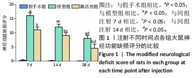
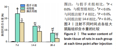
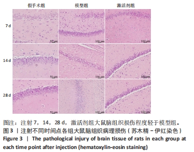
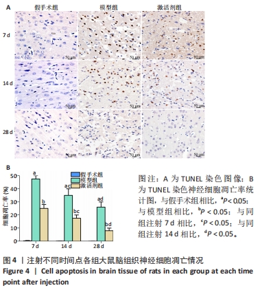
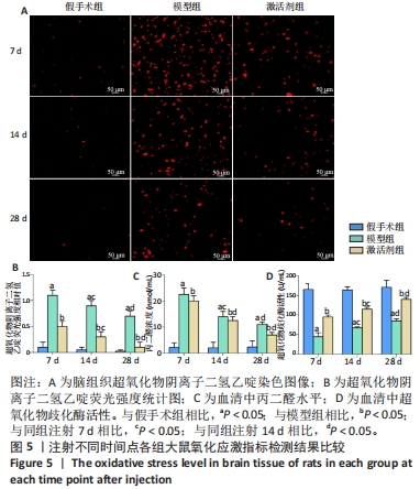
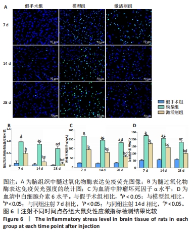


 综上所述,腹腔注射SIRT1信号激活剂SRT1720能明显下调急性颅脑损伤大鼠脑组织的氧化和炎性应激水平、抑制脑组织的神经凋亡、促进血管新生,减轻脑组织病理损伤,但是这其中更为详尽的分子作用网络仍需要更深入的研究,以帮助疾病的新药开发。