中国组织工程研究 ›› 2023, Vol. 27 ›› Issue (19): 3017-3022.doi: 10.12307/2023.667

• 干细胞基础实验 basic experiments of stem cells • 上一篇    下一篇
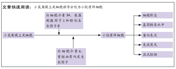
小鼠角膜上皮细胞诱导获得小胶质样细胞

许慧芳,单  萍,李俐娟   

  1. 华中科技大学同济医学院附属武汉市第一医院神经内科,湖北省武汉市   430000
  • 收稿日期:2022-08-01 接受日期:2022-09-19 出版日期:2023-07-08 发布日期:2022-11-28
  • 通讯作者: 许慧芳,博士,主治医师,华中科技大学同济医学院附属武汉市第一医院神经内科,湖北省武汉市 430000 单萍,博士,副主任医师,华中科技大学同济医学院附属武汉市第一医院神经内科,湖北省武汉市 430000
  • 作者简介:许慧芳,女,1981年生,山东省日照市人,汉族,2009年华中科技大学同济医学院毕业,博士,主治医师,主要从事脑血管病、头痛、痴呆、干细胞研究。
  • 基金资助:
    国家自然科学基金(81703870),项目负责人:单萍;武汉市卫健委重点项目(WX16B01) ,项目负责人:许慧芳;武汉市科技局面上项目(2015061701011618),项目负责人:许慧芳

Obtaining microglia-like cells after mouse corneal epithelial cell induction

Xu Huifang, Shan Ping, Li Lijuan   

  1. Department of Neurology, Wuhan No. 1 Hospital, Tongji Medical College of Huazhong University of Science and Technology, Wuhan 430000, Hubei Province, China
  • Received:2022-08-01 Accepted:2022-09-19 Online:2023-07-08 Published:2022-11-28
  • Contact: Xu Huifang, PhD, Attending physician, Department of Neurology, Wuhan No. 1 Hospital, Tongji Medical College of Huazhong University of Science and Technology, Wuhan 430000, Hubei Province, China Shan Ping, PhD, Associate Chief physician, Department of Neurology, Wuhan No. 1 Hospital, Tongji Medical College of Huazhong University of Science and Technology, Wuhan 430000, Hubei Province, China
  • About author:Xu Huifang, PhD, Attending physician, Department of Neurology, Wuhan No. 1 Hospital, Tongji Medical College of Huazhong University of Science and Technology, Wuhan 430000, Hubei Province, China
  • Supported by:
    National Natural Science Foundation of China, No. 81703870 (to SP); a grant from Wuhan Municipal Health Commission, No. WX16B01 (to XHF); the General Program of Wuhan Science and Technology Bureau, No. 2015061701011618 (to XHF)

摘要:


文题释义:

角膜上皮细胞:角膜是没有血管结构的透明组织,角膜的上皮细胞层位于角膜最外层。自然状态下角膜上皮细胞本身并不分裂为新的细胞,而是由干细胞-角膜缘细胞分化而来,在培养条件下角膜上皮细胞趋向于去分化,因而保持了一定的干细胞特征。
小胶质细胞:是一种存在于脑与脊髓中的神经胶质细胞,占脑细胞数量的10%-15%。小胶质细胞在中枢神经系统的分布区域通常与其他神经胶质细胞(如星形胶质细胞)互不重叠。小胶质细胞对维持中枢神经系统的正常功能十分重要:它们能清除中枢神经系统中的神经炎性斑、病原体以及损伤或无功能的神经元与轴突。小胶质细胞能通过细胞表面存在的一种特殊钾离子通道感知到细胞外钾离子浓度的微小变化,进而使其得以对中枢神经系统的潜在微小病变及时做出反应,以防止这样的微小病变扩大为足以威胁机体生命的疾病。

背景:小胶质细胞是中枢神经系统最主要的免疫细胞,对研究多种疾病的发生和治疗有重要意义,但体外个体化细胞的获得受限。
目的:建立一种新的小胶质细胞诱导培养体系,将小鼠角膜上皮细胞诱导成为小胶质样细胞并对其进行鉴定。
方法:裂解2周龄小鼠角膜上皮组织获得上皮细胞,先后加入含有白细胞介素34 (100 ng/mL)、集落刺激因子1(5 ng/mL)和转化生长因子β(1 ng/mL)、白细胞介素6 (1 ng/mL)、重组血管内皮生长因子(1 ng/mL) 的改良培养基,并且持续处理到16 d。通过转录水平、蛋白表达水平、亚细胞定位、流式分析和形态学观察多角度评估诱导后的细胞性质。
结果与结论:诱导后获得表达TMEM119、CXCL11、CD11b、CD68小胶质细胞特征分子的细胞,形态学观察、流式分析和荧光检验后确定其表达产物具有较高丰度,与小胶质细胞极为相似。

https://orcid.org/0000-0002-7520-1208 (许慧芳)

中国组织工程研究杂志出版内容重点:干细胞;骨髓干细胞;造血干细胞;脂肪干细胞;肿瘤干细胞;胚胎干细胞;脐带脐血干细胞;干细胞诱导;干细胞分化;组织工程

关键词: 小鼠角膜上皮细胞, 小胶质细胞, 诱导分化, TMEM119, CD68, CXCL11, CD11b

Abstract: BACKGROUND: Microglial cells are the predominant immune cells of the central nervous system and are important for the study of the development and treatment of many diseases. However, the methods for obtaining individualized cells in vitro are still lacking.  
OBJECTIVE: To establish a new microglia induction culture system to induce mouse corneal epithelial cells into microglia-like cells.
METHODS: Corneal epithelial tissues of 2-week-old mice were lysed to obtain epithelial cells, and modified media containing interleukin-34 (100 ng/mL), colony stimulating factor 1 (5 ng/mL), transforming growth factor β (1 ng/mL), interleukin-6 (1 ng/mL), and recombinant vascular endothelial growth factor (1 ng/mL) were successively added. Treatment lasted up to 16 days. Cell properties were evaluated after induction by transcriptional level, protein expression level, subcellular localization, flow cytometry and morphological observation.  
RESULTS AND CONCLUSION: After induction, microglia-like cells which express characteristic molecules of TMEM119, CXCL11, CD11b and CD68 were obtained. Morphological observation, flow cytometry and fluorescence analysis confirmed that the expression products of TMEM119, CXCL11, CD11b and CD68 microglia had high abundance, which was very similar to microglia.

Key words: mouse corneal epithelial cell, microglial cell, induced differentiation, TMEM119, CD68, CXCL11, CD11b

中图分类号: