中国组织工程研究 ›› 2020, Vol. 24 ›› Issue (32): 5108-5113.doi: 10.3969/j.issn.2095-4344.2856

• 软骨组织构建 cartilage tissue construction • 上一篇    下一篇

人甲壳质酶蛋白40通过PI3K/Akt信号通路调控膝骨性关节炎兔软骨细胞的凋亡

田胜兰1,王国延1,杨  扬2   

  1. 1武汉科技大学医院,湖北省武汉市  430065;2武汉科技大学临床学院,湖北省武汉市  430064
  • 收稿日期:2019-12-17 修回日期:2019-12-24 接受日期:2020-02-12 出版日期:2020-11-18 发布日期:2020-09-24
  • 通讯作者: 杨扬,博士,主任医师,硕士生导师,武汉科技大学临床学院,湖北省武汉市 430064
  • 作者简介:田胜兰,女,1971年生,湖南省益阳市人,汉族,武汉科技大学医院主任医师,副教授,主要从事骨关节疼痛诊疗研究。
  • 基金资助:
    湖北省卫生厅科研项目(WJ2017M178)

Mechanism of YKL-40 regulating apoptosis of rabbit osteoarthritis chondrocytes via PI3K/Akt signaling pathway

Tian Shenglan1, Wang Guoyan1, Yang Yang2   

  1. 1Wuhan University of Science and Technology Hospital, Wuhan 430065, Hubei Province, China; 2School of Clinical Medicine, Wuhan University of Science and Technology, Wuhan 430064, Hubei Province, China
  • Received:2019-12-17 Revised:2019-12-24 Accepted:2020-02-12 Online:2020-11-18 Published:2020-09-24
  • Contact: Yang Yang, MD, Chief physician, Master’s supervisor, School of Clinical Medicine, Wuhan University of Science and Technology, Wuhan 430064, Hubei Province, China
  • About author:Tian Shenglan, Chief physician, Associate professor, Wuhan University of Science and Technology Hospital, Wuhan 430065, Hubei Province, China
  • Supported by:
    the Scientific Research Project of Hubei Health Department, No. WJ2017M178

摘要:

文题释义:

膝骨关节炎(knee osteoarthritisKOA)常发生在中老年人群,其病理过程涉及软骨基质退行性病变、软骨细胞性状显著性改变和数量明显减少。在外力及内部环境改变等因素影响下,软骨细胞外基质、软骨细胞及软骨下骨的合成和降解动态平衡出现紊乱,软骨细胞大量凋亡使得软骨修复、重塑等稳定状态出现异常,上述情况均可诱导膝骨关节炎的发生。

人甲壳质酶蛋白40(YKL-40)是一种广泛存在于关节滑膜细胞、软骨细胞中的软骨糖蛋白,具有多种生物学功能作用:①促进软骨细胞、成骨细胞的增殖生长及分化;②促进新生血管组织的形成;③调控细胞外基质的重构过程;④协助细胞适应生长环境的显著性改变及缺氧等病理损伤,此外在软骨细胞凋亡方面也发挥一定作用。

背景:由于PI3K/Akt信号通路与骨组织生理代谢之间存在着密切的联系,而人甲壳质酶蛋白40(YKL-40)对乳腺癌发病机制中的PI3K/Akt信号通路具有一定调控作用,由此推测YKL-40可能通过PI3K/Akt信号通路对膝骨性关节炎软骨细胞凋亡进行调控。

目的研究YKL-40通过PI3K/Akt信号通路调控膝骨性关节炎兔软骨细胞凋亡的作用机制。
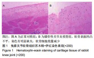
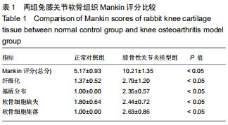
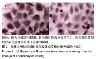
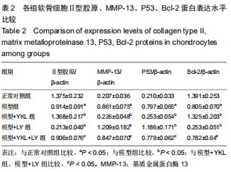
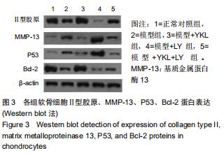
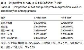
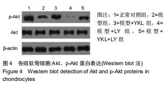
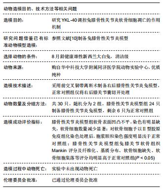
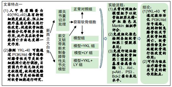
方法将新西兰大白兔随机分为2组,膝骨性关节炎模型组采用前交叉韧带离断术制作右后膝骨性关节炎动物模型,正常对照组仅切开右后膝关节囊,造模后第6周取材并分离软骨细胞,予以软骨组织苏木精-伊红染色及Mankin组织学评分,同时免疫组化染色检测软骨细胞型胶原表达;正常对照组兔第2代软骨细胞为正常对照组;将膝骨性关节炎模型组兔第2代软骨细胞分为4组,模型组仅予以含体积分数10%胎牛血清的高糖DMEM培养基培养,模型+YKL组培养基中加入100 μg/L YKL-40干预,模型+LY组培养基中加入50 µmol/L PI3K通路抑制剂LY294002干预,模型+YKL+LY组:培养基中加入100 μg/L YKL-4050 µmol/L LY294002干预,采用免疫印迹法检测各组软骨细胞型胶原、基质金属蛋白酶13Aktp-AktP53Bcl-2蛋白表达水平。

结果与结论经软骨组织苏木精-伊红染色、Mankin组织学评分及软骨细胞型胶原免疫组化染色证实,膝骨性关节炎动物模型构建和软骨细胞培养均获得成功;模型组型胶原、Bcl-2p-Akt蛋白表达明显低于正常对照组(P < 0.05),基质金属蛋白酶13P53蛋白表达明显高于正常对照组(P < 0.05);与模型组比较,模型+YKL组上述指标得到明显改善(P < 0.05),而模型+LY组上述指标则进一步恶化(P < 0.05),模型+YKL+LY组上述指标与模型组比较无显著性差异(P > 0.05),但与模型+YKL组和模型+LY组比较有显著性差异(P < 0.05);各组Akt蛋白表达水平比较无显著性差异(P > 0.05)。提示:YKL-40可通过激活活化PI3K/Akt信号通路,起到抑制兔膝骨性关节炎软骨细胞凋亡,加快软骨损伤修复和延缓软骨退行性改变的作用,可作为临床治疗膝骨性关节炎的新作用靶点。

ORCID: 0000-0002-7889-445X(田胜兰)

中国组织工程研究杂志出版内容重点:组织构建;骨细胞;软骨细胞;细胞培养;成纤维细胞;血管内皮细胞;骨质疏松;组织工程

关键词: 膝, 骨性关节炎, 人甲壳质酶蛋白40, 磷脂酰肌醇3-激酶, 通路, 软骨细胞, 凋亡, 实验

Abstract:

BACKGROUND: Due to the close relationship between the phosphatidylinositol 3-kinase (PI3K)/Akt signaling pathway and the physiological metabolism of bone tissue, human chitinase protein 40 (YKL-40) can regulate the PI3K/Akt signaling pathway related to breast cancer pathogenesis. Therefore, it is speculated that YKL-40 may regulate apoptosis in knee osteoarthritis (KOA) chondrocytes through the PI3K/Akt signaling pathway.

OBJECTIVE: To study the mechanism by which YKL-40 regulates apoptosis in rabbit KOA chondrocyte s through the PI3K/Akt signaling pathway.

METHODS: (1) New Zealand white rabbits were randomized into two groups. An animal model of KOA was made using anterior cruciate ligament dissection in the model group, whereas the right posterior knee joint capsule was cut but not dissected in the control group. Chondrocytes were extracted from the rabbits at 6 weeks after modeling. Hematoxylin-eosin staining and Mankin histological scoring of the cartilage tissue were performed, whereas immunohistochemical staining was used to detect type II collagen expression in chondrocytes. (2) The second-generation chondrocytes in the control group were used as normal control group, and those in the model group were further divided into four groups, followed by culture with high glucose DMEM medium containing 10% fetal bovine serum in KOA model group, 100 μg/L YKL-40 in KOA + YKL group,    50 µmol/L LY294002 in KOA + LY group, and 50 µmol/L LY294002 + 100 μg/L YKL-40 in KOA + YKL + LY group. The expression levels of collagen type II, matrix metalloproteinase 13, Akt, p-Akt, P53, Bcl-2 proteins in chondrocytes were detected by western blot.

RESULTS AND CONCLUSION: Hematoxylin-eosin staining, Mankin histological score, and collagen type II immunohistochemical staining confirmed the successful construction of KOA animal model and successful chondrocyte culture. Compared with the normal control group, the collagen type II, Bcl-2, p-Akt protein expression levels in chondrocytes were significantly reduced in the KOA model group (P < 0.05), and matrix metalloproteinase 13 and P53 protein expression levels were significantly increased in the KOA model group (P < 0.05). Compared with the KOA model group, these indicators were significantly improved in the KOA + YKL group (P < 0.05), significantly worsened in the KOA + LY group (P < 0.05), and had no significant changes in the KOA + YKL + LY group (P > 0.05). However, there were significant differences between the KOA + YKL group and the KOA + LY group (P < 0.05). In addition, the expression level of p-Akt protein in chondrocytes had no difference among groups (P > 0.05). To conclude, YKL-40 can inhibit the apoptosis of KOA chondrocytes, accelerate the repair of KOA cartilage damage and delay the degeneration of cartilage by activating the PI3K/Akt signaling pathway. Therefore, YKL-40 can be used as a new target for clinical treatment of KOA.

Key words: knee, osteoarthritis, human chitinase protein 40, phosphatidylinositol 3-kinase, pathway, chondrocyte, apoptosis, experiment

中图分类号: