中国组织工程研究 ›› 2020, Vol. 24 ›› Issue (12): 1881-1885.doi: 10.3969/j.issn.2095-4344.2544

• 数字化骨科 digital orthopedics • 上一篇    下一篇

个性化3D打印术前模拟与传统截骨在高位胫骨截骨中的应用与比较

陈金国,陈国仙,林宗锦,许国松,陈伟义,游丰源   

  1. 莆田市第一医院关节外科,福建省莆田市  351100
  • 收稿日期:2019-07-30 修回日期:2019-08-02 接受日期:2019-09-06 出版日期:2020-04-28 发布日期:2020-03-01
  • 通讯作者: 陈国仙,博士,副主任医师,莆田市第一医院关节外科,福建省莆田市 351100
  • 作者简介:陈金国,男,1987年生,福建省仙游县人,汉族,2013年福建医科大学毕业,硕士,主治医师,主要从事关节外科研究。
  • 基金资助:
    福建省教育厅科技项目(JAT160428);福建莆田市科技局资助项目(2016S3005)

Application and comparison of personalized 3D printing with preoperative simulation and traditional method in high tibial osteotomy 

Chen Jinguo, Chen Guoxian, Lin Zongjin, Xu Guosong, Chen Weiyi, You Fengyuan   

  1. Department of Joint Surgery, First Hospital of Putian City, Putian 351100, Fujian Province, China
  • Received:2019-07-30 Revised:2019-08-02 Accepted:2019-09-06 Online:2020-04-28 Published:2020-03-01
  • Contact: Chen Guoxian, MD, Associate chief physician, Department of Joint Surgery, First Hospital of Putian City, Putian 351100, Fujian Province, China
  • About author:Chen Jinguo, Master, Attending physician, Department of Joint Surgery, First Hospital of Putian City, Putian 351100, Fujian Province, China
  • Supported by:
    a grant from Department of Education Science and Technology of Fujian Province, No. JAT160428; a grant from Science and Technology Bureau of Putian City of Fujian Province, No. 2016S3005

摘要:

文题释义:
高位胫骨截骨:是治疗早期单纯内侧间室膝关节骨关节炎行之有效的术式,包括内侧开放与外侧闭合截骨两种术式。
3D打印技术:是将计算机三维数字成像技术和多层次连续打印技术相结合的一种新兴应用技术,与传统的材料去除加工方法不同的是,它通过分层加工、叠加成型的方式,逐层增加材料来生成3D实体模型。有研究报道3D打印截骨模块可精确指导全膝关节置换及内侧开放高位胫骨截骨术中截骨。

背景:高位胫骨截骨是治疗早期单纯内侧间室膝关节骨关节炎的经典术式,3D打印可用于制作个性化手术工具,课题组已应用3D打印技术辅助高位胫骨截骨治疗膝关节骨关节炎。

目的:对比传统截骨方法,探讨个性化3D打印截骨辅助下外侧闭合楔形高位胫骨截骨联合膝关节清理治疗合并内翻畸形膝关节骨关节炎的优势与不足。

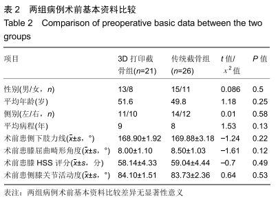
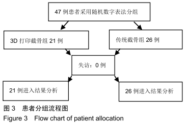
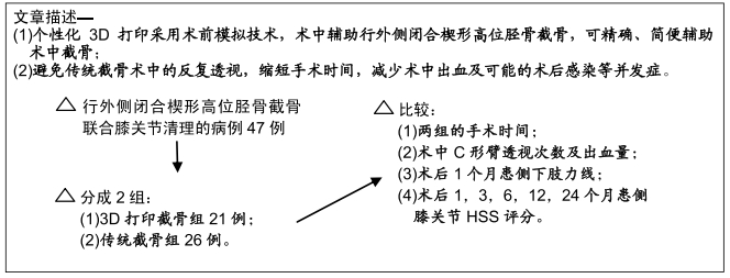
方法:行外侧闭合楔形高位胫骨截骨联合膝关节清理的病例47例,采用随机数字法分成2组:3D打印截骨组21例术中采用个性化3D打印截骨,传统截骨组26例予传统截骨。对比两组的手术时间、术中C形臂透视次数及出血量、术后1个月患侧下肢力线,以及术后1,3,6,12,24个月评估患侧膝关节美国特种外科医院(hospital of special surgery,HSS)评分。研究方案经莆田市第一医院伦理委员会审查批准,参与试验的患病个体对治疗过程完全知情同意。

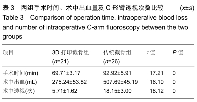
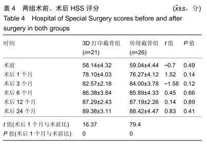
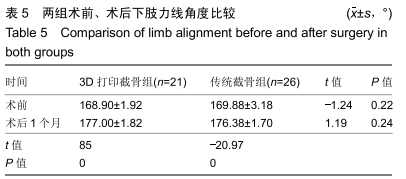
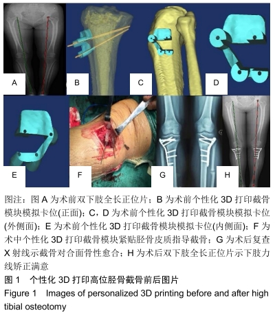
结果与结论:①术中3D打印截骨模块均与术前模拟卡位相一致;②3D打印截骨组手术时间较传统截骨组明显缩短[(69.71±3.17),(92.92±5.91)min,t=-17.21,P < 0.01];3D打印截骨组术中C形臂透视次数明显少于传统截骨组[(5.71±1.62),(18.15±3.00)次,t=-18.12,P < 0.01];3D打印截骨组出血量显著少于传统截骨组[(275.24±53.82),(507.69±45.19)mL,t=-16.10,P < 0.01];③两组术后1个月患侧下肢力线及术后24个月患侧膝关节HSS评分差异无显著性意义(均P > 0.05);④结果说明,对比传统截骨,在外侧闭合楔形高位胫骨截骨中应用个性化3D打印截骨可明显缩短手术时间,减少术中透视和出血,但二者术后2年随访患者膝关节功能恢复无明显差异。

ORCID: 0000-0001-9766-6164(陈金国)

中国组织工程研究杂志出版内容重点:人工关节;骨植入物;脊柱骨折;内固定;数字化骨科;组织工程

关键词: 高位胫骨截骨, 3D打印, 个性化截骨, 传统截骨, 膝关节, 骨关节炎

Abstract:

BACKGROUND: High tibial osteotomy is a classic operation for the treatment of early simple medial compartment osteoarthritis of the knee. Three-dimensional (3D) printing can be used to make personalized surgical tools. We have used 3D printing technology to assist high tibial osteotomy for the treatment of knee osteoarthritis.

OBJECTIVE: By comparing traditional methods, to analyze the advantages and disadvantages of the high tibial osteotomy assisted by personalized 3D printing osteotomy combined with arthroscopic debridement for the treatment of osteoarthritis of the knee with varus deformity.

METHODS: Totally 47 cases undergoing lateral closed wedge high tibial osteotomy combined with knee joint debridement were randomly assigned to two groups. The 3D printing osteotomy group (n=21) received personalized 3D printing osteotomy. The traditional osteotomy group (n=26) received traditional osteotomy. The operation time, number of intraoperative C-arm fluoroscopy and blood loss, limb alignment on the affected side 1 month after surgery, and Hospital of Special Surgery scores on the affected side knee joints at 1, 3, 6, 12 and 24 months after surgery were compared between the two groups. This study was approved by the Ethics Committee of First Hospital of Putian City. All patients signed the informed consent.

RESULTS AND CONCLUSION: (1) The intraoperative 3D printing osteotomy module was consistent with the preoperative simulated place. (2) The operation time was significantly shorter in the 3D printing osteotomy group than in the traditional osteotomy group [(69.71±3.17), (92.92±5.91) minutes, t=-17.21, P < 0.01]. The number of C-arm fluoroscopy was significantly less in the 3D printing osteotomy group than in the traditional osteotomy group [(5.71±1.62), (18.15±3.00) times, t=-18.12, P < 0.01]. Blood loss was significantly less in the 3D printing osteotomy group than in the traditional osteotomy group [(275.24±53.82), (507.69±45.19) mL, t=-16.10, P < 0.01]. (3) There was no difference between the two groups in the limb alignment on the affected side at 1 month after operation and the Hospital of Special Surgery score on the affected knee at 24 months after operation (all P > 0.05). (4) In conclusion, compared with traditional osteotomy, personalized 3D printing-assisted osteotomy in lateral closed wedge high tibial osteotomy can significantly shorten the operation time, reduce the number of intraoperative C-arm fluoroscopy and the amount of blood loss, but there is no significant difference in knee function between the two groups at 2 years after surgery.

Key words: high tibial osteotomy, 3D printing, personalized osteotomy, traditional osteotomy, knee, osteoarthritis

中图分类号: