中国组织工程研究 ›› 2019, Vol. 23 ›› Issue (23): 3630-3635.doi: 10.3969/j.issn.2095-4344.1306
• 骨组织构建 bone tissue construction • 上一篇 下一篇
泼尼松龙可抑制转化生长因子β激活激酶1诱导的成骨细胞凋亡
张 雯1,张 蕾1,任守忠1,刘日升2
- (1海南医学院第一附属医院,海南省海口市 570102;2海南省肿瘤医院,海南省海口市 570102)
Prednisolone inhibites osteoblast apoptosis induced by transforming growth factor beta activated kinase 1
Zhang Wen1, Zhang Lei1, Ren Shouzhong1, Liu Risheng2
- (1the First Affiliated Hospital of Hainan Medical University, Haikou 570102, Hainan Province, China; 2Hainan Cancer Hospital, Haikou 570102, Hainan Province, China)
摘要:
文章快速阅读:
.jpg)
文题释义:
碱性磷酸酶染色:碱性磷酸酶较多存在于成熟中性粒细胞中。在碱性条件下,碱性磷酸酶经镁离子激活后能将磷酸酯水解为磷酸钠和甘油,磷酸钠再与氯化钙、硝酸钴、硫化铵发生一系列化学反应,生成棕色的硫化钴定位于胞质内,可用于评估成骨细胞分化能力。
钙结节染色:成骨分化是干细胞的一个重要特性。在特定的诱导培养基作用一段时间后,干细胞可分化为成骨细胞,在细胞表面沉积钙盐,形成钙结节。应用茜素红的复合物,可用于鉴定干细胞是否已成功向成骨细胞转化。
文题释义:
碱性磷酸酶染色:碱性磷酸酶较多存在于成熟中性粒细胞中。在碱性条件下,碱性磷酸酶经镁离子激活后能将磷酸酯水解为磷酸钠和甘油,磷酸钠再与氯化钙、硝酸钴、硫化铵发生一系列化学反应,生成棕色的硫化钴定位于胞质内,可用于评估成骨细胞分化能力。
钙结节染色:成骨分化是干细胞的一个重要特性。在特定的诱导培养基作用一段时间后,干细胞可分化为成骨细胞,在细胞表面沉积钙盐,形成钙结节。应用茜素红的复合物,可用于鉴定干细胞是否已成功向成骨细胞转化。
摘要
背景:研究显示转化生长因子β激活激酶1(transforming growth factor beta activated kinase 1,TAK1)在骨、关节的发育以及骨形态信号转导中发挥着重要作用,与骨关节炎的发生也存在一定的相关性。
目的:探究泼尼松龙通过抑制TAK1表达对诱导成骨细胞凋亡的影响。
方法:将M3T3-E1成骨细胞经原代培养后传代培养。取第3代细胞分为3组,正常细胞组(对照组)、阴性转染+泼尼松龙组、TAK1 siRNA转染+泼尼松龙组。采用碱性磷酸酶染色和钙结节染色评估成骨细胞分化能力的变化;采用免疫印迹法(Western blot)检测细胞内磷酸化(p)-TAK1、TAK1、磷酸化c-jun氨基末端激酶(p-JNK)、JNK蛋白表达,MTT法检测M3T3-E1细胞增殖情况;流式细胞仪检测细胞周期以及细胞凋亡变化。
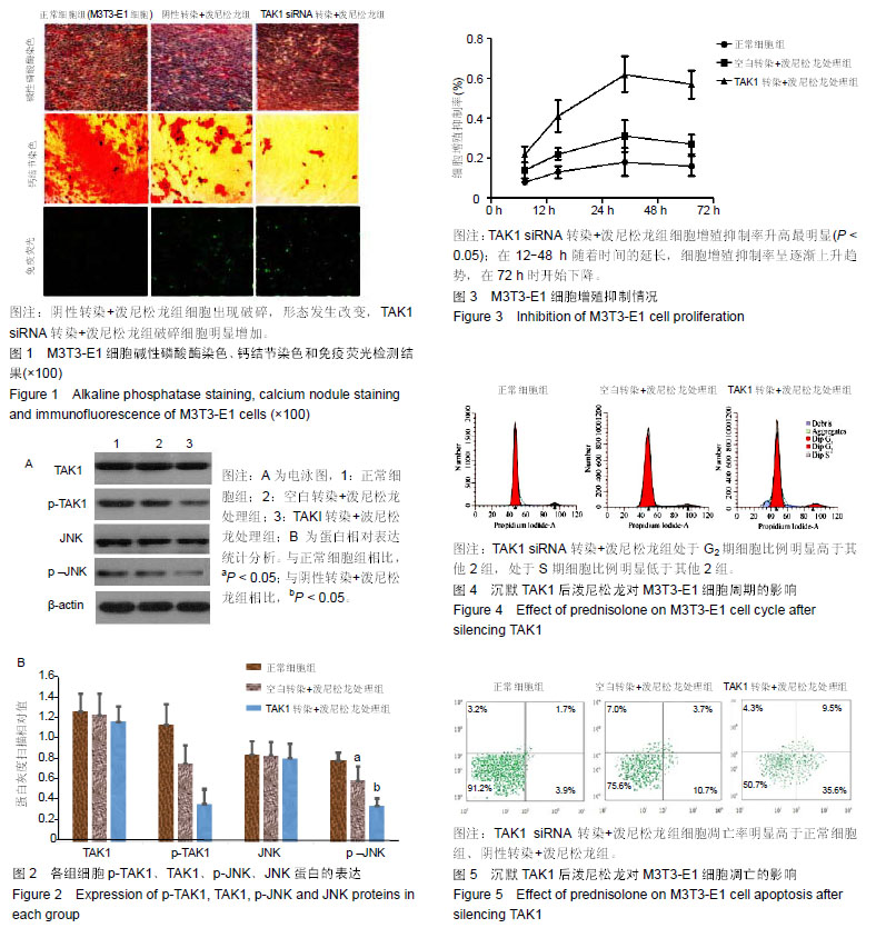
结果与结论:①TAK1 siRNA转染+泼尼松龙组细胞的碱性磷酸酶红染程度较少,阴性转染+泼尼松龙组次之,正常细胞组碱性磷酸酶染色最多;钙结节染色显示与正常细胞组相比,阴性转染+泼尼松龙组钙结节数量明显减少,TAK1 siRNA转染+泼尼松龙组结节数量最少;②荧光显微镜显示,阴性转染+泼尼松龙组细胞出现破碎,形态发生改变,TAK1 siRNA转染+泼尼松龙组破碎细胞数量明显增加;③Western Blot 显示,3组间p-TAK1、p-JNK 蛋白表达量逐渐降低(P < 0.05);④MTT检测显示,3组间TAK1 siRNA转染+泼尼松龙组细胞增殖抑制率最高(P < 0.05);在12-48 h随着时间的延长,细胞增殖抑制率呈逐渐上升趋势,在72 h时开始下降;⑤流式细胞仪检测结果显示,TAK1 siRNA转染+泼尼松龙组G2期细胞比例高于其他2组,S期细胞比例显著低于其他2组(P < 0.05);⑥TAK1 siRNA转染+泼尼松龙组细胞凋亡率明显高于正常细胞组、阴性转染+泼尼松龙组(P < 0.05);⑦结果说明,沉默TAK1表达后能够增强泼尼松龙诱导成骨细胞凋亡的作用,可能与JNK信号通路相关。
背景:研究显示转化生长因子β激活激酶1(transforming growth factor beta activated kinase 1,TAK1)在骨、关节的发育以及骨形态信号转导中发挥着重要作用,与骨关节炎的发生也存在一定的相关性。
目的:探究泼尼松龙通过抑制TAK1表达对诱导成骨细胞凋亡的影响。
方法:将M3T3-E1成骨细胞经原代培养后传代培养。取第3代细胞分为3组,正常细胞组(对照组)、阴性转染+泼尼松龙组、TAK1 siRNA转染+泼尼松龙组。采用碱性磷酸酶染色和钙结节染色评估成骨细胞分化能力的变化;采用免疫印迹法(Western blot)检测细胞内磷酸化(p)-TAK1、TAK1、磷酸化c-jun氨基末端激酶(p-JNK)、JNK蛋白表达,MTT法检测M3T3-E1细胞增殖情况;流式细胞仪检测细胞周期以及细胞凋亡变化。
结果与结论:①TAK1 siRNA转染+泼尼松龙组细胞的碱性磷酸酶红染程度较少,阴性转染+泼尼松龙组次之,正常细胞组碱性磷酸酶染色最多;钙结节染色显示与正常细胞组相比,阴性转染+泼尼松龙组钙结节数量明显减少,TAK1 siRNA转染+泼尼松龙组结节数量最少;②荧光显微镜显示,阴性转染+泼尼松龙组细胞出现破碎,形态发生改变,TAK1 siRNA转染+泼尼松龙组破碎细胞数量明显增加;③Western Blot 显示,3组间p-TAK1、p-JNK 蛋白表达量逐渐降低(P < 0.05);④MTT检测显示,3组间TAK1 siRNA转染+泼尼松龙组细胞增殖抑制率最高(P < 0.05);在12-48 h随着时间的延长,细胞增殖抑制率呈逐渐上升趋势,在72 h时开始下降;⑤流式细胞仪检测结果显示,TAK1 siRNA转染+泼尼松龙组G2期细胞比例高于其他2组,S期细胞比例显著低于其他2组(P < 0.05);⑥TAK1 siRNA转染+泼尼松龙组细胞凋亡率明显高于正常细胞组、阴性转染+泼尼松龙组(P < 0.05);⑦结果说明,沉默TAK1表达后能够增强泼尼松龙诱导成骨细胞凋亡的作用,可能与JNK信号通路相关。
中国组织工程研究杂志出版内容重点:组织构建;骨细胞;软骨细胞;细胞培养;成纤维细胞;血管内皮细胞;骨质疏松;组织工程
ORCID: 0000-0002-2395-5948(张雯)
中图分类号:
.jpg)
.jpg) #br#
文题释义:#br#
碱性磷酸酶染色:碱性磷酸酶较多存在于成熟中性粒细胞中。在碱性条件下,碱性磷酸酶经镁离子激活后能将磷酸酯水解为磷酸钠和甘油,磷酸钠再与氯化钙、硝酸钴、硫化铵发生一系列化学反应,生成棕色的硫化钴定位于胞质内,可用于评估成骨细胞分化能力。#br#
钙结节染色:成骨分化是干细胞的一个重要特性。在特定的诱导培养基作用一段时间后,干细胞可分化为成骨细胞,在细胞表面沉积钙盐,形成钙结节。应用茜素红的复合物,可用于鉴定干细胞是否已成功向成骨细胞转化。#br#
#br#
#br#
文题释义:#br#
碱性磷酸酶染色:碱性磷酸酶较多存在于成熟中性粒细胞中。在碱性条件下,碱性磷酸酶经镁离子激活后能将磷酸酯水解为磷酸钠和甘油,磷酸钠再与氯化钙、硝酸钴、硫化铵发生一系列化学反应,生成棕色的硫化钴定位于胞质内,可用于评估成骨细胞分化能力。#br#
钙结节染色:成骨分化是干细胞的一个重要特性。在特定的诱导培养基作用一段时间后,干细胞可分化为成骨细胞,在细胞表面沉积钙盐,形成钙结节。应用茜素红的复合物,可用于鉴定干细胞是否已成功向成骨细胞转化。#br#
#br#