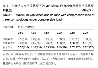
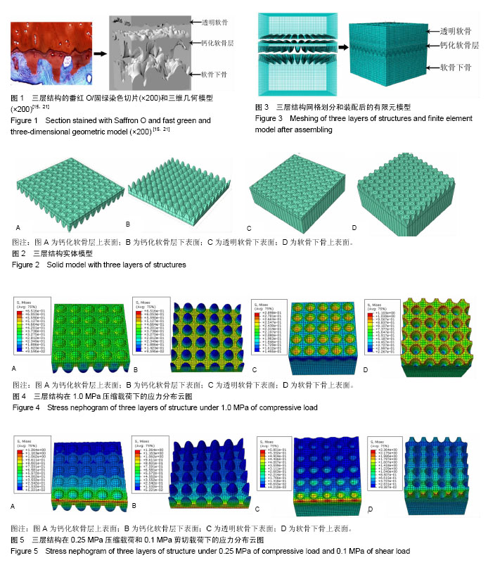
| [1] Burr DB.Anatomy and physiology of the mineralized tissues: role in the pathogenesis of osteoarthrosis. Osteoarthritis Cartilage.2004;12 Suppl A: S20-30.[2] Koszyca B, Fazzalari NL, Vernon-Roberts B.Quantitative analysis of the bone-cartilage interface within the knee.The Knee.1996;3(1-2):23-31.[3] Arkill KP, Winlove CP.Solute transport in the deep and calcified zonesof articular cartilage. Osteoarthritis Cartilage. 2008;16(6): 708-714.[4] Pan J, Zhou X, Li W, et al. In situ measurement of transport between subchondral bone and articular cartilage.J Orthop Res.2009;27(10):1347-1352.[5] Guévremont M, Martel-Pelletier J, Massicotte F, et al. Human adult chondrocytes express hepatocyte growth factor (HGF) isoforms but not HgF: potential implication of osteoblasts on the presence of HGF in cartilage.J Bone Miner Res.2003;18(6): 1073-1081.[6] 宋伟,王富友,杨柳.关节软骨钙化层研究进展[J].中国修复重建外科杂志,2011,25(11): 1339-1342. [7] Lories RJ, Luyten FP. The bone-cartilage unit in osteoarthritis. Nat Rev Rheumatol.2011;7(1): 43-49.[8] Hoemann CD,Lafantaisie-Favreau CH,Lascau-Coman V,et al.The cartilage-bone interface.J Knee Surg. 2012;25(2): 85-97.[9] Wang F,Ying Z,Duan X,et al.Histomorphometric analysis of adult articular calcified cartilage zone.J Struct Biol.2009;168 (3):359-365.[10] Mansfield JC,Winlove CP.A multi-modal multiphoton investigation of microstructure in the deep zone and calcified cartilage.J Anat.2012;220(4):405-416.[11] Zizak I,Roschger P,Paris O,et al.Characteristics of mineral particles in the human bone/cartilage interface.J Struct Biol. 2003;141(3): 208-217.[12] Norrdin RW,Kawcak CE,Capwell BA,et al.Calcified Cartilage Morphometry and Its Relation to Subchondral Bone Remodeling in Equine Arthrosis.Bone.1999;24(2): 109-114.[13] Allan KS,Pilliar RM,Wang J,et al.Formation of biphasic constructs containing cartilage with a calcified zone interface. Tissue Eng.2007;13(1):167-177.[14] Hargrave-Thomas E,van Sloun F,Dickinson M,et al. Multi-scalar mechanical testing of the calcified cartilage and subchondral bone comparing healthy vs early degenerative states. Osteoarthritis Cartilage.2015;23(10):1755-1762.[15] 王富友,杨柳,段小军,等.正常膝关节软骨钙化层形态结构研究[J].中国修复重建外科杂志,2008,22(5):524-527.[16] Stender ME, Carpenter RD, Regueiro RA, et al. An evolutionary model of osteoarthritis including articular cartilage damage, and bone remodeling in a computational study.J Biomech.2016;49(14):3502-3508.[17] Anderson DD,Brown TD,Radin EL.The influence of basal cartilage calcification on dynamic juxtaarticular stress transmission.Clin Orthop.1993;286: 298-307.[18] Malekipour F,Oetomo D,Lee PV.Equine subchondral bone failure threshold under impact compression applied through articular cartilage.J Biomech.2016;49(10): 2053-2059.[19] Goldring SR,Goldring MB.Changes in the osteochondral unit during osteoarthritis: structure, function and cartilage-bone crosstalk.Nat Rev Rheumatol.2016;12(11): 632-644.[20] Lee WD,Hurtig MB,Pilliar RM,et al.Engineering of hyaline cartilage with a calcified zone using bone marrow stromal cells.Osteoarthritis Cartilage.2015;23(8):1307-1315.[21] 王富友,杨柳,段小军,等.人体正常膝关节钙化软骨层组成成分研究[J].第三军医大学学报,2008,30(8): 687-690.[22] Simha NK,Jin H,Hall ML,et al.Effect of indenter size on elastic modulus of cartilage measured by indentation.J Biomech Eng. 2007;129(5): 767-775.[23] Mente PL,Lewis JL.Elastic modulus of calcified cartilage is an order of magnitude less than that of subchondral bone.J Orthop Res.1994;12(5): 637-647.[24] St-Pierre JP, Gan L, Wang J, et al. The incorporation of a zone of calcified cartilage improves the interfacial shear strength between in vitro-formed cartilage and the underlying substrate [J]. Acta Biomater.2012;8(4):1603-1615.[25] Turley SM, Thambyah A, Riggs CM, et al. Microstructural changes in cartilage and bone related to repetitive overloading in an equine athlete model.J Anat.2014;224(6): 647-658.[26] Burr DB, Radin EL. Microfractures and microcracks in subchondral bone: are they relevant to osteoarthrosis? Rheum Dis Clin North Am.2003;29(4): 675-685.[27] Herman BC,Cardoso L,Majeska RJ,et al.Activation of bone remodeling after fatigue: differential response to linear microcracks and diffuse damage.Bone.2010;47(4): 766-772.[28] Dar FH,Aspden RM.A finite element model of an idealized diarthrodial joint to investigate the effects of variation in the mechanical properties of the tissues. Proc Inst Mech Eng H.2003;217(5): 341-348.[29] 陈凯,张德坤,戴祖明,等.牛膝关节软骨的力学承载特性及其有限元仿真分析[J].医用生物力学,2007, 27(6): 675-680.[30] Warner MD,Taylor WR,Cliff SE.Cyclic loading moves the peak stress to the cartilage surface in a biphasic model with isotropic solid phase properties.Med Eng Phys.2004;26(3): 247-249. |
.jpg)


.jpg)