中国组织工程研究 ›› 2017, Vol. 21 ›› Issue (36): 5812-5817.doi: 10.3969/j.issn.2095-4344.2017.36.013
• 组织构建实验造模 experimental modeling in tissue construction • 上一篇 下一篇
新生小鼠皮肤发育与毛囊发生的组织形态学变化及分期
施 恩1,刘英芹2
- 1青岛黄海学院护理与健康学院,山东省青岛市 266427;2桂林医学院基础医学院,广西壮族自治区桂林市 541000
Histomorphological changes and stage development of the skin and hair follicle in neonatal mice
Shi En1, Liu Ying-qin2
- 1School of Nursing and Health, Qingdao Huaihai University, Qingdao 264273, Shandong Province, China; 2School of Basic Medicine, Guilin Medical University, Guilin 541000, Guangxi Zhuang Autonomous Region, China
摘要:
文章快速阅读:
.jpg) 文题释义:
角质细胞(Keratinocyte):所形成的天然高分子纤苎是头发的主要成分,角质细胞存在于人的头部表皮,可以产生多种角质细胞,角质层细胞起到非常重要的保护作用。角质层的细胞含有8-10 nm的角蛋白丝,形成一种天然屏障,可以防止大自然中的各种物理、化学、微生物和水的入侵,也能防止紫外线摄入皮肤深层。
毛囊:在真皮乳头层里,毛根被内、外毛鞘包围,外毛鞘又被结缔组织细胞包围,形成口袋状,称毛囊。毛囊是包围在毛发根部的囊状组织,内层是上皮组织性毛囊,外层是结缔组织性毛囊,内层与表皮相连,外层则与真皮相连。毛囊由毛杆,毛凸,毛球,毛乳头,毛基质等部分组成。神经纤维末梢使毛囊具有感觉的功能,动脉和静脉毛细血管丛给毛囊提供血液。毛囊的最深处是位于角质层3-7 mm的毛乳头,它含有神经和血管,向毛杆提供养分。毛囊的发育经过表皮和真皮之间一系列复杂的相互作用而形成,而具有周期性。
文题释义:
角质细胞(Keratinocyte):所形成的天然高分子纤苎是头发的主要成分,角质细胞存在于人的头部表皮,可以产生多种角质细胞,角质层细胞起到非常重要的保护作用。角质层的细胞含有8-10 nm的角蛋白丝,形成一种天然屏障,可以防止大自然中的各种物理、化学、微生物和水的入侵,也能防止紫外线摄入皮肤深层。
毛囊:在真皮乳头层里,毛根被内、外毛鞘包围,外毛鞘又被结缔组织细胞包围,形成口袋状,称毛囊。毛囊是包围在毛发根部的囊状组织,内层是上皮组织性毛囊,外层是结缔组织性毛囊,内层与表皮相连,外层则与真皮相连。毛囊由毛杆,毛凸,毛球,毛乳头,毛基质等部分组成。神经纤维末梢使毛囊具有感觉的功能,动脉和静脉毛细血管丛给毛囊提供血液。毛囊的最深处是位于角质层3-7 mm的毛乳头,它含有神经和血管,向毛杆提供养分。毛囊的发育经过表皮和真皮之间一系列复杂的相互作用而形成,而具有周期性。摘要
背景:虽然小鼠皮肤在胚胎期的形态学形成和生长发育的规律已有较多报道,但对出生后小鼠皮肤发育与毛囊发生的组织形态学演变过程的差异性尚未被完全揭示。
目的:分析新生小鼠皮肤发育与毛囊发生的组织形态学变化过程及其规律。
方法:采用常规石蜡包埋切片和苏木精-伊红染色技术,应用Image J软件测量分析小鼠出生后第1-12天背部皮肤厚度;在光学显微镜下观察毛囊的组织形态结构,统计毛囊及“囊泡样”结构的数量;根据毛囊发生特点分为4期,并与Ralf Paus分期对比分析。
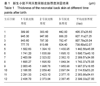
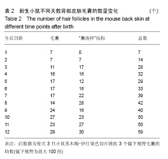
结果与结论:①第1-4天为毛囊形成早期,小鼠出生后当天皮肤最薄为(405.36±26.60) μm,皮下无脂肪组织,表皮层可见少许基板毛球;②第2天皮肤明显增厚为(927.11±27.25) μm,真皮成纤维细胞增多,基板角质细胞从表皮基底膜下移,形成毛乳头;③第3,4天皮肤变薄(738.60±82.07)μm,毛囊增多,毛球膨大,真皮根鞘覆盖真皮层;④第5-7天毛囊形成中期,第5天皮肤再次增厚,第7天达峰值(2 369.57±34.06) μm,毛囊及“囊泡样”结构数量增多,毛球增多,其近端部分突出,真皮内根鞘伸入表皮层形成毛根,真皮根鞘分化形成外根鞘,内根鞘延伸到深层,形成完全封闭的毛发矩阵;⑤第8,9天毛囊形成后期,皮肤厚度分别下降至(1 743.37±75.05) μm和(1 987.07±18.65) μm,毛乳头被毛母质所包围,毛管、毛干逐渐形成,毛干顶端通过毛管长出内根鞘,内根鞘进入毛发深层,形成更多毛根;⑥第10-12天毛囊成熟期,皮肤厚度增加至(2 399.33±27.00) μm,毛囊及“囊泡样”结构数量均达到近30个,毛干长度加大,达到皮下肌肉层,毛干通过毛管向上生长并穿过表皮层形成肉眼可见的完整毛发;⑦结果表明,新生小鼠背部皮肤发育与毛囊发生存在同步性和非同步性,呈向上的曲线形。毛囊发生分化过程存在非均衡性和差异性,毛囊分期对观察毛囊发生分化过程有实用价值。
中国组织工程研究杂志出版内容重点:组织构建;骨细胞;软骨细胞;细胞培养;成纤维细胞;血管内皮细胞;骨质疏松;组织工程
ORCID: 0000-0001-5888-9146(施恩)
中图分类号:
.jpg) 文题释义:
角质细胞(Keratinocyte):所形成的天然高分子纤苎是头发的主要成分,角质细胞存在于人的头部表皮,可以产生多种角质细胞,角质层细胞起到非常重要的保护作用。角质层的细胞含有8-10 nm的角蛋白丝,形成一种天然屏障,可以防止大自然中的各种物理、化学、微生物和水的入侵,也能防止紫外线摄入皮肤深层。
毛囊:在真皮乳头层里,毛根被内、外毛鞘包围,外毛鞘又被结缔组织细胞包围,形成口袋状,称毛囊。毛囊是包围在毛发根部的囊状组织,内层是上皮组织性毛囊,外层是结缔组织性毛囊,内层与表皮相连,外层则与真皮相连。毛囊由毛杆,毛凸,毛球,毛乳头,毛基质等部分组成。神经纤维末梢使毛囊具有感觉的功能,动脉和静脉毛细血管丛给毛囊提供血液。毛囊的最深处是位于角质层3-7 mm的毛乳头,它含有神经和血管,向毛杆提供养分。毛囊的发育经过表皮和真皮之间一系列复杂的相互作用而形成,而具有周期性。
文题释义:
角质细胞(Keratinocyte):所形成的天然高分子纤苎是头发的主要成分,角质细胞存在于人的头部表皮,可以产生多种角质细胞,角质层细胞起到非常重要的保护作用。角质层的细胞含有8-10 nm的角蛋白丝,形成一种天然屏障,可以防止大自然中的各种物理、化学、微生物和水的入侵,也能防止紫外线摄入皮肤深层。
毛囊:在真皮乳头层里,毛根被内、外毛鞘包围,外毛鞘又被结缔组织细胞包围,形成口袋状,称毛囊。毛囊是包围在毛发根部的囊状组织,内层是上皮组织性毛囊,外层是结缔组织性毛囊,内层与表皮相连,外层则与真皮相连。毛囊由毛杆,毛凸,毛球,毛乳头,毛基质等部分组成。神经纤维末梢使毛囊具有感觉的功能,动脉和静脉毛细血管丛给毛囊提供血液。毛囊的最深处是位于角质层3-7 mm的毛乳头,它含有神经和血管,向毛杆提供养分。毛囊的发育经过表皮和真皮之间一系列复杂的相互作用而形成,而具有周期性。