中国组织工程研究 ›› 2017, Vol. 21 ›› Issue (36): 5769-5774.doi: 10.3969/j.issn.2095-4344.2017.36.006
• 口腔组织构建 oral tissue construction • 上一篇 下一篇
三维数字化建立恒牙期安氏Ⅱ类错牙合模型对牙弓和基骨弓的测量分析
武 杰1,王超然2,赵 伟1,孙梦姣1,李洪发1
- 1天津医科大学口腔医院正畸科,天津市 300070;2天津解放军第二五四医院口腔科,天津市 300142
Characteristics of dental arch and basal bone in permanent dentition Angle Class II malocclusion based on three-dimensional digital models
Wu Jie1, Wang Chao-ran2, Zhao Wei1, Sun Meng-jiao1, Li Hong-fa1
- 1Department of Orthodontics, Stomatological Hospital of Tianjin Medical University, Tianjin 300070, China; 2Department of Stomatology, No. 254 Hospital of Chinese PLA, Tianjin 300142, China
摘要:
文章快速阅读:
.jpg) 文题释义:
正畸三维数字化模型的获取:是通过扫描仪器将患者口内牙颌形态转化为虚拟数字信息,然后在相应软件中生成三维立体图像。该技术能够实现非接触测量,且具有速度快、精度高的优点,为实物数字化提供了方便快捷的手段。
锥型束计算机体层扫描(cone-beam computed tomography,CBCT):CBCT是目前口腔医疗领域应用最成熟的获取颌面部软硬组织三维图像的方法,在正畸临床诊断、矫治中发挥着重要作用。CBCT其数据格式为DICOM,能够与数字化模型准确地匹配。数字化模型包含了牙列中牙冠部分的全部信息,CBCT的信息包括了软硬组织的三维影像,二者的结合可以实现信息互补。
文题释义:
正畸三维数字化模型的获取:是通过扫描仪器将患者口内牙颌形态转化为虚拟数字信息,然后在相应软件中生成三维立体图像。该技术能够实现非接触测量,且具有速度快、精度高的优点,为实物数字化提供了方便快捷的手段。
锥型束计算机体层扫描(cone-beam computed tomography,CBCT):CBCT是目前口腔医疗领域应用最成熟的获取颌面部软硬组织三维图像的方法,在正畸临床诊断、矫治中发挥着重要作用。CBCT其数据格式为DICOM,能够与数字化模型准确地匹配。数字化模型包含了牙列中牙冠部分的全部信息,CBCT的信息包括了软硬组织的三维影像,二者的结合可以实现信息互补。
文题释义:
正畸三维数字化模型的获取:是通过扫描仪器将患者口内牙颌形态转化为虚拟数字信息,然后在相应软件中生成三维立体图像。该技术能够实现非接触测量,且具有速度快、精度高的优点,为实物数字化提供了方便快捷的手段。
锥型束计算机体层扫描(cone-beam computed tomography,CBCT):CBCT是目前口腔医疗领域应用最成熟的获取颌面部软硬组织三维图像的方法,在正畸临床诊断、矫治中发挥着重要作用。CBCT其数据格式为DICOM,能够与数字化模型准确地匹配。数字化模型包含了牙列中牙冠部分的全部信息,CBCT的信息包括了软硬组织的三维影像,二者的结合可以实现信息互补。摘要
背景:正畸医生使用模型测量能够获得关于牙颌的全面数据,三维数字化模型显示能比传统模型获得更准确的信息。
目的:通过对恒牙期安氏Ⅱ类错牙合三维数字化模型进行测量,探讨其牙弓和基骨弓的特点。
方法:实验组为恒牙期安氏Ⅱ1类组(30例)、安氏Ⅱ2类组(30例),对照组为恒牙期个别正常牙合(30例)。使用3shape R700扫描仪将3组石膏模型扫描生成三维数字化模型,使用三维测量软件Orthoanalyzer 2013对数字化模型进行测量。采用SPSS 19.0软件对上述3组测量数据进行统计学分析,组间两两比较采用方差分析LSD法。
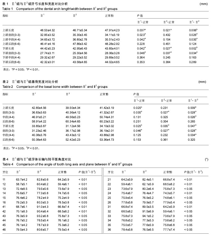
结果与结论:①上下颌牙弓长度、上颌牙弓前中段、下颌基骨中段宽度:Ⅱ2>个别正常颌>Ⅱ1 (P < 0.05);下颌牙弓前段宽度、上下颌基骨长度:个别正常颌>Ⅱ1>Ⅱ2 (P < 0.05);上颌牙弓后段、上颌前中段、下颌前段基骨宽度:个别正常颌>Ⅱ2 >Ⅱ1 (P < 0.05);②牙体长轴与牙合平面角度:安氏Ⅱ1、Ⅱ2、个别正常颌前牙区该角度差异显著;上颌后牙区(冠):Ⅱ2 >Ⅱ1>个别正常颌(P < 0.05);下颌后牙区(冠):Ⅱ2>个别正常颌>Ⅱ1 (P < 0.05);③安氏Ⅱ1弓形有狭而长的趋势,安氏Ⅱ2弓形有宽而短的趋势;④安氏Ⅱ1牙列前段(3-3)存在矢状向不调,表现为上下颌唇倾;中(4-4)后(6-6)段存在冠状向不调,上牙列表现为腭倾,下牙列表现为颊倾。安氏Ⅱ2牙列前段存在矢状向不调,表现为上下颌舌倾;后牙区无不调。
中国组织工程研究杂志出版内容重点:组织构建;骨细胞;软骨细胞;细胞培养;成纤维细胞;血管内皮细胞;骨质疏松;组织工程
ORCID: 0000-0002-3680-6685(武杰)
中图分类号:

.jpg)
.jpg)
.jpg)
.jpg)
.jpg)
.jpg) 文题释义:
正畸三维数字化模型的获取:是通过扫描仪器将患者口内牙颌形态转化为虚拟数字信息,然后在相应软件中生成三维立体图像。该技术能够实现非接触测量,且具有速度快、精度高的优点,为实物数字化提供了方便快捷的手段。
锥型束计算机体层扫描(cone-beam computed tomography,CBCT):CBCT是目前口腔医疗领域应用最成熟的获取颌面部软硬组织三维图像的方法,在正畸临床诊断、矫治中发挥着重要作用。CBCT其数据格式为DICOM,能够与数字化模型准确地匹配。数字化模型包含了牙列中牙冠部分的全部信息,CBCT的信息包括了软硬组织的三维影像,二者的结合可以实现信息互补。
文题释义:
正畸三维数字化模型的获取:是通过扫描仪器将患者口内牙颌形态转化为虚拟数字信息,然后在相应软件中生成三维立体图像。该技术能够实现非接触测量,且具有速度快、精度高的优点,为实物数字化提供了方便快捷的手段。
锥型束计算机体层扫描(cone-beam computed tomography,CBCT):CBCT是目前口腔医疗领域应用最成熟的获取颌面部软硬组织三维图像的方法,在正畸临床诊断、矫治中发挥着重要作用。CBCT其数据格式为DICOM,能够与数字化模型准确地匹配。数字化模型包含了牙列中牙冠部分的全部信息,CBCT的信息包括了软硬组织的三维影像,二者的结合可以实现信息互补。