中国组织工程研究 ›› 2017, Vol. 21 ›› Issue (16): 2552-2557.doi: 10.3969/j.issn.2095-4344.2017.16.016

• 神经组织构建 nerve tissue construction • 上一篇    下一篇

移植活化小胶质细胞改善急性脑梗死小鼠神经功能的作用机制

项  薇1,2,潘速跃1,谭  焱2,武肖娜2,张  洧2,邓  镇1,田灿辉1   

  1. 1南方医科大学附属南方医院神经内科,广东省广东市  510515;2解放军广州军区广州总医院神经内科,广东省广东市  510010
  • 修回日期:2017-01-30 出版日期:2017-06-08 发布日期:2017-07-06
  • 通讯作者: 潘速跃,博士,教授,主任医师,南方医科大学附属南方医院神经内科,广东省广东市 510515
  • 作者简介:项薇,女,1981年生,江西省宜春市人,汉族,主治医师,南方医科大学在读博士,主要从事缺血性脑卒中的研究。
  • 基金资助:

    广东省医学科学技术研究基金项目(A2016067),课题名称:let7i在rt-PA溶栓后脑水肿及出血转化中的作用及机制研究;广东省自然科学基金项目(2014A030313598),课题名称:阿托伐他汀钙对钙化性主动脉瓣狭窄作用机制的实验研究

Activated microglia transplantation improves the neural function following acute cerebral infarction in mice

Xiang Wei1, 2, Pan Su-yue1, Tan Yan2, Wu Xiao-na2, Zhang Wei2, Deng Zhen1, Tian Can-hui1   

  1. 1Department of Neurology, Nanfang Hospital, Nanfang Medical University, Guangzhou 510515, Guangdong Province, China; 2Department of Neurology, General Hospital of Guangzhou Military Command of PLA, Guangzhou 510010, Guangdong Province, China
  • Revised:2017-01-30 Online:2017-06-08 Published:2017-07-06
  • Contact: Pan Su-yue, M.D., Professor, Chief physician, Department of Neurology, Nanfang Hospital, Nanfang Medical University, Guangzhou 510515, Guangdong Province, China
  • About author:Xiang Wei, Studying for doctorate, Attending physician, Department of Neurology, Nanfang Hospital, Nanfang Medical University, Guangzhou 510515, Guangdong Province, China; Department of Neurology, General Hospital of Guangzhou Military Command of PLA, Guangzhou 510010, Guangdong Province, China
  • Supported by:

    the Medical Science and Technology Research Foundation of Guangdong Province, No. A2016067; the Natural Science Foundation of Guangdong Province, No. 2014A030313598

摘要:

文章快速阅读:

文题释义:
小胶质细胞:
是神经胶质细胞的一种,相当于脑和脊髓中的巨噬细胞,是中枢神经系统中的第一道也是最主要的一道免疫防线。小胶质细胞大约占大脑中的神经胶质细胞的20%。小胶质细胞不停的清除着中枢神经系统中的损坏的神经,斑块及感染性物质。无数临床上和神经病理学研究表明激活的小胶质细胞在神经退化类疾病的发病机制中起到十分重要的作用,如帕金森病,多发性硬化和阿兹海默症等。但是过多激活或失控的小胶质细胞会引起神经毒性。他们是促炎因子和氧化应激的重要来源,如肿瘤坏死因子,一氧化氮,白细胞介素等有神经毒性的物质。
脑源性神经营养因子:是1982年Barde等首先在猪脑中发现的一种具有神经营养作用的蛋白质。脑源性神经营养因子及其受体在神经系统广泛表达。一种小分子二聚体蛋白质脑源性神经营养因子结构、分布及信号转导脑源性神经营养因子分子单体是由119个氨基酸残基组成的分泌型成熟多肽,蛋白等电点为9.99,相对分子质量为3.5×10³,主要由β折叠和无规则卷曲二级结构组成,含有3个二硫键,为一种碱性蛋白质。脑源性神经营养因子分布在中枢神经系统、周围神经系统、内分泌系统、骨和软骨组织等广泛区域内,但主要是在中枢神经系统内表达,其中海马和皮质的含量最高。
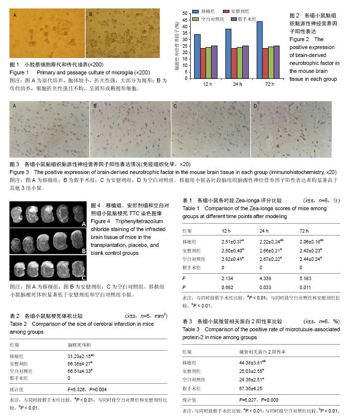
 

摘要
背景:
静止状态的小胶质细胞担负免疫监视的重任,而对活化后小胶质细胞的作用则存在较大争议。
目的:分析活化小胶质细胞在急性脑梗死中的作用机制。
方法:选取96只雄性昆明小鼠,随机分为4组。其中移植组、安慰剂组及空白对照组小鼠经线栓栓塞大脑中动脉法建立永久性局灶性脑梗死动物模型,移植组建模后12 h经锁骨下静脉注入小胶质细胞悬液,安慰剂组注入同体积小胶质细胞培养基;空白对照组仅建模;假手术组小鼠经假手术12 h后注入同移植组同体积的小胶质细胞悬液。观察建模后12,24,72 h时神经功能Zea-longa评分、脑源性神经营养因子表达情况及72 h时脑梗死体积、存活神经细胞(即微管相关蛋白2阳性率)差异。
结果与结论:①假手术组小鼠各时段Zea-longa评分均为0分,显著低于其他3组小鼠(P < 0.01);小胶质细胞移植24,72 h时,移植组Zea-longa评分显著低于安慰剂组和空白对照组小鼠(P < 0.01);②移植组各时段脑组织脑源性神经营养因子阳性表达率均显著高于其他3组小鼠(P < 0.01);③假手术组未见脑梗死,小胶质细胞移植72 h时,移植组小鼠脑梗死体积为显著低于安慰剂组和空白对照组(P < 0.01),微管相关蛋白2阳性率显著高于安慰剂组和空白对照组(P < 0.01);④结果表明,活化小胶质细胞对提高神经细胞存活率、促进脑神经功能恢复、抑制脑梗死体积扩大等具有积极影响。


ORCID: 0000-0001-9865-1036(项薇)

关键词: 组织构建, 组织工程, 活化小胶质细胞, 急性脑梗死, 作用机制, 广东省自然科学基金

Abstract:

BACKGROUND: Microglia play an important role in immune surveillance in their quiescent state, but the role of the activated microglia is under discussion.
OBJECTIVE: To analyze the mechanism of activated microglia in acute cerebral infarction.
METHODS: Totally 96 male Kunming mice were selected and randomly divided into four groups, including transplantation, placebo, blank control and sham operation groups. Permanent occlusion of the middle cerebral artery was performed using suture method in the mice of the transplantation, placebo and blank control groups, followed by injection of microglia suspension via subclavian vein, medium containing the same volume of microglia, and nothing, respectively, at 12 hours after modeling. In the meanwhile, the same amount of microglia suspension was injected into the mice of the sham operation group. The Zea-longa scale and brain-derived neurotrophic factor expression at 12, 24 and 72 hours after modeling, the volume of cerebral infarction and the number of nerve cells positive for microtubule-associated protein-2 at 72 hours after modeling were detected.
RESULTS AND CONCLUSION: The Zea-longa scale score was 0 point in the sham operation group, which was significantly lower than that in the other three groups at each time point after modeling (P < 0.01). The Zea-longa scores in the transplantation group were significantly lower than those in the placebo and blank control groups at 24 and 72 hours after transplantation (P < 0.01). The positive expression rate of brain-derived neurotrophic factor in the transplantation group was significantly higher than that in the other three groups after transplantation (P < 0.01). The sham group showed no infarction, while the size of cerebral infarction in the transplantation group was significantly lower than that in the placebo and blank control groups (P < 0.01), and the microtubule-associated protein-2 positive rate was significantly higher than that in the placebo and blank control groups (P < 0.01). These results manifest that the activated microglia can improve the survival rate of nerve cells, promote the recovery of cerebral nerve function and reduce the size of cerebral infarction.


Key words: Glioma, Brain Infarction, Brain-Derived Neurotrophic Factor, Tissue Engineering

中图分类号: