| [1] Pelletier CA, Dumont FS, Leblond J, et al. Self-report of one-year fracture incidence and osteoporosis prevalence in a community cohort of canadians with spinal cord injury. Top Spinal Cord Inj Rehabil. 2014; 20(4):302-309. [2] Coupaud S, McLean AN, Purcell M, et al. Decreases in bone mineral density at cortical and trabecular sites in the tibia and femur during the first year of spinal cord injury. Bone. 2015;74:69-75. [3] Gifre L, Vidal J, Carrasco J, et al. Incidence of skeletal fractures after traumatic spinal cord injury: a 10-year follow-up study. Clin Rehabil. 2014;28(4):361-369. [4] Jiang SD, Jiang LS, Dai LY. Mechanisms of osteoporosis in spinal cord injury. Clin Endocrinol (Oxf). 2006;65(5):555-565.[5] Galea MP. Spinal cord injury and physical activity: preservation of the body. Spinal Cord. 2012;50(5): 344-351.[6] Dolbow DR, Gorgey AS, Daniels JA, et al. The effects of spinal cord injury and exercise on bone mass: a literature review. NeuroRehabilitation. 2011;29(3): 261-269. [7] Chang KV, Hung CY, Chen WS, et al. Effectiveness of bisphosphonate analogues and functional electrical stimulation on attenuating post-injury osteoporosis in spinal cord injury patients- a systematic review and meta-analysis. PLoS One. 2013;8(11):e81124. [8] Nyandege AN, Slattum PW, Harpe SE. Risk of fracture and the concomitant use of bisphosphonates with osteoporosis-inducing medications. Ann Pharmacother. 2015;49(4):437-447.[9] McGreevy C, Williams D. Safety of drugs used in the treatment of osteoporosis. Ther Adv Drug Saf. 2011; 2(4):159-172. [10] Beggs LA, Ye F, Ghosh P, et al. Sclerostin inhibition prevents spinal cord injury-induced cancellous bone loss. J Bone Miner Res. 2015;30(4):681-689. [11] Maïmoun L, Couret I, Mariano-Goulart D, et al. Changes in osteoprotegerin/RANKL system, bone mineral density, and bone biochemicals markers in patients with recent spinal cord injury. Calcif Tissue Int. 2005;76(6):404-411.[12] Cummings SR, San Martin J, McClung MR, et al. Denosumab for prevention of fractures in postmenopausal women with osteoporosis. N Engl J Med. 2009;361(8):756-765.[13] Morse LR, Sudhakar S, Danilack V, et al. Association between sclerostin and bone density in chronic spinal cord injury. J Bone Miner Res. 2012;27(2):352-359. [14] Garland DE, Stewart CA, Adkins RH, et al. Osteoporosis after spinal cord injury. J Orthop Res. 1992;10(3):371-378.[15] Jiang SD, Yang YH, Chen JW, et al. Isolated osteoblasts from spinal cord-injured rats respond less to mechanical loading as compared with those from hindlimb-immobilized rats. J Spinal Cord Med. 2013; 36(3):220-224. [16] Karsenty G, Yadav VK. Regulation of bone mass by serotonin: molecular biology and therapeutic implications. Annu Rev Med. 2011;62:323-331.[17] Amireault P, Sibon D, Côté F. Life without peripheral serotonin: insights from tryptophan hydroxylase 1 knockout mice reveal the existence of paracrine/autocrine serotonergic networks. ACS Chem Neurosci. 2013;4(1):64-71. [18] Ghosh M, Pearse DD. The role of the serotonergic system in locomotor recovery after spinal cord injury. Front Neural Circuits. 2015;8:151. [19] Haney EM, Warden SJ. Skeletal effects of serotonin (5-hydroxytryptamine) transporter inhibition: evidence from clinical studies. J Musculoskelet Neuronal Interact. 2008;8(2):133-145.[20] Ducy P. 5-HT and bone biology. Curr Opin Pharmacol. 2011;11(1):34-38. [21] Karsenty G, Gershon MD. The importance of the gastrointestinal tract in the control of bone mass accrual. Gastroenterology. 2011;141(2):439-442.[22] Westbroek I, van der Plas A, de Rooij KE, et al. Expression of serotonin receptors in bone. J Biol Chem. 2001;276(31):28961-28968.[23] Cosentino M, Marino F, Maestroni GJ. Sympathoadrenergic modulation of hematopoiesis: a review of available evidence and of therapeutic perspectives. Front Cell Neurosci. 2015;9:302.[24] Lambert AM, Bonkowsky JL, Masino MA. The conserved dopaminergic diencephalospinal tract mediates vertebrate locomotor development in zebrafish larvae. J Neurosci. 2012;32(39): 13488-13500.[25] Cobacho N, de la Calle JL, Paíno CL. Dopaminergic modulation of neuropathic pain: analgesia in rats by a D2-type receptor agonist. Brain Res Bull. 2014;106:62-71.[26] Ondo WG, He Y, Rajasekaran S, et al. Clinical correlates of 6-hydroxydopamine injections into A11 dopaminergic neurons in rats: a possible model for restless legs syndrome. Mov Disord. 2000;15(1): 154-158.[27] Miyake H, Nagashima K, Onigata K, et al. Allelic variations of the D2 dopamine receptor gene in children with idiopathic short stature. J Hum Genet. 1999;44(1): 26-29.[28] Yamada Y, Ando F, Niino N, et al. Association of a polymorphism of the dopamine receptor D4 gene with bone mineral density in Japanese men. J Hum Genet. 2003;48(12):629-633. [29] Hanami K, Nakano K, Saito K, et al. Dopamine D2-like receptor signaling suppresses human osteoclastogenesis. Bone. 2013;56(1):1-8. [30] Hanami K, Nakano K, Tanaka Y. Dopamine receptor signaling regulates human osteoclastogenesis. Nihon Rinsho Meneki Gakkai Kaishi. 2013;36(1):35-39.[31] Rizzoli R, Cooper C, Reginster JY, et al. Antidepressant medications and osteoporosis. Bone. 2012;51(3): 606-613. [32] Eom CS, Lee HK, Ye S, et al. Use of selective serotonin reuptake inhibitors and risk of fracture: a systematic review and meta-analysis. J Bone Miner Res. 2012; 27(5): 1186-1195.[33] Tsapakis EM, Gamie Z, Tran GT, et al. The adverse skeletal effects of selective serotonin reuptake inhibitors. Eur Psychiatry. 2012;27(3):156-169.[34] Meyer JM, Lehman D. Bone mineral density in male schizophrenia patients: a review. Ann Clin Psychiatry. 2006;18(1):43-48.[35] Wang M, Hou R, Jian J, et al. Effects of antipsychotics on bone mineral density and prolactin levels in patients with schizophrenia: a 12-month prospective study. Hum Psychopharmacol. 2014;29(2):183-189.[36] O'Keane V. Antipsychotic-induced hyperprolactinaemia, hypogonadism and osteoporosis in the treatment of schizophrenia. J Psychopharmacol. 2008;22(2 Suppl): 70-75.[37] Kunimatsu T, Kimura J, Funabashi H, et al. The antipsychotics haloperidol and chlorpromazine increase bone metabolism and induce osteopenia in female rats. Regul Toxicol Pharmacol. 2010;58(3):360-368. [38] Lapointe NP, Guertin PA. Synergistic effects of D1/5 and 5-HT1A/7 receptor agonists on locomotor movement induction in complete spinal cord-transected mice. J Neurophysiol. 2008;100(1):160-168.[39] Lapointe NP, Rouleau P, Ung RV, et al. Specific role of dopamine D1 receptors in spinal network activation and rhythmic movement induction in vertebrates. J Physiol. 2009;587(Pt 7):1499-1511. [40] Jiang W, Huang Y, He F, et al. Dopamine D1 Receptor Agonist A-68930 Inhibits NLRP3 Inflammasome Activation, Controls Inflammation, and Alleviates Histopathology in a Rat Model of Spinal Cord Injury. Spine (Phila Pa 1976). 2016;41(6):E330-334.[41] Ganzer PD, Manohar A, Shumsky JS, et al. Therapy induces widespread reorganization of motor cortex after complete spinal transection that supports motor recovery. Exp Neurol. 2016;279:1-12. [42] Cornide-Petronio ME, Fernández-López B, Barreiro-Iglesias A, et al. Traumatic injury induces changes in the expression of the serotonin 1A receptor in the spinal cord of lampreys. Neuropharmacology. 2014;77:369-378.[43] Murray KC, Stephens MJ, Ballou EW, et al. Motoneuron excitability and muscle spasms are regulated by 5-HT2B and 5-HT2C receptor activity. J Neurophysiol. 2011;105(2):731-748. [44] S?awińska U, Miazga K, Jordan LM. 5-HT? and 5-HT? receptor agonists facilitate plantar stepping in chronic spinal rats through actions on different populations of spinal neurons. Front Neural Circuits. 2014;8:95. [45] Demirel G, Yilmaz H, Paker N, et al. Osteoporosis after spinal cord injury. Spinal Cord. 1998;36(12):822-825.[46] Ducy P, Karsenty G. The two faces of serotonin in bone biology. J Cell Biol. 2010;191(1):7-13. [47] Otake T, Ieshima H, Ishida H, et al. Bone atrophy in complex regional pain syndrome patients measured by microdensitometry. Can J Anaesth. 1998;45(9): 831-838. |
.jpg) 文题释义:
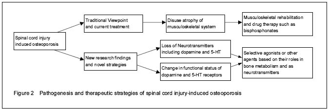
脊髓损伤导致的骨质疏松症:发生于脊髓损伤后的继发性骨质疏松症,既往有观点认为是由于神经损伤后长期肌肉骨骼系统功能障碍导致的失用性疾病,近年研究则提示参与骨代谢调节的神经递质可能在疾病进展中作用更为重要。该类继发性骨质疏松症在脊髓损伤患者中其实际发病率高,但诊断率低,临床治疗手段有限。
多巴胺和5-羟色胺受体系统:多巴胺和5-羟色胺为中枢神经系统内的重要神经递质,同时均对机体骨代谢具有调控作用,在脊髓损伤后多巴胺和5-羟色胺的正常传输及相应受体系统的功能状态变化与多项病理生理改变和临床并发症相关。
文题释义:
脊髓损伤导致的骨质疏松症:发生于脊髓损伤后的继发性骨质疏松症,既往有观点认为是由于神经损伤后长期肌肉骨骼系统功能障碍导致的失用性疾病,近年研究则提示参与骨代谢调节的神经递质可能在疾病进展中作用更为重要。该类继发性骨质疏松症在脊髓损伤患者中其实际发病率高,但诊断率低,临床治疗手段有限。
多巴胺和5-羟色胺受体系统:多巴胺和5-羟色胺为中枢神经系统内的重要神经递质,同时均对机体骨代谢具有调控作用,在脊髓损伤后多巴胺和5-羟色胺的正常传输及相应受体系统的功能状态变化与多项病理生理改变和临床并发症相关。.jpg) 文题释义:
脊髓损伤导致的骨质疏松症:发生于脊髓损伤后的继发性骨质疏松症,既往有观点认为是由于神经损伤后长期肌肉骨骼系统功能障碍导致的失用性疾病,近年研究则提示参与骨代谢调节的神经递质可能在疾病进展中作用更为重要。该类继发性骨质疏松症在脊髓损伤患者中其实际发病率高,但诊断率低,临床治疗手段有限。
多巴胺和5-羟色胺受体系统:多巴胺和5-羟色胺为中枢神经系统内的重要神经递质,同时均对机体骨代谢具有调控作用,在脊髓损伤后多巴胺和5-羟色胺的正常传输及相应受体系统的功能状态变化与多项病理生理改变和临床并发症相关。
文题释义:
脊髓损伤导致的骨质疏松症:发生于脊髓损伤后的继发性骨质疏松症,既往有观点认为是由于神经损伤后长期肌肉骨骼系统功能障碍导致的失用性疾病,近年研究则提示参与骨代谢调节的神经递质可能在疾病进展中作用更为重要。该类继发性骨质疏松症在脊髓损伤患者中其实际发病率高,但诊断率低,临床治疗手段有限。
多巴胺和5-羟色胺受体系统:多巴胺和5-羟色胺为中枢神经系统内的重要神经递质,同时均对机体骨代谢具有调控作用,在脊髓损伤后多巴胺和5-羟色胺的正常传输及相应受体系统的功能状态变化与多项病理生理改变和临床并发症相关。
.jpg)
.jpg) 文题释义:
脊髓损伤导致的骨质疏松症:发生于脊髓损伤后的继发性骨质疏松症,既往有观点认为是由于神经损伤后长期肌肉骨骼系统功能障碍导致的失用性疾病,近年研究则提示参与骨代谢调节的神经递质可能在疾病进展中作用更为重要。该类继发性骨质疏松症在脊髓损伤患者中其实际发病率高,但诊断率低,临床治疗手段有限。
多巴胺和5-羟色胺受体系统:多巴胺和5-羟色胺为中枢神经系统内的重要神经递质,同时均对机体骨代谢具有调控作用,在脊髓损伤后多巴胺和5-羟色胺的正常传输及相应受体系统的功能状态变化与多项病理生理改变和临床并发症相关。
文题释义:
脊髓损伤导致的骨质疏松症:发生于脊髓损伤后的继发性骨质疏松症,既往有观点认为是由于神经损伤后长期肌肉骨骼系统功能障碍导致的失用性疾病,近年研究则提示参与骨代谢调节的神经递质可能在疾病进展中作用更为重要。该类继发性骨质疏松症在脊髓损伤患者中其实际发病率高,但诊断率低,临床治疗手段有限。
多巴胺和5-羟色胺受体系统:多巴胺和5-羟色胺为中枢神经系统内的重要神经递质,同时均对机体骨代谢具有调控作用,在脊髓损伤后多巴胺和5-羟色胺的正常传输及相应受体系统的功能状态变化与多项病理生理改变和临床并发症相关。