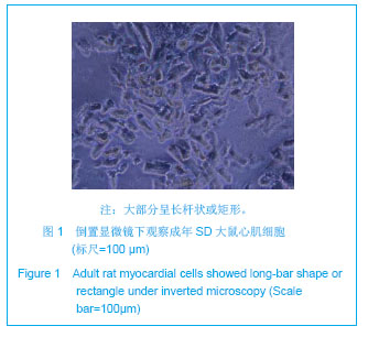
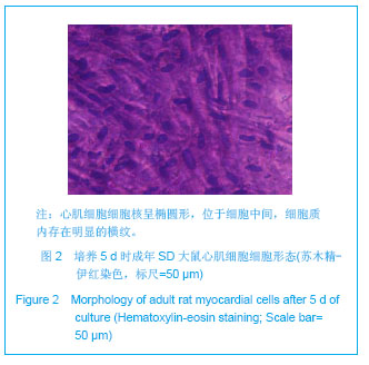
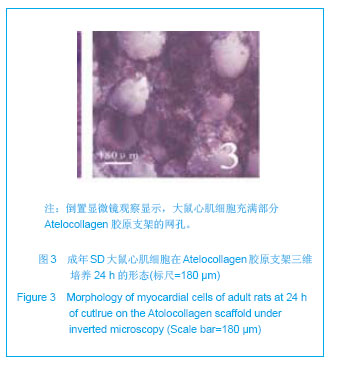
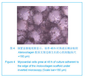
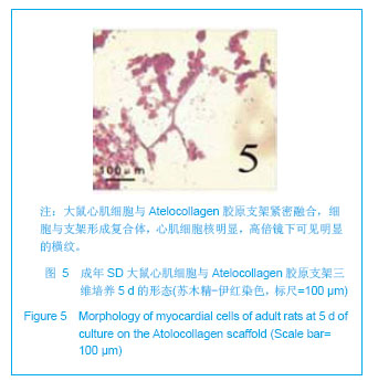
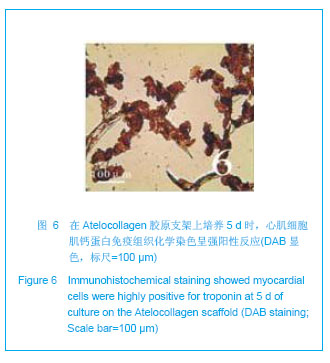
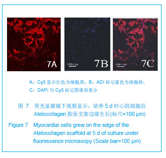
| [1]Alcon A,Cagavi BE,Qyang Y.Regenerating functional heart tissue for myocardial repair.Cell Mol Life Sci.2012;69(16): 2635-2656.[2]Curtis MW,Russell B.Cardiac tissue engineering. Cardiovasc Nurs.2009; 24(2): 87-92.[3]Nunes SS,Song H,Chiang CK,et al.Stem cell-based cardiac tissue engineering. Cardiovasc Transl Res.2011;4(5): 592-602.[4]Beitnes JO,Lunde K,Brinchmann JE,et al.Stem cells for cardiac repair in acute myocardial infarction.Expert Rev Cardiovasc Ther. 2011;9(8): 1015-1025.[5]Teng W,Guo ZK,Li Q,et al.Jiepou Xuebao. 2010;41(6): 885-890.滕伟,郭志坤,李琼,等. 在Atelocollagen胶原支架上体外三维培养乳大鼠心肌[J].解剖学报,2010,41(6): 885-890.[6]Guo Z,Iku S,Zheng X,et al.Three-Dimensional Geometry of Honeycomb Collagen Promotes Higher Beating Rate of Myocardial Cells in Culture. Artif Organs. 2012;36(9):816-819.[7]Huang ZB,Xiao JF,Shen JX.Jiguang Shengwu Xuebao. 2010; 19(3): 307-313.黄泽炳,肖剑锋,沈建新.胞外镁离子对SD成年大鼠心肌细胞胞内钙信号的影响[J].激光生物学报,2010,19(3): 307-313.[8]Wei LL,Mo SR.Zhongguo Zuzhi Gongcheng Yanjiu. 2012; 16(11): 1969-1972.韦丽兰,莫书荣.成年大鼠心肌细胞的急性分离方法[J].中国组织工程研究,2012,16(11): 1969-1972.[9]Li D,Tang B,Yang DC,et al.Zhongguo Shiyan Dongwu Xuebao. 2011;19(2): 150-152.李德,唐兵,杨大春,等.Ⅱ型胶原酶加压灌流提高成年大鼠心肌细胞分离效率[J].中国实验动物学报,2011,19(2): 150-152.[10]Liao H,Mi T,Tu ZY,et al. Zhongguo Zuzhi Gongcheng Yanjiu yu Linchuang Kangfu. 2009;13(33): 6536-6539.廖华,糜涛,涂志业,等.成年大鼠心肌细胞分离方法的改良[J].中国组织工程研究与临床康复,2009,13(33): 6536-6539.[11]Li H,Xiao YB.Disan Junyi Daxue Xuebao. 2004;26(7): 644-646.李洪,肖颖彬 .成年大鼠心室肌细胞的分离、培养与鉴定[J].第三军医大学学报,2004,26(7): 644-646.[12]Zhu XX,Niu XL,Zhu XL,et al.Jiefangjun Yixue Zazhi.2008; 33(9): 1089-1091.朱肖星,牛小麟,朱萧玲,等.不同活性的胶原酶对大鼠心肌细胞分离的影响[J].解放军医学杂志,2008,33(9): 1089-1091.[13]Chang H,Zhang L,Yu ZB.Zhongguo Yingyong Shenglixue Zazhi. 3011;27(1): 57-61,136.常惠,张琳,余志斌.培养成年大鼠心肌细胞存活的形态标志[J].中国应用生理学杂志,3011,27(1): 57-61,136.[14]Gong HB,Wang J,Wang L.Zhongguo Weixunhuan. 2009; 13(1):55-59.宫海滨,王洁,王雷.不同培养基对成年大鼠心肌细胞形态、存活率、收缩功能的影响[J].中国微循环,2009,13(1): 55-59.[15]Pavlovic D,McLatchie LM,Shattock MJ.The rate of loss of T-tubules in cultured adult ventricular myocytes is species dependent. Exp Physiol.2010; 95(4): 518-527.[16]Smyrnias I,Mair W,Harzheim D,et al. Comparison of the T-tubule system in adult rat ventricular and atrial myocytes, and its role in excitation-contraction coupling and inotropic stimulation. Cell Calcium. 2010; 47(3): 210-223.[17]Zhai YJ,Dong XH,Zhou LN,et al.Disi Junyi Daxue Xuebao. 2005;25(8): 705-707.翟迎九, 董雪红,周丽诺,等.成年SD大鼠心肌细胞的培养[J].第四军医大学学报,2005,25(8): 705-707.[18]Wang T,Fu Y,Fang XS,et al.Lingnan Jizhen Yixue Zazhi. 2007; 12(5): 323-325.王彤,符岳,方向韶,等.普通大鼠心肌细胞的分离培养和收缩舒张观察[J].岭南急诊医学杂志,2007,12(5): 323-325.[19]Wang Y,Sun H,Fan YM,et al.Xuzhou Yixueyuan Xuebao. 2005; 25(5): 393-396.王影,孙红,范乐明,等.成年大鼠心肌细胞的分离和培养技术[J].徐州医学院学报,2005,25(5): 393-396.[20]Li YG,Shi YK,Chen HW,et al.Huaxi Yixue. 2005;20(2): 308-309.李勇刚, 石应康,陈焕文,等.成年大鼠心肌细胞的分离和培养[J].华西医学,2005,20(2): 308-309.[21]Xu XF,Li WB,Chen BT,et al.Shoudu Yike Daxue Xuebao. 2000;21(2): 104-107.许秀芳,李温斌,陈宝田,等.成年大鼠心肌细胞培养方法的建立和形态学观察[J].首都医科大学学报,2000,21(2): 104-107.[22]Kato S,Takemura G,Maruyama R,et al. Apoptosis, rather than oncosis, is the predominant mode of spontaneous death of isolated adult rat cardiac myocytes in culture.Jpn Circ J. 2001;65(8): 743-748.[23]Hein S,Kostin S,Schaper J.Adult rat cardiac myocytes in culture: 'Second-floor' cells and coculture experiments. Exp Clin Cardiol.2006;11(3): 175-182. [24]Bugaisky LB,Zak R.Differentiation of adult rat cardiac myocytes in cell culture. Circ Res.1989;64(3): 493-500.[25]Guo ZK.Beijing:Renmin Junyi Chbanshe.2005:6.郭志坤.正常心脏组织学图谱[M].北京:人民军医出版社,2005: 6.[26]Ohno S,Hirano S,Kanemaru S,et al.Implantation of an atelocollagen sponge with autologous bone marrow-derived mesenchymal stromal cells for treatment of vocal fold scarring in a canine model. Ann Otol Rhinol Laryngol. 2011;120(6): 401-408.[27]Lee KI,Moon SH,Kim H,et al. Tissue engineering of the intervertebral disc with cultured nucleus pulposus cells using atelocollagen scaffold and growth factors. Spine (Phila Pa 1976). 2012;37(6): 452-458.[28]Ko EC,Fujihara Y,Ogasawara T,et al.BMP-2 Embedded Atelocollagen Scaffold for Tissue-Engineered Cartilage Cultured in the Medium Containing Insulin and Triiodothyronine-A New Protocol for Three-Dimensional In Vitro Culture of Human Chondrocytes. Tissue Eng Part C Methods.2012;18(5):374-386.[29]Zheng XJ,Guo ZK,Chang KS,et al.Jiepou Xuebao. 2008;39(4): 582-585.郑先杰,郭志坤,常联生,等.Atelocoilagen胶原支架在三维培养大鼠心肌中的生物相容性[J].解剖学报,2008,39(4): 582-585.[30]Akhyari P,Fedak PW,Weisel RD,et al.Mechanical stretch regimenenhances the formation of bioengineered autologous cardiac muscle grafts. Circulation. 2002;106(Supp l1): Ⅰ137-Ⅰ142.[31]Egorova MV,Afanas'ev SA,Popov SV.A simple method for isolation of cardiomyocytes from adult rat heart. Bull Exp Biol Med. 2005;140(3): 370-373.[32]Xu X, Colecraft HM.Primary culture of adult rat heart myocytes. Vis Exp. 2009;(28): pii: 1308.[33]Eckel J,van EG,Reinauer H. Adult cardiac myocytes in primary culture: cell characteristics and insulin-receptor interaction. Am J Physiol.1985;249(2 Pt 2): H212-221.[34]Jacobson SL,Piper HM.Cell cultures of adult cardiomyocytes as models of the myocardium.Mol Cell Cardiol.1986;18(7): 661-678.[35]Dubus I,Samuel JL,Marotte F,et al.Beta-adrenergic agonists stimulate the synthesis of noncontractile but not contractile proteins in cultured myocytes isolated from adult rat heart. Circ Res. 1990;66(3): 867-874.[36]Schwarzfeld TA,Jacobson SL.Isolation and development in cell culture of myocardial cells of the adult rat. Mol Cell Cardiol. 1981;13(6): 563-575.[37]Yamamoto S,Yasui K,Palade PT,et al.Spontaneous death of isolated adult rat cardiocytes in culture in association with internucleosomal cleavage of genomic DNA. Apoptosis.1997; 2(2): 178-188.[38]Louch WE,Bito V,Heinzel FR,et al.Reduced synchrony of Ca2+ release with loss of T-tubules-a comparison to Ca2+ release in human failing cardiomyocytes.Cardiovasc Res. 2004;62(1): 63-73.[39]Leach RN, Desai JC,Orchard CH. Effect of cytoskeleton disruptors on L-type Ca channel distribution in rat ventricular myocytes. Cell Calcium. 2005;38(5): 515-526.[40]Jacobson SL,Piper HM.Cell cultures of adult cardiomyocytes as models of the myocardium. Mol Cell Cardiol.1986;18(7): 661-678. |
.jpg)