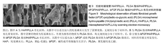
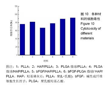
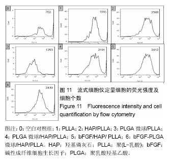
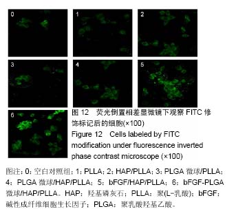
| [1]Parsons B, Strauss E. Surgical management of chronic osteomyelitis. Am J Surg. 2004;188(1A Suppl):57-66.[2]Kovacevic D, Fox AJ, Bedi A, et al. Calcium-phosphate matrix with or without TGF-β3 improves tendon-bone healing after rotator cuff repair. Am J Sports Med. 2011;39(4):811-819.[3]Servin-Trujillo MA, Reyes-Esparza JA, Garrido-Fariña G, et al. Use of a graft of demineralized bone matrix along with TGF-β1 leads to an early bone repair in dogs. J Vet Med Sci. 2011;73(9): 1151-1161.[4]Bichara DA, O'Sullivan NA, Pomerantseva I, et al. The tissue-engineered auricle: past, present, and future. Tissue Eng Part B Rev. 2012;18(1):51-61.[5]Eppley BL, Pietrzak WS, Blanton MW. Allograft and alloplastic bone substitutes: a review of science and technology for the craniomaxillofacial surgeon. J Craniofac Surg. 2005;16(6): 981-989.[6]Goldberg VM. Selection of bone grafts for revision total hip arthroplasty. Clin Orthop Relat Res. 2000;(381):68-76.[7]Karaoglu S, Baktir A, Kabak S, et al. Experimental repair of segmental bone defects in rabbits by demineralized allograft covered by free autogenous periosteum. Injury. 2002;33(8): 679-683.[8]Clayer M. Injectable form of calcium sulphate as treatment of aneurysmal bone cysts. ANZ J Surg. 2008;78(5):366-370.[9]Yang J, Yamato M, Nishida K, et al. Cell delivery in regenerative medicine: the cell sheet engineering approach. J Control Release. 2006;116(2):193-203.[10]Ouyang HW, Toh SL, Goh J, et al. Assembly of bone marrow stromal cell sheets with knitted poly (L-lactide) scaffold for engineering ligament analogs. J Biomed Mater Res B Appl Biomater. 2005;75(2):264-271.[11]Castoldi M, Pistone M, Caruso C, et al. Osteoblastic cells from rat long bone. II: Adhesion to substrata and integrin expression in primary and propagated cultures. Cell Biol Int. 1997;21(1):7-16.[12]Manduca P, Sanguineti C, Pistone M, et al. Differential expression of alkaline phosphatase in clones of human osteoblast-like cells. J Bone Miner Res. 1993;8(3):291-300.[13]Huibregtse BA, Johnstone B, Goldberg VM, et al. Effect of age and sampling site on the chondro-osteogenic potential of rabbit marrow-derived mesenchymal progenitor cells. J Orthop Res. 2000;18(1):18-24.[14]Service RF. Tissue engineers build new bone. Science. 2000; 289(5484):1498-1500.[15]Srouji S, Blumenfeld I, Rachmiel A, et al. Bone defect repair in rat tibia by TGF-beta1 and IGF-1 released from hydrogel scaffold. Cell Tissue Bank. 2004;5(4):223-230.[16]Schnettler R, Alt V, Dingeldein E, et al. Bone ingrowth in bFGF-coated hydroxyapatite ceramic implants. Biomaterials. 2003;24(25):4603-4608.[17]Tan HC, Lei J, Jiang DZ. Effect of bFGF on hematopoietic regulation of bone marrow stromal cells. Hunan Yi Ke Da Xue Xue Bao. 2003;28(1):83-84.[18]Varkey M, Kucharski C, Haque T, et al. In vitro osteogenic response of rat bone marrow cells to bFGF and BMP-2 treatments. Clin Orthop Relat Res. 2006;443:113-123.[19]Li Y, Chen HQ, Cheng M, et al. Bone marrow stromal cells co-modified with BMP-2 and bFGF gene.Xi Bao Yu Fen Zi Mian Yi Xue Za Zhi. 2006;22(2):133-136.[20]Kilic MA, Ozlu E, Calis S. A novel protein-based anticancer drug encapsulating nanosphere: apoferritin-doxorubicin complex. J Biomed Nanotechnol. 2012;8(3):508-514.[21]Batheja P, Sheihet L, Kohn J, et al. Topical drug delivery by a polymeric nanosphere gel: Formulation optimization and in vitro and in vivo skin distribution studies. J Control Release. 2011;149(2):159-167.[22]Estevanato L, Cintra D, Baldini N, et al. Preliminary biocompatibility investigation of magnetic albumin nanosphere designed as a potential versatile drug delivery system. Int J Nanomedicine. 2011;6:1709-1717.[23]Vukomanovi? M, Skapin SD, Poljanšek I, et al. Poly(D,L-lactide-co-glycolide)/hydroxyapatite core-shell nanosphere. Part 2: Simultaneous release of a drug and a prodrug (clindamycin and clindamycin phosphate). Colloids Surf B Biointerfaces. 2011;82(2):414-421.[24]Holmes TC. Novel peptide-based biomaterial scaffolds for tissue engineering. Trends Biotechnol. 2002;20(1):16-21.[25]Tsuchiya A, Sotome S, Asou Y, et al. Effects of pore size and implant volume of porous hydroxyapatite/collagen (HAp/Col) on bone formation in a rabbit bone defect model. J Med Dent Sci. 2008;55(1):91-99.[26]Zhang H, Wang W, Chu D, et al. Preliminary study on chitosan/HAP bilayered scaffold. Zhongguo Xiu Fu Chong Jian Wai Ke Za Zhi. 2008;22(11):1358-1363.[27]Han XQ, Han XQ, Dong ZH, et al. Experimental study of tissue-engineered bone constructed with simvastatin carried by PLGA/CPC and bone marrow stromal cells. Shanghai Kou Qiang Yi Xue. 2014;23(1):7-14.[28]Lee K, Weir MD, Lippens E, et al. Bone regeneration via novel macroporous CPC scaffolds in critical-sized cranial defects in rats. Dent Mater. 2014;30(7):e199-207. |
.jpg)
.jpg)