中国组织工程研究 ›› 2017, Vol. 21 ›› Issue (27): 4373-4378.doi: 10.3969/j.issn.2095-4344.2017.27.019

• 骨与关节图像与影像 bone and joint imaging • 上一篇    下一篇

设计国人直肠后间隙入路轴向腰骶椎融合内固定的影像学测量

曾德辉,张 卫,张 彬,向 亮,侯 威   

  1. 南华大学附属南华医院骨科,湖南省衡阳市   421001
  • 出版日期:2017-09-28 发布日期:2017-10-24
  • 作者简介:曾德辉,男,1977年生,湖南省新化县人,汉族,2009年南华大学毕业,硕士,副主任医师,副教授,主要从事微创脊柱与创伤方面的研究。
  • 基金资助:

    湖南省科学技术厅资助项目(2012SK3150)

Imaging measurement for internal fixation design of axial lumbosacral vertebral fusion via posterior rectal space  

Zeng De-hui, Zhang Wei, Zhang Bin, Xiang Liang, Hou Wei   

  1. Department of Orthopedics, Nanhua Hospital Affiliated to University of South China, Hengyang 421001, China
  • Online:2017-09-28 Published:2017-10-24
  • About author:Zeng De-hui, Master, Associate chief physician, Associate professor, Department of Orthopedics, Nanhua Hospital Affiliated to University of South China, Hengyang 421001, China
  • Supported by:

    the Project of China Hunan Provincial Science & Technology Department, No. 2012SK3150

摘要:

文章快速阅读:


文题释义:
经皮腰骶椎前柱内固定系统(AxiaLIF):是经骶骨前入路轴向行腰骶椎前柱融合的内固定系统,其手术入路经直肠后间隙,沿腰骶椎的中轴进行。因直肠后间隙内主要为脂肪组织,又是于腰椎中轴进行手术,故该系统对手术入路周围组织、脊柱周围组织、脊柱本身结构(如韧带、小关节、椎板等)的损伤程度较小,从而可以较大限度地减少手术损伤,降低脊柱融合病的发生率。
腰骶部:是L5和S1间软、硬组织,包括腰骶部韧带、肌腱、腱膜、筋膜、椎间盘、椎骨关节及椎体,腰骶部正常位于活动度较大的腰椎与甚少活动的骨盆交接处,同时又位于腰椎生理前凸与腰椎生理后凸的交接处,杠杆作用特别大,容易受到损伤。
 
摘要
背景:经骶骨前入路的经皮腰骶椎前柱内固定系统即AxiaLIF系统先后在美国及欧洲应用于临床,取得较好的临床疗效。但是国人与欧美人腰骶椎的解剖差异较大,国外内固定系统能否可以直接用于国人尚不明确,且价格昂贵,不适合中国国情,同时该内固定系统在应用过程中出现了一系列的问题,如能优化轴向螺钉设计,该术式在国内将会有更大的推广价值。
目的:通过测量国人正常人群腰椎侧位片及腰椎CT L5、S1椎体横断面,为设计适合国人的轴向螺钉提供解剖学数据。
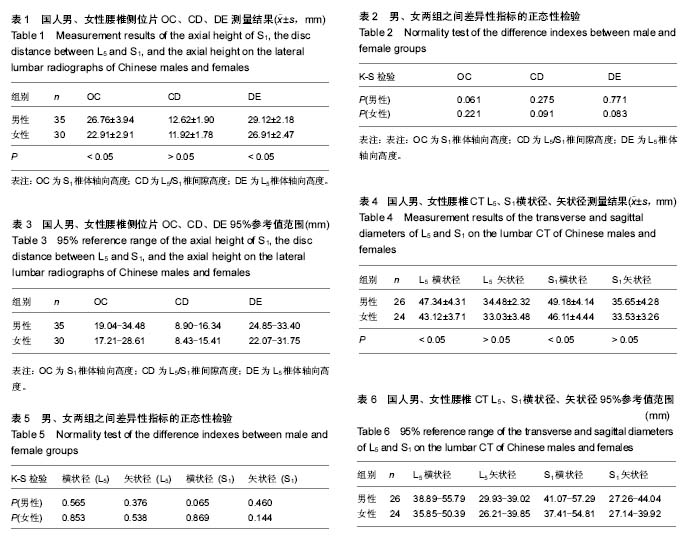
方法:①选择国人正常成人腰椎侧位片男35例,女30例,分别测量S1椎体轴向高度、L5/S1椎间隙高度、L5椎体轴向高度,为轴向螺钉的长度设计提供解剖学数据;②选择成人腰椎CT片男26例,女24例,测量腰椎CT L5、S1椎体经椎弓根层面的横状径与矢状径,为轴向螺钉的直径设计提供解剖学数据。
结果与结论:①国人男、女性腰椎侧位片S1椎体轴向高度为(26.76±3.94) mm和(22.91±2.91) mm(P < 0.05)、L5/S1椎间隙高度为(12.62±1.90) mm和(11.92±1.78) mm(P > 0.05)、L5椎体轴向高度为(29.12±2.18) mm和(26.91±2.47) mm(P < 0.05);②国人男、女性腰椎CT L5横状径为(47.34±4.31) mm和(43.12±3.71) mm(P < 0.05),S1横状径为(49.18±4.14) mm和(46.11±4.44) mm(P < 0.05),L5矢状径为(34.48±2.32) mm和(33.03±3.48) mm(P > 0.05),S1矢状径为(35.65±4.28) mm和(33.53±3.26) mm(P > 0.05);③综上,通过国人腰椎侧位片中轴线及腰椎CT L5、S1椎体横断面的测量,可为设计出适合国人腰骶椎融合的轴向螺钉提供解剖数据;根据该影像学测量方法对患者术前影像学资料进行分析,可以预判患者行该手术方式的可行性,并对内固定型号选择做出预估,进行个性化置钉。

中国组织工程研究杂志出版内容重点:人工关节;骨植入物;脊柱骨折;内固定;数字化骨科;组织工程
ORCID: 0000-0003-4737-6294(曾德辉)

关键词: 骨科植入物, 脊柱植入物, 直肠后间隙, 腰骶椎, 轴向螺钉, 置钉, 影像学

Abstract:

BACKGROUND: The minimally invasive axial lumbar interbody approach (AxiaLIF) for L4–S1 fusion has been applied in America and Europe, and has obtained satisfactory curative efficacy. Because of significant anatomical differences between Chinese and Europeans and Americans, whether AxiaLIF is appropriate for Chinese remains unclear. Moreover, there are some problems in the application of AxiaLIF, so how to optimize AxiaLIF is a key to its promotion in China.

OBJECTIVE: To provide anatomical data for the design of axial screws suitable for Chinese through measuring the mid axial line of the lateral lumbar radiograph and cross sections of L5 and S1 on lumbar CT in normal Chinese population.
METHODS: The lateral lumbar radiographs from Chinese healthy population were selected, including 35 males and 30 females, the axial height of S1, the disc distance between L5 and S1, and the axial height of L5 were measured so as to provide anatomical data for designing the length of the axial screw. The transverse and sagittal diameters of L5 and S1 in the lumbar CT of 26 adult healthy males and 24 healthy females were measured to provide anatomical data for designing the diameter of the axial screw.
RESULTS AND CONCLUSION: (1) The axial height of S1 in males and females was (26.76±3.94) mm and (22.91±2.91) mm, respectively (P < 0.05). The disc distance between L5 and S1 was (12.62±1.90) mm for males and (11.92±1.78) mm for females (P > 0.05). The axial height of L5 was (29.12±2.18) mm for males and (26.91±2.47) mm for females (P < 0.05). (2) The transverse diameter of S1 was (49.14±4.14) mm for males and (46.11±4.44) mm for females (P < 0.05). The transverse diameter of L5 was (41.34±4.31) mm for males and (43.12±3.71) mm for females (P < 0.05). The sagittal diameter of L5 was (34.48±2.32) mm for males and (33.03±3.48) mm for females, and the sagittal diameter of S1 was (35.65±4.28) mm for males and (33.53±3.26) for females, (both P > 0.05). (3) That is to say, this study provides the anatomical data for designing the axial screws suitable for the lumbar fusion of Chinese by measuring the mid axial line of the lateral lumbar radiographs and the cross sections of L5 and S1 on lumbar CT. The image measurement method can be used to analyze the preoperative images of the patients to predict the feasibility of the surgical approach and pre-select the internal fixation model for personalized screw positioning.

中国组织工程研究杂志出版内容重点:人工关节;骨植入物;脊柱骨折;内固定;数字化骨科;组织工程

Key words: Lumbar Vertebrae, Sacral Vertebrae, Bone Nails, Tissue Engineering

中图分类号: