中国组织工程研究 ›› 2015, Vol. 19 ›› Issue (51): 8276-8280.doi: 10.3969/j.issn.2095-4344.2015.51.014
• 组织构建细胞学实验 cytology experiments in tissue construction • 上一篇 下一篇
婴幼儿增生期血管瘤内皮细胞观察:培养、鉴定及生长状况
李攀登1,陈润芳2,张 莉3,张远贵1
- 徐州医学院附属医院,1烧伤整形外科,2检验科,江苏省徐州市 221002;3蚌埠医学院第一附属医院整形外科,安徽省蚌埠市 233004
Endothelial cells in infantile hemangioma at proliferative phase: culture, identification and growth
Li Pan-deng1, Chen Run-fang2, Zhang Li3, Zhang Yuan-gui1
- 1Department of Burn Plastic Surgery, 2Clinical Laboratory, Affiliated Hospital of Xuzhou Medical College, Xuzhou 221002, Jiangsu Province, China; 3Department of Plastic Surgery, First Affiliated Hospital of Bengbu Medical College, Bengbu 233004, Anhui Province, China
摘要:
背景:目前婴幼儿血管瘤的发病机制尚不明确,通过体外培养的血管瘤内皮细胞进行发病机制的研究为当前研究的热点。 目的:探讨血管瘤内皮细胞体外培养的方法并观察其生物学特性。 方法:采用组织块贴壁法原代培养血管瘤内皮细胞, 第Ⅷ因子相关抗原、CD31、CD34免疫组织化学SP法鉴定内皮细胞并测出内皮细胞纯度;观察血管瘤内皮细胞的生物学特性。 结果与结论:成功培养出血管瘤内皮细胞,第Ⅷ因子相关抗原、CD31、CD34均为阳性。检测纯度:CD34(+)数为(68.35±3.58)%。细胞形态为较规则的圆形、椭圆形或多角形,呈现特有“铺路石”特征,而且还具有血管瘤内皮细胞特有的“管腔形成”现象。结果表明,组织块贴壁法能成功培养较纯的婴幼儿血管瘤内皮细胞,传代后第3,4代最适于体外实验研究。
中图分类号:
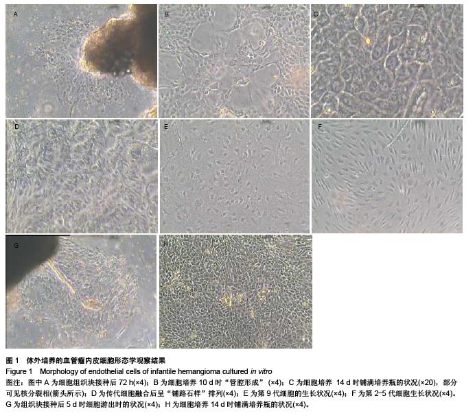
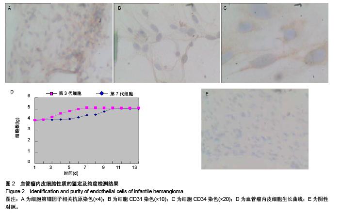
.jpg)