中国组织工程研究 ›› 2013, Vol. 17 ›› Issue (11): 1951-1958.doi: 10.3969/j.issn.2095-4344.2013.11.008

• 组织构建与生物活性因子 tissue construction and bioactive factors • 上一篇    下一篇

低氧训练与低氧大鼠心肌细胞凋亡及凋亡因子的表达

林喜秀1, 2,周 桔3,罗自强1,瞿树林3,赵用强2,邱继旺2   

  1. 1 中南大学基础医学院博士后流动站,湖南省长沙市 410000
    2 湖南工业大学体育学院,湖南省株洲市 412000
    3 湖南师范大学医学院生理应用教研室,湖南省长沙市 410000
  • 收稿日期:2012-09-20 修回日期:2012-01-31 出版日期:2013-03-12 发布日期:2013-03-12
  • 通讯作者: 罗自强,博士,教授,中南大学基础医学院博士后流动站,湖南省长沙市 410000 luozq1962@160.com 并列通讯作者:瞿树林,硕士,教授,湖南师范大学医学院生理应用教研室,湖南省长沙市 410000 qushulin2007@yahoo.com
  • 作者简介:林喜秀☆,女,1973年生,湖南省辰溪县人,苗族,2011年湖南师范大学生命科学院毕业,博士,讲师,主要从事低氧细胞生理研究。 Linxixiu1010@163.com

Hypoxia training regulates myocardial apoptosis and apoptotic factor expression in rats

Lin Xi-xiu1, 2, Zhou Ju3, Luo Zi-qiang1, Qu Shu-lin3, Zhao Yong-qiang2, Qiu Ji-wang2   

  1. 1 Postdoctoral Research Station, School of Basic Medical Sciences, Central South University, Changsha 410000, Hunan Province, China
    2 Sports Academy of Hunan University of Technology, Zhuzhou 412000, Hunan Province, China
    3 Department of Physiological Applications, School of Medical Science, Hunan Normal University, Changsha 410012, Hunan Province, China
  • Received:2012-09-20 Revised:2012-01-31 Online:2013-03-12 Published:2013-03-12
  • Contact: Luo Zi-qiang, Doctor, Professor, Postdoctoral Research Station, School of Basic Medical Sciences, Central South University, Changsha 410000, Hunan Province, China luozq1962@160.com Qu Shu-lin, Master, Professor, Department of Physiological Applications, School of Medical Science, Hunan Normal University, Changsha 410012, Hunan Province, China qushulin2007@yahoo.com
  • About author:Lin Xi-xiu☆, Doctor, Lecturer, Postdoctoral Research Station, School of Basic Medical Sciences, Central South University, Changsha 410000, Hunan Province, China; Sports Academy of Hunan University of Technology, Zhuzhou 412000, Hunan Province, China Linxixiu1010@163.com

摘要:

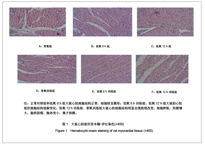
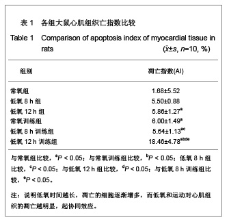
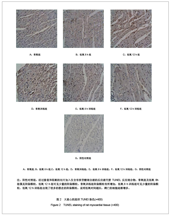
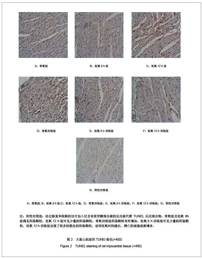
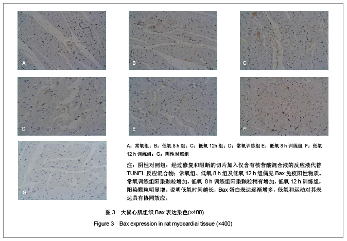
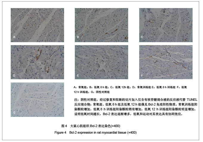
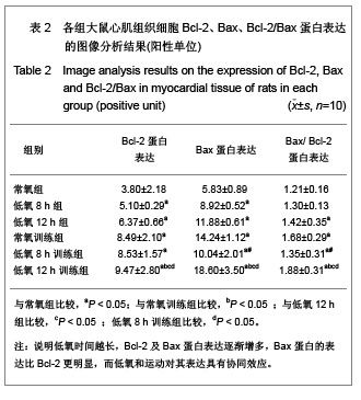
背景:低氧训练时,机体既要承受运动负荷,同时处于外界的低氧环境,此时, 心组织将如何适应其变化?其机制研究国内外较少。
目的:观察低氧与低氧训练对大鼠心肌细胞凋亡及Bax及Bcl-2表达的影响。
方法:SD大鼠共60只随机分为6组,常氧组、低氧8 h组、低氧12 h组、常氧训练组、低氧8 h训练组和低氧12 h训练组,每组10只。后3组大鼠每天在坡度为0的动物跑台上以25 m/min的速度训练1 h。训练完后,将低氧8 h组、低氧8 h训练组和低氧12 h组、低氧12 h训练组放入氧体积分数为12.5%(相当于海拔4 000 m)的低氧舱内8 h和12 h。实验期为4周,5 d/周。最后1次实验结束后24 h,大鼠均实施速眠新Ⅱ腹腔麻醉后取材,采用苏木精-伊红染色、原位末端脱氧核糖核苷酸转移酶介导的dUTP缺口末端标记法及蛋白免疫组织化学法检测各组大鼠心肌细胞凋亡和Bcl-2、Bax蛋白表达。
结果与结论:①与常氧组相比, 低氧12h组、常氧训练组、低氧训练组心肌细胞凋亡指数均显著增加 (P < 0.05) ;低氧12 h训练组心肌细胞凋亡指数显著多于常氧训练组和低氧8h训练组(P < 0.05) 。②与常氧组比较,其他各组Bcl-2、Bax、Bcl-2/Bax均显著性增高(P < 0.05) ;常氧训练组Bcl-2、Bax、Bcl-2/Bax表达显著高于低氧 8 h组,显著低于低氧12 h训练组(P < 0.05) ;低氧12 h训练组Bcl-2、Bax、Bcl-2/Bax表达比低氧12 h组、低氧8 h训练组显著增加(P < 0.05)。提示低氧、低氧训练可诱导大鼠心肌细胞Bcl-2、Bax蛋白表达, 运动时低氧刺激与细胞凋亡率、凋亡指数及病理损伤有关,其中以低氧12 h后运动训练组最明显,心肌细胞的凋亡调控与Bcl-2和Bax相关。

关键词: 组织构建, 组织构建与生物活性因子, 细胞凋亡, 低氧, 低氧运动, 缺氧诱导因子1a, Bcl-2, Bax, 心肌细胞, 省级基金, 组织构建图片文章

Abstract:

BACKGROUND: During hypoxia training, the body should bear the motor load and stay in the hypoxic environment outside.
OBJECTIVE: To observe the effect of hypoxia and hypoxia training on myocardial apoptosis and the expression of Bax and Bcl-2 in rats.
METHODS: Sixty Sprague-Dawley rats were randomly divided into six groups: normoxia group, 8 hours hypoxia group, 12 hours hypoxia group, normoxia training group, 8 hours hypoxia training group and 12 hours hypoxia training group, 10 rats in each group. The rats of normoxia training group, 8 hours hypoxia training group and 12 hours hypoxia training group were introduced to treadmill running on an incline of 0° at 25 m/min for 1 hour. After training, the rats of group 8 hours hypoxia group, 8 hours hypoxia training group, 12 hours hypoxia group and 12 hours hypoxia training group were exposed to hypoxic chamber with 12.5% volume fraction of oxygen (equal to altitude 4000 m) for 8 hours and 12 hours every day respectively. The course of the experiment was 4 weeks, 5 days every week. At 24 hours after the final experiment, the rats were treated with intraperitoneal anesthesia of sumianxin Ⅱ to obtain the samples, then hematoxylin-eosin staining, in situ terminal deoxyribonucleotide transferase-mediated dUTP nick end labeling method and protein immunohistochemistry methods were used to detect the myocardial apoptosis and the expression of Bax and Bcl-2 in rats.
RESULTS AND CONCLUSION: Compared with the normoxia group, the myocardial apoptosis index was significantly increased in the 24 hours hypoxia group, normoxia training group and hypoxia training group (P < 0.05); the myocardial apoptosis index in the 12 hours hypoxia training group was significantly higher than that in the normoxia training group and 8 hours hypoxia training group (P < 0.05). Compared with normoxia group, the expressions of Bcl-2, Bax and Bcl-2/Bax in the other five groups were significantly increased (P < 0.05); the expressions of Bcl-2, Bax and Bcl-2/Bax in normoxia training group were significantly higher than those in the 8 hours hypoxia group, and significantly lower than those in the 12 hours hypoxia group (P < 0.05); the expressions of Bcl-2, Bax and Bcl-2/Bax in the 12 hours hypoxia training group were significantly higher than those in the 12 hours hypoxia group and the 8 hours hypoxia training group (P < 0.05). Hypoxia and hypoxia training could induce the protein expressions of Bcl-2 and Bax in myocardial tissue. Hypoxia training was related with apoptosis rate and apoptotic index and pathological damage, especially the 12 hours hypoxia training. Bcl-2 and Bax participated in regulating the myocardial apoptosis.

Key words: tissue construction, tissue construction and bioactive factors, apoptosis, hypoxia, hypoxia training, hypoxia-inducible factor 1a, Bcl-2, Bax, myocardial cells, provincial grants-supported paper, tissue construction photographs-containing paper

中图分类号: