中国组织工程研究 ›› 2021, Vol. 25 ›› Issue (1): 38-43.doi: 10.3969/j.issn.2095-4344.2119

• 骨髓干细胞 bone marrow stem cells • 上一篇    下一篇

两种棘球绦虫原头节促进骨髓间充质干细胞钙化的差异性分析

桂显伟 1 ,姜慧娇 1 ,武 杰 1 ,梁学奇 1 ,徐小丹 2 ,王二强 2 ,邹海亮 1 ,陈贺捷 1 ,陈雪玲 2 ,吴向未 1
  

  1. 1 石河子大学医学院第一附属医院普外科,新疆维吾尔自治区石河子市 832008;2 石河子大学医学院免疫学教研室,新疆维吾尔自治区石河子市 832008
  • 收稿日期:2019-12-04 修回日期:2019-12-06 接受日期:2020-01-13 出版日期:2021-01-08 发布日期:2020-10-28
  • 通讯作者: 吴向未,博士,教授,主任医师,石河子大学医学院第一附属医院普外科,新疆维吾尔自治区石河子市 832008
  • 作者简介:桂显伟,男,1991年生,四川省合江县人,汉族,2020年石河子大学毕业,硕士,医师,主要从事普通外科疾病基础及干细胞转化医学相关研究。
  • 基金资助:
    国家自然科学基金(81760570);国家自然科学基金(81760371);兵团中青年科技创新领军人才计划项目(2018CB017);兵团重点领域科技公关项目(2019AB031)

Mesenchymal stem cell calcification induced by protoscolex of two species of Echinococcus: a differential analysis

Gui Xianwei1 , Jiang Huijiao1 , Wu Jie1 , Liang Xueqi1 , Xu Xiaodan2 , Wang Erqiang2 , Zou Hailiang1 , Chen Hejie1 , Chen Xueling2 , Wu Xiangwei1
  

  1. 1 Department of General Surgery, the First Affiliated Hospital, School of Medicine, Shihezi University, Shihezi 832008, Xinjiang Uygur Autonomous Region, China;
    2 Department of Immunology, School of Medicine, Shihezi University, Shihezi 832008, Xinjiang Uygur Autonomous Region, China
  • Received:2019-12-04 Revised:2019-12-06 Accepted:2020-01-13 Online:2021-01-08 Published:2020-10-28
  • Contact: Wu Xiangwei, MD, Professor, Chief physician, Department of General Surgery, the First Affiliated Hospital, School of Medicine, Shihezi University, Shihezi 832008, Xinjiang Uygur Autonomous Region, China
  • About author:Gui Xianwei, Master, Physician, Department of General Surgery, the First Affiliated Hospital, School of Medicine, Shihezi University, Shihezi 832008, Xinjiang Uygur Autonomous Region, China
  • Supported by:
    he National Natural Science Foundation of China, No. 81760570  and 81760371; the Young and Middle-Aged Innovation Talents Project of the Xinjiang Corps, No. 2018CB017; the Key Area Science and Technology Public Relations Project of the Corps, No. 2019AB031

摘要:

文题释义:
纤维钙化囊壁:囊型肝包虫病的囊壁分为内外2层,2层之间有可分离间隙存在,近肝侧外层纤维性囊壁主要以受挤压和纤维化的Glisson系统和肝静脉系统为主;内层纤维性钙化囊壁以肉芽肿反应为主,囊壁钙化主要集中分布于内层纤维性囊壁,且程度、形态各异,与近肝侧外层比较有显著性差异。
靶向药物治疗:是在细胞分子水平上针对已经明确的特异性位点的治疗方式,该位点可以是细胞内部的一个蛋白分子,也可以是一个基因片段,相应的治疗药物进入体内会与特异性位点相结合发生效用,而不会波及正常组织细胞,所以分子靶向治疗又被称为“生物导弹”。

摘要
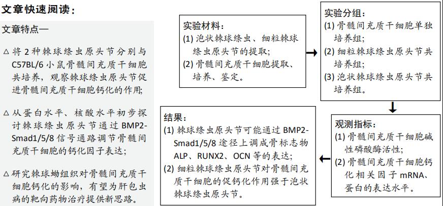
背景:细粒棘球蚴与泡状棘球蚴的生长方式不同,肝细粒棘球蚴病可以形成完整的纤维钙化囊壁,肝泡状棘球蚴病呈浸润性生长,不能形成完整的纤维钙化囊壁。骨髓间充质干细胞参与棘球蚴病纤维钙化囊壁的形成,但细粒棘球蚴和泡状棘球蚴钙化特征不同与骨髓间充质干细胞的作用尚不清楚。
目的:比较2种棘球绦虫原头节对骨髓间充质干细胞钙化的作用,初步探讨2种棘球蚴钙化灶差异的形成机制。
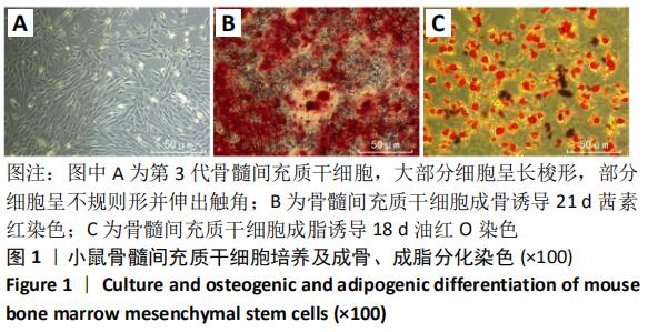
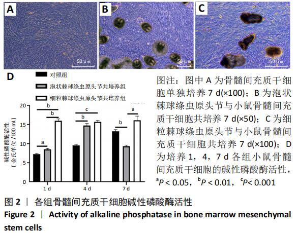
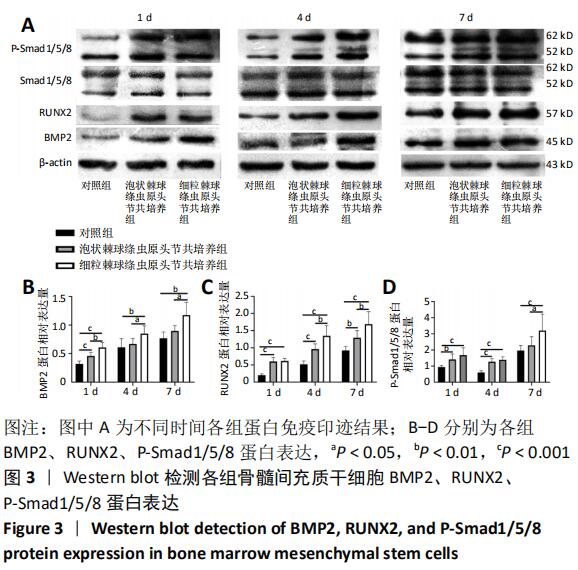
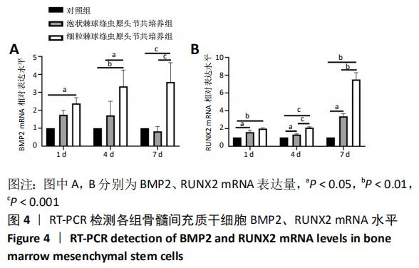
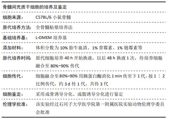
方法:提取、培养、鉴定C57BL/6小鼠骨髓间充质干细胞。将骨髓间充质干细胞分别与泡状棘球绦虫原头节、细粒棘球绦虫原头节共培养,以骨髓间充质干细胞单独培养为对照组。共培养1,4,7 d取骨髓间充质干细胞通过微量酶标法检测成骨标记物碱性磷酸酶活性,RT-qPCR检测BMP2和RUNX2 mRNA的表达,Western blot 检测BMP2、RUNX2和P-Smad1/5/8的蛋白表达。
结果与结论:①细粒棘球绦虫原头节共培养组、泡状棘球绦虫原头节共培养组1,4 d的碱性磷酸酶活性均高于对照组(P < 0.05),且细粒棘球绦虫原头节共培养组1,4,7 d 的碱性磷酸酶活性高于泡状棘球绦虫原头节共培养组(P < 0.05)。②Western blot 结果显示:细粒棘球绦虫原头节共培养组和泡状棘球绦虫原头节共培养组1,4 d的BMP2、RUNX2、P-Smad1/5/8蛋白表达高于对照组(P < 0.05),细粒棘球绦虫原头节共培养组高于泡状棘球绦虫原头节共培养组(P < 0.05)。③RT-qPCR结果显示:细粒棘球绦虫原头节共培养组1,4,7 d的BMP2、RUNX2 mRNA表达水平显著高于对照组(P < 0.05),细粒棘球绦虫原头节共培养组4,7 d的BMP2、RUNX2 mRNA表达水平显著高于泡状棘球绦虫原头节共培养组(P < 0.05)。泡状棘球绦虫原头节共培养组1,4,7 d的RUNX2 mRNA表达水平显著高于对照组(P < 0.05)。④结果表明,棘球绦虫原头节与骨髓间充质干细胞共培养通过上调BMP-Smad1/5/8通路促进骨髓间充质干细胞钙化因子碱性磷酸酶和RUNX2的表达,共培养后期泡状棘球蚴促钙化作用明显减弱,细粒棘球蚴促钙化作用保持不变,提示该机制可能与2种棘球蚴生长方式不同相关。

关键词: 骨髓间充质干细胞, 泡状棘球蚴,  细粒棘球蚴,  钙化,  纤维钙化囊壁,  棘球绦虫原头节

Abstract: Abstract
BACKGROUND: The growth pattern of Echinococcus granulosus is different from that of Echinococcus alveolaris. Hepatic echinococcosis can form a complete fibrous calcified cyst wall, while hepatic alveolar echinococcosis can grow infiltratively and cannot form a complete fibrous calcified cyst wall. Bone marrow mesenchymal stem cells (BMSCs) are involved in the formation of calcified wall of hydatidosis, but the calcification characteristics of Echinococcus granulosus and Echinococcus alveolaris are different and the role of BMSCs is still unclear.
OBJECTIVE: To compare the effects of Echinococcus granulosus and Echinococcus alveolaris on the calcification of BMSCs and to preliminarily investigate the formation mechanism of echinococcosis calcifications.
METHODS:  BMSCs of C57BL/6 mice were extracted, cultured and identified, followed by co-culture with the protoscolex of Echinococcus granulosus (BMSC+CE group) and Echinococcus alveolaris (BMSC+AE group), respectively. BMSCs cultured alone were used as control group. After 1, 4, and 7 days of co-culture, alkaline phosphatase activity was detected by a microplate reader, the expression of BMP2 and RUNX2 mRNA was detected by RT-q PCR, and the expression of BMP2, RUNX2 and phosphorylated Smad1/5/8 (P-Smad1/5/8) proteins was detected by western blot assay.  
RESULTS AND CONCLUSION: (1) The alkaline phosphatase activity of the BMSC+CE group and BMSC+AE group was significantly higher than that of the control group at 1 and 4 days after culture (P < 0.05), and the alkaline phosphatase activity of the BMSC+CE group was significantly higher than that of BMSC+AE group (P < 0.05). (2) Western blot results showed that the expression of BMP2, RUNX2, and P-Smad1/5/8 protein in the BMSC+CE group and BMSC+AE group was significantly higher than that in the control group at 1 and 4 days after culture (P < 0.05), while the expression of BMP2, RUNX2, and P-Smad1/5/8 protein in the BMSC+CE group was significantly higher than that in the BMSC+AE group (P < 0.05). (3) RT-qPCR results showed that the expression of BMP2 and RUNX2 mRNA in the BMSC+CE group was significantly higher than that in the control group at 1, 4 and 7 days after culture (P < 0.05), and was significantly higher than that in the BMSC+AE group at 4 and 7 days after culture (P < 0.05). The expression of RUNX2 mRNA in the BMSC+AE group was significantly higher than that in the control group at 1, 4, and 7 days after culture (P < 0.05). (4) To conclude, co-culture of the protoscolex of Echinococcus alveolaris and BMSCs promotes the expression of alkaline phosphatase and RUNX2 in BMSCs by up-regulating BMP-Smad1/5/8 pathway. At the later stage of co-culture, the effect of Echinococcus alveolaris on BMSCs calcification is significantly weakened, while the effect of Echinococcus granulosus on BMSCs calcification remains unchanged, suggesting that this mechanism may be related to the different growth patterns of two kinds of hydatids.


Key words: bone marrow mesenchymal stem cells,  Echinococcus alveolaris,  Echinococcus granulosus,  calcification,  fibrous calcified wall,   protoscolex of Echinococcus

中图分类号: