中国组织工程研究 ›› 2017, Vol. 21 ›› Issue (27): 4385-4390.doi: 10.3969/j.issn.2095-4344.2017.27.021

• 骨与关节图像与影像 bone and joint imaging • 上一篇    下一篇

Micro-CT月骨矢状面有限元模型建立及应力分析

杜传超1,2,熊 革1,任 爽3,荣起国4,张 衡2   

  1. 1北京积水潭医院手外科,北京市 100035;2清华大学第一附属医院,北京市 100016;3北京大学工学院生物力学实验室,北京市 100871;4北京大学工学院,北京市 100871
  • 出版日期:2017-09-28 发布日期:2017-10-24
  • 通讯作者: 熊革,教授,北京积水潭医院手外科,北京市 100035 荣起国,教授,北京大学工学院,北京市 100871
  • 作者简介:杜传超,男,1985年生,汉族,2014年7月北京大学医学部第四临床医学院暨北京积水潭医院毕业,硕士,医师。
  • 基金资助:

    国家临床重点专科建设项目-手外科[财社(2010)305号];北京市卫生系统高层次卫生技术人才培养计划(2009-3-17)

Construction of a finite element model based on lunate sagittal Micro-CT images and its stress analysis

Du Chuan-chao1, 2, Xiong Ge1, Ren Shuang3, Rong Qi-guo4, Zhang Heng2   

  1. 1Department of Hand Surgery, Beijing Jishuitan Hospital, Beijing 100035, China; 2the First Hospital of Tsinghua University, Beijing 100016, China; 3Biomechanical Laboratory of College of Engineering, Peking University, Beijing 100871, China; 4College of Engineering, Peking University, Beijing 100871, China
  • Online:2017-09-28 Published:2017-10-24
  • Contact: Xiong Ge, Professor, Department of Hand Surgery, Beijing Jishuitan Hospital, Beijing 100035, China; Rong Qi-guo, Professor, College of Engineering, Peking University, Beijing 100871, China
  • About author:Du Chuan-chao, Master, Physician, Department of Hand Surgery, Beijing Jishuitan Hospital, Beijing 100035, China; the First Hospital of Tsinghua University, Beijing 100016, China
  • Supported by:

    the National Clinical Key Construction Project-Hand Surgery, No. (2010)305; the High-Level Health Technology Talent Training Program of Beijing Health System, No. 2009-3-17

摘要:

文章快速阅读:


文题释义:
Micro-CT:也称为显微 CT,是基于螺旋CT断层扫描三维重建技术而研发的,其成像原理和普通X线成像原理大致相同,区别是采用微焦点X线球管技术,将图像的分辨率提高到微米级别。
有限元分析:是利用数学近似的方法对真实连续的物理系统(几何和载荷工况)进行分析、设计和建模的一种高效数值计算方法,将无限未知的真实物理系统分成简单而又相互作用的元素,即单元,就可以用有限数量的未知量去逼近无限未知量的真实系统。
 
摘要
背景:腕关节生物力学机制复杂,过去的研究多着眼于腕骨间应力的传导,但对腕骨内部骨小梁的应力分布及形变特点研究极少。
目的:建立人月骨矢状面的二维有限元模型,并探讨在月骨矢状面上施加轴向应力时应力的分布规律及形变特点。
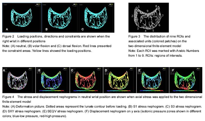
方法:运用Micro-CT扫描离体的正常人月骨标本,将处理后的矢状面图像导入有限元分析软件,建立起月骨矢状面的二维有限元模型。在二维有限元模型上均匀地选取9个兴趣区,计算每个兴趣区的第一主应力、第三主应力及节点等效应力、平面剪切力及纵向位移,比较其大小和方向,并生成月骨矢状面二维有限元模型的形变图、各应力及位移云图。
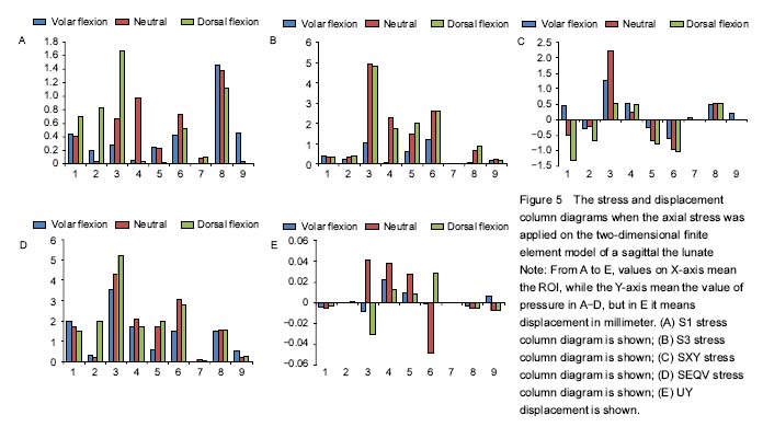
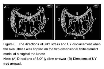
结果与结论:①人月骨矢状面的二维有限元模型分析:月骨矢状面近端兴趣区所受的应力大于远端兴趣区所受的应力;月骨矢状面掌侧兴趣区所受的应力大于背侧兴趣区所受的应力;月骨矢状面近端和远端的掌、背侧兴趣区所受的应力分别大于对应的月骨中间兴趣区所受的应力;月骨矢状面远端兴趣区被压向近端,近端中间区域的兴趣区被压向远端,而近端掌侧和背侧兴趣区的纵向位移方向随着腕关节姿势的不同而不同;屈腕位和背伸位时各兴趣区的应力和位移明显增加。此外,实验还发现应力集中区域是平面剪切力方向发生变化及轴向位移方向发生变化的主要区域。②结果证实:月骨矢状面在施加轴向应力时,其应力分布具有一定的规律,在月骨矢状面近端的掌、背侧及远端的掌、背侧形成4个应力集中区。而且,腕关节的姿势可以明显地影响月骨矢状面上所受的应力大小和分布。

中国组织工程研究杂志出版内容重点:人工关节;骨植入物;脊柱骨折;内固定;数字化骨科;组织工程
orcid: 0000-0002-7893-8846(Du Chuan-chao)

关键词: 骨科植入物, 数字化骨科, 骨关节图像与影像, 月骨, 腕关节, 腕关节位置, 关节接触面, Micro-CT, 二维有限元模型, 有限元分析, 应力分布, 轴向位移, 形变, 应力集中区

Abstract:

BACKGROUND: Biomechanical mechanisms are complex, and previous studiers focus on the stress conduction in the carpus. However, the stress distribution and characteristics of trabecula in the carpus are rarely reported.

OBJECTIVE: To investigate the stress distribution and deformation characteristics of the normal lunate through a two-dimensional sagittal finite element model.
METHODS: A normal cadaveric lunate sample was scanned with Micro-CT and the central sagittal image was chosen for further finite element analysis (FEA). The chosen image was processed and imported into the finite element analysis software (Ansys 14.0). A two-dimensional sagittal finite element model of the lunate bone was established. Axial pressure was applied to the model with the wrist held in different positions, and nine regions of interests (ROIs) were identified, for which stress and displacement nephograms were created. These included the first principal stress (S1, the maximum stress in a principal plane), the third principal stress (S3, the minimal stress in a principal plane), shear stress (SXY, the component of stress coplanar with a material cross section), von Mises stress (SEQV, yielding begins when the elastic energy of distortion reaches a critical value) and displacement of each ROI (UY, displacement on the vertical plane of the lunate) which were calculated and compared.
RESULTS AND CONCLUSION: (1) The stresses on ROIs located in the proximal and volar cortices of the lunate bone were much higher than those in the distal and dorsal cortices. At the proximal lunate, S1 was less than S3; however at the distal lunate, S1 was greater than S3. The ROIs of the distal and proximal ends of the lunate bone received much higher stress than the ROIs of the middle part. As for axial trabecular displacement, both distal and proximal ROIs were compressed by axial pressure. However, the dorsal and the volar parts of the proximal lunate moved in different directions at different wrist postures. Besides, the stress values and magnitudes of displacement were elevated in wrist flexion and extension compared to neutral position. Furthermore, the stress concentration zones (the proximal volar ROI, the proximal dorsal ROI, the distal volar ROI, and the distal dorsal ROI) had different directions of shear stress and displacement in different wrist postures. (2) These results suggest that when stress is loaded on a normal lunate model, four stress concentration zones, the proximal volar ROI, the proximal dorsal ROI, the distal volar ROI, and the distal dorsal ROI are found. The wrist postures can significantly affect the value and distribution of axial stress on the sagittal lunate.

中国组织工程研究杂志出版内容重点:人工关节;骨植入物;脊柱骨折;内固定;数字化骨科;组织工程

Key words: Tissue Engineering, Wrist Joint, Fracture, Bone

中图分类号: