中国组织工程研究 ›› 2017, Vol. 21 ›› Issue (26): 4106-4112.doi: 10.3969/j.issn.2095-4344.2017.26.002

• 组织工程口腔材料 tissue-engineered oral materials • 上一篇    下一篇

碱性成纤维细胞生长因子/壳聚糖/聚乳酸支架用于牙周组织再生工程的体外实验

陈立红1,雷志敏1,王莉莉2,3
  

  1. 1武汉大学人民医院口腔科,湖北省武汉市  430060;2天津市口腔医院修复科,天津市  300000;3锦州医科大学附属口腔医院修复科,辽宁省锦州市  121000
  • 收稿日期:2017-07-23 出版日期:2017-09-18 发布日期:2017-09-28
  • 通讯作者: 雷志敏,副主任医师,武汉大学人民医院口腔科,湖北省武汉市 430060
  • 作者简介:陈立红,女,1975年生,辽宁省本溪市人,汉族,武汉大学在读硕士,医师,主要从事骨生物材料在口腔的应用研究。
  • 基金资助:
    辽宁省大学生创新训练项目(201510160000045);辽宁省科学计划项目(2014022003)

In vitro experimental research of basic fibroblast growth factor/chitosan/polylactic acid scaffolds in periodontal tissue regeneration

Chen Li-hong1, Lei Zhi-min1, Wang Li-li2, 3
  

  1. 1Department of Stomatology, Renmin Hospital of Wuhan University, Wuhan 430060, Hubei Province, China; 2Department of Prosthodontics, Tianjin Stomatological Hospital, Tianjin 300000, China; 3Department of Prosthodontics, Stomatological Hospital of Jinzhou Medical University, Jinzhou 121000, Liaoning Province, China
  • Received:2017-07-23 Online:2017-09-18 Published:2017-09-28
  • Contact: Lei Zhi-min, Associate chief physician, Department of Stomatology, Renmin Hospital of Wuhan University, Wuhan 430060, Hubei Province, China
  • About author:Chen Li-hong, Studying for master’s degree, Physician, Department of Stomatology, Renmin Hospital of Wuhan University, Wuhan 430060, Hubei Province, China
  • Supported by:
    the Innovation Training Project for Undergraduate Students in Liaoning Province, No. 201510160000045; the Science Plan Project of Liaoning Province, No. 2014022003

摘要:

文章快速阅读:


文题释义:
碱性成纤维细胞生长因子:属于肝素结合家族,为一种多肽型生长因子,具有广泛的生物学作用,能够促进牙周膜成纤维细胞的有丝分裂和趋化,有利于细胞外基质及牙周膜新生血管网的生成,但其易降解,半衰期短,且生物活性的发挥具有剂量依赖性,单独局部应用代谢迅速。
壳聚糖/聚乳酸复合支架:聚乳酸是人工合成半结晶高分子性物质,溶解性、共混性均较好,但其机械强度不足,脆性亦高,单独应用效果不甚理想;壳聚糖属于碱性多糖,具有良好的生物相容性、生物降解性及机械强度,若将壳聚糖加入聚乳酸中,两种高分子物质相结合,壳聚糖可表现出良好的黏度和韧性,明显改善聚乳酸脆性大、易碎裂的性质。
 
背景:碱性成纤维细胞生长因子(basic fibroblast growth factor,bFGF)可促进牙周膜成纤维细胞的有丝分裂和趋化,有利于细胞外基质及牙周膜新生血管网的生成,但其易降解、半衰期短,单独局部应用代谢迅速。
目的:探讨bFGF/壳聚糖/聚乳酸复合支架对牙周膜细胞生长和增殖的影响。
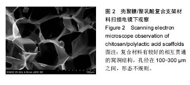
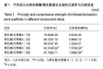
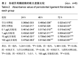
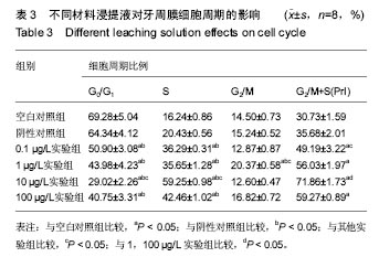
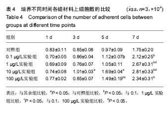
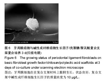
方法:采用冷冻干燥法制备不同比例(聚乳酸与壳聚糖的质量比分别为4∶1、3∶2、1∶1、2∶3、1∶4)的壳聚糖/聚乳酸复合支架,以孔隙率、力学强度筛选最优配比的复合支架,用于制备含0,0.1,1,10,100 µg/L bFGF的壳聚糖/聚乳酸支架。①细胞毒性实验:分别以含0,0.1,1,10,100 µg/L bFGF的壳聚糖/聚乳酸支架浸提液(实验组)及DMEM培养液(空白对照组)培养大鼠牙周膜细胞,24,48,72 h后,MTT法检测细胞增殖;培养5 d后,流式细胞仪检测细胞周期;②细胞相容性实验:将含0.1,1,10,100 µg/L bFGF的壳聚糖/聚乳酸支架分别与大鼠牙周膜细胞共培养,1,3,5,7 d后进行细胞计数;培养3 d后,扫描电镜观察细胞生长情况。
结果与结论:①从孔隙率、力学强度指标综合考虑,聚乳酸与壳聚糖的最佳质量比为2∶3;②随着培养时间的延长,各组细胞增殖A值逐渐增加;培养48,72 h时,1,10,100 µg/L实验组细胞增殖A值高于空白对照组(P < 0.05),且10 µg/L实验组细胞增殖A值最高(P < 0.05);③0.1,1,10,100 µg/L实验组G0/G1期细胞百分比低于空白对照组(P < 0.05),以10 µg/L实验组最低;0.1,1,10,100 µg/L实验组S期、G2/M+S期细胞比例均高于空白对照组(P < 0.05),以10 µg/L实验组最高(P < 0.05);④随着培养时间的延长,各组细胞计数逐渐增多;培养3,5 d时,10 µg/L实验组细胞计数高于其余各组(P < 0.05)。培养3 d后,大鼠牙周膜细胞在10 µg/L实验组支架上黏附生长,状态良好;⑤结果表明,bFGF/壳聚糖/聚乳酸复合支架可促进牙周膜细胞的增殖。

关键词: 生物材料, 材料相容性, 牙周组织工程, 牙周膜细胞, 壳聚糖, 聚乳酸, 碱性成纤维细胞生长因子

Abstract:

BACKGROUND: Basic fibroblast growth factor (bFGF) can promote mitosis and chemotaxis of periodontal ligament fibroblasts (PDLCs), and help the generation of extracellular matrix and new blood capillaries. But it is easy to degrade, has short half-life, and metabolizes rapidly.
OBJECTIVE: To evaluate the influence of bFGF/chitosan/polylactic acid scaffolds on PDLCs growth and proliferation.
METHODS: Polylactic acid/chitosan composite scaffolds in different proportions (4:1, 3:2, 1:1, 2:3, 1:4) were prepared through freeze-drying method, to study their microstructure, porosity and mechanical strength and then choose the optimal ratio of chitosan/polylactic acid scaffold that was used to prepare the composite scaffolds which contained different concentrations (0, 0.1, 1, 10, 100 µg/L) of bFGF. (1) Cytotoxicity test: PDLCs were cultured with leaching liquid of polylactic acid/chitosan scaffolds which contained different concentrations (0, 0.1, 1, 10, 100 µg/L) of bFGF and DMEM culture medium respectively. The effects on cell proliferation were tested by MTT after 24, 48, 72 hours. Cell cycles were tested using flow cytometry at 5 days of culture. (2) Cytocompatibility test: PDLCs were co-cultured with the polylactic acid/chitosan scaffolds which contained different concentrations (0, 0.1, 1, 10, 100 µg/L) of bFGF. The number of PDLCs was counted at 1, 3, 5 and 7 days. The growing status of PDLCs on the scaffolds was observed by scanning electron microscope at 3 days of culture.
RESULTS AND CONCLUSION: (1) The best mass ratio of polylactic acid and chitosan was 2:3 by test of porosity and mechanical strength. (2) The absorbance value of each group was increased over time. The absorbance values of 0.1, 1, 10, 100 µg/L bFGF groups were higher than that of the control group (0 µg/L bGFG), and the A value of 10 µg/L group was highest in all groups at 48 and 72 hours after co-culture (P < 0.05). (3) Cell percentages at G0/G1 phase of 0.1, 1, 10, 100 µg/L bFGF groups were higher than that of the control group, and the percentage of 10 µg/L group was lowest in all groups (P < 0.05). Cell percentage at S phase and G2/M+S of 0.1, 1, 10, 100 µg/L bFGF groups were higher than that of the control group, and the percentage of 10 µg/L group was highest in all groups (P < 0.05). (4) The number of cells in each group was increased with time. The cell number in the 10 µg/L was most in all groups at 3 and 5 days of co-culture (P < 0.05). Observation of scanning electron microscopy showed that PDLCs adhered and grew well on the 10 µg/L bFGF/polylactic acid/chitosan composite scaffold when co-cultured for 3 days. Overall, these findings indicate that the bFGF/polylactic acid/chitosan composite scaffold contributes to PDLCs proliferation.

Key words: Periodontium, Chitosan, Fibroblast Growth Factor 2

中图分类号: