中国组织工程研究 ›› 2017, Vol. 21 ›› Issue (8): 1202-1208.doi: 10.3969/j.issn.2095-4344.2017.08.010
• 脊柱组织构建 spinal tissue construction • 上一篇 下一篇
胰岛素样生长因子1通过PI3K/Akt信号通路促进髓核细胞聚集蛋白聚糖及Ⅱ型胶原的表达
李大鹏1,吴 燕2,岳佳伟2,王家伦2,胡 浪1,黄永辉1
- 1江苏大学附属医院,江苏省镇江市 212001;2江苏大学医学院,江苏省镇江市 212013
Insulin-like growth factor-1 upregulates the expression of aggrecan and collagen type II in nucleus pulposus cells via PI3K/Akt signaling pathway
Li Da-peng1, Wu Yan2, Yue Jia-wei2, Wang Jia-lun2, Hu Lang1, Huang Yong-hui1
- 1Affiliated Hospital of Jiangsu University, Zhenjiang 212001, Jiangsu Province, China; 2Medical College of Jiangsu University, Zhenjiang 212013 Jiangsu Province, China
摘要:
文章快速阅读:
.jpg) 文题释义:
胰岛素样生长因子1:是含有70 个氨基酸的多肽,在分子结构上与胰岛素类似,因此被称为胰岛素样生长因子1。胰岛素样生长因子1通过内分泌、自分泌和旁分泌的3种途径产生的,在正常机体内主要由肝脏合成。胰岛素样生长因子1可明显的促进多种来源的软骨细胞分裂增殖及软骨基质的合成,椎间盘髓核细胞类似于软骨细胞,能合成分泌聚集蛋白聚糖及Ⅱ型胶原等髓核基质,因而,胰岛素样生长因子1逐步应用于椎间盘退变的体内外研究。
文题释义:
胰岛素样生长因子1:是含有70 个氨基酸的多肽,在分子结构上与胰岛素类似,因此被称为胰岛素样生长因子1。胰岛素样生长因子1通过内分泌、自分泌和旁分泌的3种途径产生的,在正常机体内主要由肝脏合成。胰岛素样生长因子1可明显的促进多种来源的软骨细胞分裂增殖及软骨基质的合成,椎间盘髓核细胞类似于软骨细胞,能合成分泌聚集蛋白聚糖及Ⅱ型胶原等髓核基质,因而,胰岛素样生长因子1逐步应用于椎间盘退变的体内外研究。
文题释义:
胰岛素样生长因子1:是含有70 个氨基酸的多肽,在分子结构上与胰岛素类似,因此被称为胰岛素样生长因子1。胰岛素样生长因子1通过内分泌、自分泌和旁分泌的3种途径产生的,在正常机体内主要由肝脏合成。胰岛素样生长因子1可明显的促进多种来源的软骨细胞分裂增殖及软骨基质的合成,椎间盘髓核细胞类似于软骨细胞,能合成分泌聚集蛋白聚糖及Ⅱ型胶原等髓核基质,因而,胰岛素样生长因子1逐步应用于椎间盘退变的体内外研究。摘要
背景:采用生长因子等生物学方法修复退变椎间盘是目前椎间盘退变治疗的研究热点,胰岛素样生长因子1能促进髓核细胞增殖、促进功能性细胞外基质的合成,但其机制仍未阐明。
目的:观察胰岛素样生长因子1对髓核细胞聚集蛋白聚糖及Ⅱ型胶原表达的促进作用,并探讨其信号转导机制。
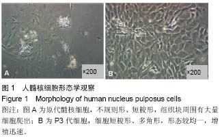
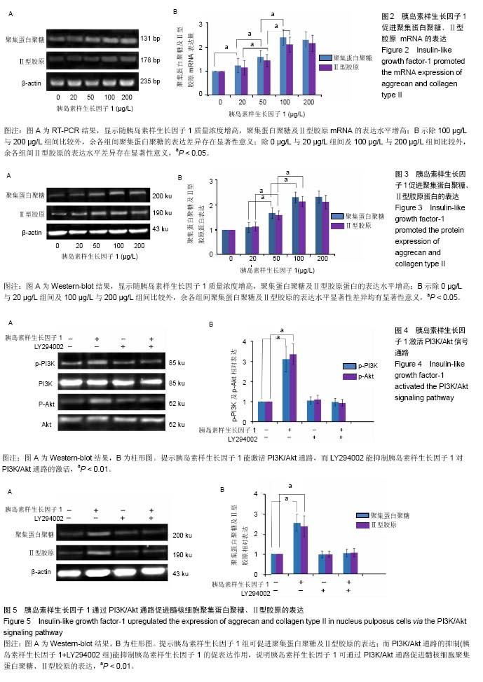
方法:分离培养人髓核细胞,取第3代细胞给予不同质量浓度的胰岛素样生长因子1(0,20,50,100, 200 μg/L)进行刺激,采用RT-PCR、Western-blot检测聚集蛋白聚糖及Ⅱ型胶原的表达。100 μg/L的胰岛素样生长因子1作用于髓核细胞,采用Western-blot检测胰岛素样生长因子1对PI3K/Akt信号通路的激活作用,并通过LY294002的应用检测PI3K/Akt通路的抑制对聚集蛋白聚糖及Ⅱ型胶原表达的影响。
结果与结论:①随胰岛素样生长因子1质量浓度增高,聚集蛋白聚糖及Ⅱ型胶原的表达水平逐渐升高,该作用呈剂量依赖性。胰岛素样生长因子1(100 μg/L)能显著促进p-PI3K和p-Akt的表达(P < 0.01),而LY294002能抑制胰岛素样生长因子1的促进作用(P < 0.01);②胰岛素样生长因子1(100 μg/L)能促进髓核细胞聚集蛋白聚糖及Ⅱ型胶原的表达(P < 0.01),而LY294002能抑制胰岛素样生长因子1 对聚集蛋白聚糖及Ⅱ型胶原表达的促进作用(P < 0.01);③因此认为胰岛素样生长因子1可通过PI3K/Akt通路促进髓核细胞聚集蛋白聚糖及Ⅱ型胶原的表达。
中国组织工程研究杂志出版内容重点:组织构建;骨细胞;软骨细胞;细胞培养;成纤维细胞;血管内皮细胞;骨质疏松;组织工程
ORCID: 0000-0002-1595-9682(黄永辉)
中图分类号:
.jpg) 文题释义:
胰岛素样生长因子1:是含有70 个氨基酸的多肽,在分子结构上与胰岛素类似,因此被称为胰岛素样生长因子1。胰岛素样生长因子1通过内分泌、自分泌和旁分泌的3种途径产生的,在正常机体内主要由肝脏合成。胰岛素样生长因子1可明显的促进多种来源的软骨细胞分裂增殖及软骨基质的合成,椎间盘髓核细胞类似于软骨细胞,能合成分泌聚集蛋白聚糖及Ⅱ型胶原等髓核基质,因而,胰岛素样生长因子1逐步应用于椎间盘退变的体内外研究。
文题释义:
胰岛素样生长因子1:是含有70 个氨基酸的多肽,在分子结构上与胰岛素类似,因此被称为胰岛素样生长因子1。胰岛素样生长因子1通过内分泌、自分泌和旁分泌的3种途径产生的,在正常机体内主要由肝脏合成。胰岛素样生长因子1可明显的促进多种来源的软骨细胞分裂增殖及软骨基质的合成,椎间盘髓核细胞类似于软骨细胞,能合成分泌聚集蛋白聚糖及Ⅱ型胶原等髓核基质,因而,胰岛素样生长因子1逐步应用于椎间盘退变的体内外研究。