中国组织工程研究 ›› 2017, Vol. 21 ›› Issue (2): 197-201.doi: 10.3969/j.issn.2095-4344.2017.02.007

• 组织工程骨及软骨材料 tissue-engineered bone and cartilage materials • 上一篇    下一篇

致密层骨软骨复合支架的制备及其修复关节骨软骨缺损

魏  戎1,武军龙1,吴飞翔1,王  超1,刘娟娟1,吴晴缘2
  

  1. 1郑州大学附属洛阳中心医院,河南省洛阳市  471000;2河南大学第一临床学院,河南省开封市  475001
  • 收稿日期:2016-12-13 出版日期:2017-01-18 发布日期:2017-02-27
  • 作者简介:魏戎,男,1966年生,汉族,河南省洛阳市人,副主任医师,主要从事骨科关节外科研究。
  • 基金资助:

    2016年洛阳市科技计划医疗卫生项目(1603002A-15);河南省重点实验室研究项目(J01501153)

Preparation and application of layered osteochondral composite scaffold in the repair of articular cartilage defects

Wei Rong1, Wu Jun-long1, Wu Fei-xiang1, Wang Chao1, Liu Juan-juan1, Wu Qing-yuan2 
  

  1. 1Luoyang Central Hospital Affiliated to Zhengzhou University, Luoyang 471000, Henan Province, China; 2the First Affiliated Hospital of Henan University, Kaifeng 475001, Henan Province, China
  • Received:2016-12-13 Online:2017-01-18 Published:2017-02-27
  • About author:Wei Rong, Associate chief physician, Luoyang Central Hospital Affiliated to Zhengzhou University, Luoyang 471000, Henan Province, China
  • Supported by:

    the Luoyang Science and Technology Medicine and Health Program in 2016, No. 1603002A-15; the Key Laboratory Research Project of Henan Province, No. J01501153

摘要:

文章快速阅读:


文题释义:
致密层骨软骨复合支架
:参考关节骨软骨的解剖结构、生理功能制备致密层骨软骨复合支架,采用低温沉积制造工艺在三维立体包芯结构骨架上形成致密层,并且利用“溶解粘连”技术将定向微管结构骨支架与致密层进行严密的结合,从而成功制备了致密层骨软骨复合支架。
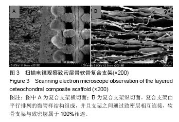
致密层骨软骨复合支架的表征:支架直径为5 mm,厚度为2 mm,孔径为(9.2±2.3) μm,孔隙率为(3.2±0.5)%,吸水率为(2.8±0.2)%,扫描电镜显示制备的致密层骨软骨复合支架由平行排列的微管样结构组成,并且支架之间通过致密层相互连接,软骨支架与致密层属于100%相连。

背景:理想的骨软骨组织工程支架应巧妙地模拟人体正常关节骨软骨结构。
目的:基于关节骨软骨的解剖结构、生理功能,制备致密层骨软骨复合支架,同时观察其修复兔关节骨软骨缺损的效果。
方法:利用快速成型技术在三维立体包芯结构骨架表面喷涂乳酸-羟基乙酸共聚物/β-磷酸三钙有机溶液,形成  0.5 mm的致密层;利用“溶解-粘连”工艺能将定向微管结构软骨支架与致密层相互连接,形成致密层骨软骨复合支架。取60只家兔,制备左膝关节软骨全层缺损,随机分3组,实验组植入致密层骨软骨复合支架,对照组植入定向微管结构软骨支架,空白对照组不植入任何材料,修复后12,24周进行缺损部位大体与组织学观察。
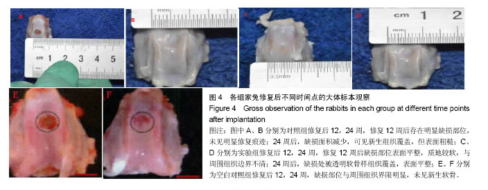
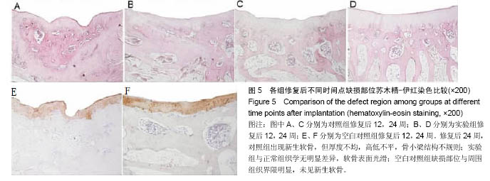
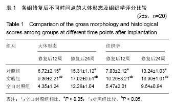
结果与结论:①大体观察结果:对照组修复后12周存在明显缺损部位,未见明显修复痕迹;24周缺损面积减少,可见新生组织覆盖,但表面粗糙。实验组修复12周后缺损部位表面平整,质地较软,与周围组织边界不清;修复后24周,被透明软骨样组织覆盖,表面平整。空白对照组修复效果较差;②组织学观察结果:修复后12周,对照组缺损部位出现形态不规则的骨痂,但未形成骨小梁;实验组出现新生骨,软骨厚度与正常软骨接近,并且软骨下存在不规则骨小梁。修复后24周,对照组出现新生软骨,但厚度不均,高低不平,骨小梁结构不规则;实验组组织与正常组织无明显差异,软骨表面光滑;空白对照组修复效果较差;③结果表明:致密层骨软骨复合支架接近人体正常关节骨软骨结构,可促进关节软骨缺损的修复。

关键词: 生物材料, 软骨生物材料, 致密层骨软骨复合支架, 关节软骨, 快速成型技术, 定向微管结构软骨支架, 膝关节软骨缺损, “溶解-粘连”工艺, 修复效果

Abstract:

BACKGROUND: Ideal osteochondral tissue-engineered scaffolds should be able to mimic the normal structure of human articular cartilage.
OBJECTIVE: To prepare a layered osteochondral composite scaffold based on the anatomical and physiological functions of osteochondral articular cartilage and to observe its repair effect on osteochondral defects in rabbits.
METHODS: The poly (lactide-glycolide acid)/β-tricalcium phosphate organic solution was sprayed on the surface of cartilage scaffold using rapid prototyping technology. The layered osteochondral composite scaffold was formed by the “dissolving-adhesion” process. Sixty rabbits were enrolled, modeled into left knee articular cartilage defects, and then randomly divided into three groups. The layered osteochondral composite scaffold and cartilage scaffold were implanted into experimental and control groups, respectively. Those without any treatment served as controls. Gross and histological observations of the defect region were performed at 12 and 24 weeks after implantation.
RESULTS AND CONCLUSION: Gross observation: At 12 weeks after implantation, the defects in the control group were obvious and not repaired at all; the 24-week defect area was decreased, covered by newly formed tissues, but with rough surface. In the experimental group, the defect surface was flat after 12 weeks of implantation, the texture was soft, and the boundary with the surrounding tissues was unclear; at 24 weeks, the defect was covered with transparent cartilaginous tissues and the surface was smooth. Histological observation: At 12 weeks after implantation, the irregular callus appeared in the control group, but the trabeculae were not formed; in the experimental group, the thickness of the new cartilage was similar with that of the normal cartilage and there was irregular trabecular bone under the cartilage. After 24 weeks of implantation, there were new tissues in the control group, but the thickness was irregular and uneven and the trabecular structure was irregular; while the cartilage surface was smooth and repaired well in the experimental group. In contrast, repair effect in the control group was poor as assessed by gross and histological observations. These results show that the layered composite scaffold holds a similar structure with human articular cartilage and can promote the repair of articular cartilage defects.

Key words: Calcium Phosphates, Knee Joint, Tissue Engineering

中图分类号: