中国组织工程研究 ›› 2016, Vol. 20 ›› Issue (44): 6620-6628.doi: 10.3969/j.issn.2095-4344.2016.44.012

• 数字化骨科 digital orthopedics • 上一篇    下一篇

三维数字化重建10-12岁儿童胸椎关节突的形态特征

刘祥伟1,2,王 星3,4,张少杰3,4,李志军3,4,刘 颖3,史 君5 
  

  1. 1赤峰市医院骨科,内蒙古自治区赤峰市 024000;内蒙古医科大学,2研究生学院,3基础医学院人体解剖学教研室;4数字医学中心;5基础医学院生理学教研室,内蒙古自治区呼和浩特市 010059
  • 修回日期:2016-08-29 出版日期:2016-10-28 发布日期:2016-10-28
  • 通讯作者: 李志军,教授,博士生导师,内蒙古医科大学基础医学院人体解剖学教研室,内蒙古自治区呼和浩特市 010059
  • 作者简介:刘祥伟,1982年生,内蒙古自治区赤峰市人,2015年内蒙古医科大学毕业,硕士,主治医师,主要从事脊柱创伤方面的研究。 并列第一作者:王星,1979年生,内蒙古自治区乌拉特中旗人,2010年内蒙古医科大学毕业,硕士,讲师,主要从事脊柱脊髓的数字化研究。
  • 基金资助:

    国家自然科学基金资助项目(81260269,81560348,81660358);内蒙古自治区高等学校科学研究资助项目(NJZC16108);内蒙古医科大学科技百万项目(2015YKDKJBW03)

Morphological characteristics of thoracic vertebrae in children aged from 10 to 12 years with three-dimensional digital reconstruction

Liu Xiang-wei1, 2, Wang Xing3, 4, Zhang Shao-jie3, 4, Li Zhi-jun3, 4, Liu Ying3, Shi Jun5
  

  1. 1Department of Orthopedics, Chifeng Hospital, Chifeng 024000, Inner Mongolia Autonomous Region, China; 2Postgraduate College, Inner Mongolia Medical University, Hohhot 010059, Inner Mongolia Autonomous Region, China; 3Department of Human Anatomy, Basic Medical College, Inner Mongolia Medical University, Hohhot 010059, Inner Mongolia Autonomous Region, China; 4Digital Medical Center, Inner Mongolia Medical University, Hohhot 010059, Inner Mongolia Autonomous Region, China; 5Department of Physiology, Basic Medical College, Inner Mongolia Medical University, Hohhot 010059, Inner Mongolia Autonomous Region, China
  • Revised:2016-08-29 Online:2016-10-28 Published:2016-10-28
  • Contact: Li Zhi-jun, Professor, Doctoral supervisor, Department of Human Anatomy, Basic Medical College, Inner Mongolia Medical University, Hohhot 010059, Inner Mongolia Autonomous Region, China; Digital Medical Center, Inner Mongolia Medical University, Hohhot 010059, Inner Mongolia Autonomous Region, China
  • About author:Liu Xiang-wei, Master, Attending physician, Department of Orthopedics, Chifeng Hospital, Chifeng 024000, Inner Mongolia Autonomous Region, China; Postgraduate College, Inner Mongolia Medical University, Hohhot 010059, Inner Mongolia Autonomous Region, China Wang Xing, Master, Lecturer, Department of Human Anatomy, Basic Medical College, Inner Mongolia Medical University, Hohhot 010059, Inner Mongolia Autonomous Region, China; Digital Medical Center, Inner Mongolia Medical University, Hohhot 010059, Inner Mongolia Autonomous Region, China Liu Xiang-wei and Wang Xing contributed equally to this paper.
  • Supported by:

    the National Natural Science Foundation of China, No. 81260269, 81560348, 81660358; the Scientific Research Funding Project of Inner Mongolia Autonomous Region Colleges and Universities, No. NJZC16108; the Science and Technology Million Project of Inner Mongolia Medical University, No. 2015YKDKJBW03

摘要:

文章快速阅读:


文题释义:
关节突关节:关节突作为脊柱后柱的重要组成部分,其关节直接承受着脊柱前屈、后伸、压缩、牵拉、剪切、扭转等负荷,关节突关节的方向从颈椎段“近水平位”,逐渐到胸椎段“近冠状位”,再到腰椎段的“近矢状位”,此结构特征的变化是脊柱所特有,而且其作为运动枢纽,对维持脊柱稳定及正常生理活动起着至关重要的作用。同时脊柱结构的稳定性对于维持脊柱正常形态和生理功能也具有重要意义,椎体、椎间盘和脊柱韧带对维持小关节的稳定和平衡为静力平衡。
数字化医学:数字医学已经广泛应用于医学领域,其打破过去传统且单一的研究手段,利用医学重建软件可清晰地将影像原始资料予以重建,在三维立体的效果下区观测脊柱各相关结构间的关系,为后期的手术治疗等提供了有利的保障。
 
摘要
背景:关节突关节是维持整个脊柱稳定和正常生理活动的重要骨性结构。在临床研究中发现,双侧关节突形态不对称也是引起椎体退行性变的原因之一,但现有的研究主要集中于成人群体,且多以颈椎和腰椎为主。
目的:探索10-12岁儿童胸椎关节突关节相关骨性结构的形态特征和增龄变化规律,并与成人相关数据比较。
方法:选取无骨质破坏、畸形、骨折、肿瘤等椎骨形态结构未发生改变及既往未行脊柱相关手术的10-12岁儿童30例,行多排螺旋CT薄层扫描(0.625-1.25 mm),范围T1-T12,将原始数据以DICOM格式导入三维重建软件进行相关指标测量及统计分析。
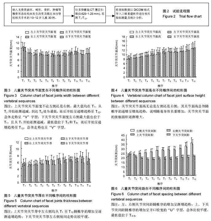
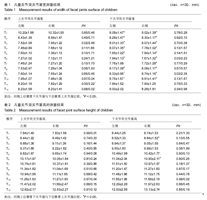
结果与结论:①关节突关节面宽总体走势呈“V字型”;关节突关节面高从T1至T12间随椎序的增加呈逐渐递增趋势;②上关节突关节厚从T1至T12间随椎序增加呈逐渐递增趋势,下关节突关节厚总体走势则较平缓;左、右侧别中上、下关节突关节面宽、高、厚除在个别椎体间存在显著性差异外,其余差异均无显著性意义;关节突间距在侧别和上、下间差异均无显著性意义,关节突间距侧别间随着椎序的增加呈递增趋势,上、下关节突间距随椎序增加均呈开口较宽的“U”字型;③结果表明,利用三维重建技术可清晰、直观地展示各椎骨形态特征,在三维条件下提高了测量的精确度;10-12岁儿童关节突关节面宽和高总体较成人小,关节突间距在上关节突间距与下关节突间距总体变化不明显,而左右侧关节突间距则随着椎序的增加呈渐增趋势,符合儿童脊柱生长发育规律。

中国组织工程研究杂志出版内容重点:人工关节;骨植入物;脊柱骨折;内固定;数字化骨科;组织工程

ORCID:
0000-0001-6025-8554 (李志军)

关键词: 骨科植入物, 数字化骨科, 胸椎, 关节突关节, 形态特征, 10-12岁儿童, 国家自然科学基金

Abstract:

BACKGROUND: Articular process of joint is an important bony structure to maintain the spinal stability and normal physiological activities. Clinical study found that bilateral articular process asymmetry is also one of the causes of vertebral degeneration, but the existing research mainly focuses on the adult, and give priority to with cervical vertebra and lumbar vertebra.

OBJECTIVE: To explore the morphological and developmental characteristics of thoracic facet joints and bony structures of 10 to 12-year-old children and compare with data of adults.
METHODS: A total of 30 normal cases aging from 10 to 12 years were admitted into this study. No cases experienced bone destruction, deformity, fractures, or tumors, and spine surgery was not involved in. DICOM 3.0 data of multi-slice spiral CT (0.625-1.25 mm), ranging from T1 to T12, were used for three-dimensional reconstruction, measurement and statistical analysis.
RESULTS AND CONCLUSION: (1) Joint width overall trends in “V”. Joint surface height from T1 to T12 increased gradually with the increase of the operation sequence. (2) The joint thickness from T1 to T12 showed a gradually increasing trend with the increase of the job sequence; articular process under the joint thickness was gentle. In the left and right sides of the upper and lower joints, joint surface width, height, and thickness did not show significant difference in addition to the significant difference between the individual vertebral body. Joints spacing on the side and there was no significant difference between upper and lower, articular process spacing between sides showed an increasing trend with the increase of operation sequence; upper and lower joints spacing increased with the job sequence, and showed wide “U” shape. (3) These results confirmed that the application of three-dimensional reconstruction techniques can display shape and characteristics of each vertebral body clearly and intuitively and improve measurement accuracy. Width and height of facet joints in 10-12-year-old school children were overall smaller than those of adults. The distance between upper and lower facet joints changed a little. Distance between left and right facet joints increased from T1 to T12, which complies with the growth and development of children.

中国组织工程研究杂志出版内容重点:人工关节;骨植入物;脊柱骨折;内固定;数字化骨科;组织工程

Key words: Thoracic Vertebrae, Zygapophyseal Joint, Child , Tissue Engineering

中图分类号: