中国组织工程研究 ›› 2016, Vol. 20 ›› Issue (38): 5629-5635.doi: 10.3969/j.issn.2095-4344.2016.38.001

• 组织工程骨及软骨材料 tissue-engineered bone and cartilage materials •    下一篇

羟基丁酸-羟基辛酸共聚体/胶原骨软骨一体化支架修复膝关节软骨损伤

段新虎   

  1. 山西医科大学,山西省太原市   030001
  • 收稿日期:2016-06-20 出版日期:2016-09-16 发布日期:2016-09-16
  • 作者简介:段新虎,男,1988年生,山西省稷山县人,汉族,2016年山西医科大学毕业,硕士,主要从事骨与软骨组织工程研究。

Poly(hydroxybutyrate-co-hydroxyoctanoate/collagen) osteochondral tissue-engineered scaffold used for repair of knee cartilage injury

Duan Xin-hu   

  1. Shanxi Medical University, Taiyuan 030001, Shanxi Province, China
  • Received:2016-06-20 Online:2016-09-16 Published:2016-09-16
  • About author:Duan Xin-hu, Master, Shanxi Medical University, Taiyuan 030001, Shanxi Province, China

摘要:

文章快速阅读:

文题释义:
关节软骨损伤
:由炎症、创伤、退行性病变引起关节表面透明软骨层部分或全层损伤,出现疼痛、活动功能障碍等一系列临床症状。
骨软骨一体化支架:该支架是组织工程领域应用的载体构架,以某种生物材料为基础原料,仿照关节结构及生理功能,采用一定的技术手段制备的具有骨层、骨软骨中间界面层、软骨层3层结构的载体结构。

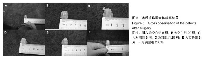
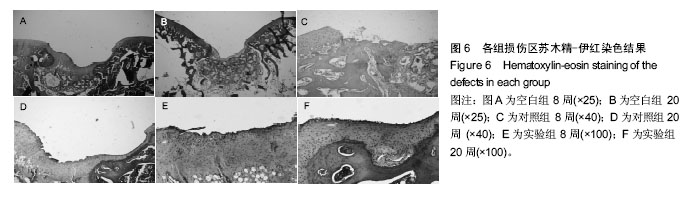
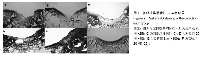
背景:由于骨软骨组织自身复杂的生理学特性,关节软骨损伤的临床修复效果难以令人满意,组织工程学方法和手段修复膝关节软骨缺损成为一种新的治疗模式,为临床修复骨软骨缺损提供了新的思路。
目的:观察羟基丁酸-羟基辛酸共聚体/胶原蛋白仿生一体化支架对兔膝关节软骨损伤的修复情况。
方法:以羟基丁酸与羟基辛酸为基础材料,混合一定比例胶原蛋白,采用溶剂浇铸/颗粒沥滤法,制备骨软骨一体化支架,接种分离、培养的种子细胞。选取4周龄健康新西兰大白兔24只,复合麻醉后,股骨髁关节面部位用电动骨钻造成关节软骨缺损(直径4.5 mm,深度5 mm),处理后随机分为3组,空白组损伤处直接缝合;对照组损伤处植入单纯支架;实验组损伤处植入支架-软骨细胞复合体。分别于术后8,20周取出股骨髁后,通过大体及组织学观察缺损处修复情况。
结果与结论:①8周:实验组缺损处见透明薄膜组织覆盖,细胞小、不规则;对照组缺损较明显,纤维组织为主;空白组缺损无明显修复痕迹;②20周:实验组软骨缺损处以透明软骨样组织覆盖,细胞排列规则;对照组缺损处白色膜状组织覆盖,基底部分及边缘可见较多软骨细胞存在;空白组缺损可见少量新生组织;③结果表明:复合种子细胞的羟基丁酸-羟基辛酸共聚体/胶原骨软骨一体化支架有利于关节软骨缺损修复。
ORCID: 0000-0002-8223-2758(段新虎)

中国组织工程研究杂志出版内容重点:生物材料;骨生物材料; 口腔生物材料; 纳米材料; 缓释材料; 材料相容性;组织工程

关键词: 生物材料, 软骨生物材料, 羟基丁酸-羟基辛酸共聚体, 软骨, 胶原蛋白, 组织工程支架, 膝关节, 关节软骨缺损

Abstract:

BACKGROUND: Due to the complex physiological characteristics of the osteochondral tissue, the clinical repair of knee cartilage injury often has dissatisfied outcomes. Tissue engineering methods and tools provide a new idea for osteochondral repair.
OBJECTIVE: To observe the effect of poly(hydroxybutyrate-co-hydroxyoctanoate/collagen) osteochondral tissue-engineered scaffold on the repair of articular cartilage injury in a rabbit.
METHODS: The poly(hydroxybutyrate-co-hydroxyoctanoate/collagen) osteochondral tissue-engineered scaffold was prepared by solvent casting/particle leaching method. Then, seed cells were isolated and cultured on the scaffold. Twenty-four healthy New Zealand white rabbits, 4 weeks of age, were used for the study. Under balanced anesthesia, an articular cartilage defect (4.5 mm in diameter, 5 mm in depth) was created on the rabbit’s femoral condyle using a bone drill. After modeling, rabbits were randomized into three groups and given direct suture in blank group, pure scaffold implantation in control group and implantation of the scaffold-cell complex in experimental group. Femoral condyle of each rabbit was taken out for gross and histological observations at 8, 20 weeks after surgery.
RESULTS AND CONCLUSION: At 8 weeks after surgery, transparent film-covered defects and small/irregular cells were found in the experimental group; the defects were filled with fibrous tissues in the control group; while there was no repair in the blank group. Until the 20th week, the defects were covered with hyaline cartilage-like tissues, accompanied by regular cell arrangement in the experimental group; in the control group, the defects were covered with white membranous tissues, and many chondrocytes were found at the basement and edge; in the blank group, some newborn tissues were visible at the defect region. These findings suggest that the poly (hydroxybutyrate-co- hydroxyoctanoate/collagen) osteochondral tissue-engineered scaffold carrying seed cells contributes to articular cartilage repair.


Key words: Cartilage, Collagen Type I, Stents, Tissue Engineering

中图分类号: