中国组织工程研究 ›› 2016, Vol. 20 ›› Issue (17): 2540-2545.doi: 10.3969/j.issn.2095-4344.2016.17.015

• 数字化骨科 digital orthopedics • 上一篇    下一篇

胫骨远端骨折有限元模型的建立及稳定性分析

杨志刚1,甘 霖2,叶俊星1   

  1. 1南通大学第三附属医院,无锡市第三人民医院,无锡市中西医结合医院骨科,江苏省无锡市 214000;2南京医科大学附属无锡市第二医院,江苏省无锡市 214000
  • 收稿日期:2016-02-14 出版日期:2016-04-22 发布日期:2016-04-22
  • 作者简介:杨志刚,男,1978年生,江苏省南通市人,汉族,2007年南通大学毕业,硕士,主治医师,主要从事关节周围骨折方面的研究。

Construction and stability of finite element models of distal tibial fractures

Yang Zhi-gang1, Gan Lin2, Ye Jun-xing1   

  1. 1Department of Orthopedics, Wuxi Integrated Traditional Chinese and Western Medicine Hospital, Wuxi Third People’s Hospital, Third Affiliated Hospital of Nantong University, Wuxi 214000, Jiangsu Province, China; 2the Second Hospital of Wuxi City Affiliated to Nanjing Medical University, Wuxi 214000, Jiangsu Province, China
  • Received:2016-02-14 Online:2016-04-22 Published:2016-04-22
  • About author:Yang Zhi-gang, Master, Attending physician, Department of Orthopedics, Wuxi Integrated Traditional Chinese and Western Medicine Hospital, Wuxi Third People’s Hospital, Third Affiliated Hospital of Nantong University, Wuxi 214000, Jiangsu Province, China

摘要:

文章快速阅读:


文题释义:
踝关节有限元模型:参照既往相关研究方法,试验建立了可靠、有效的踝关节有限元模型,对于骨骼的模拟应对皮质骨、松质骨、骨髓腔分别赋予材料参数,但实际操作中却给单元网格的划分增加了难度,为了简化计算在准静态载荷下,骨及软骨可定义为均一、各向同性材料。足踝部韧带及腱膜在关节稳定及平衡中起了重要作用,大多将其看成线弹性、各向同性材料,以杆单元模拟连接骨骼端起止点。
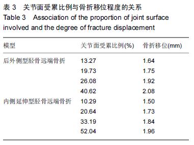
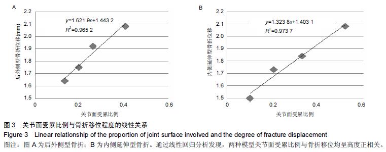
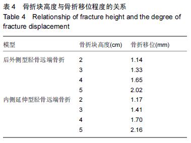
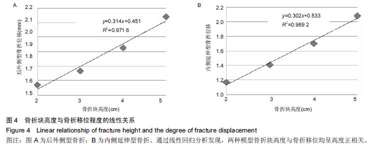
胫骨远端骨折有限元模型:胫骨远端骨折较重,多出现骨折移位,关节面多受累。试验在踝关节有限元模型基础上,对不同种类胫骨远端骨折(Pilon骨折)进行模拟,分析不同关节面受累比例及骨折块高度与骨折移位程度的关系。
 
背景:传统足踝部生物力学研究具有局限性,由于踝关节结构极为复杂、活动度大,故对于踝关节有限元模型的建立及不同骨折类型的分析也极为困难。
目的:建立胫骨远端骨折模型并进行稳定性分析。
方法:收集成年人正常足踝CT数据,采用Minics软件对志愿者右足踝部关节进行三维重建,并进行有效性验证。通过Solidwork软件假设不同关节面受累比例及骨折块高度,并制作出相应的胫骨远端骨折模型。通过ANSYS软件应用有限元法进行骨折稳定性分析。
结果与结论:胫骨远端骨折模型符合相关文献数据,可进一步行有限元分析,关节面受累比例与骨折块高度与骨折移位呈显著正相关,即与骨折稳定性呈负相关。提示踝关节及胫骨远端骨折三维有限元模型建立成功、有效,胫骨远端骨折稳定性与关节面受累比例及骨折高度相关。

中国组织工程研究杂志出版内容重点:人工关节;骨植入物;脊柱;骨折;内固定;数字化骨科;组织工程
ORCID:0000-0002-2453-8126(杨志刚)
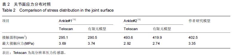
关键词: 骨科植入物, 数字化骨科, 胫骨远端骨折, 踝关节, 有限元分析, 稳定性

Abstract:

BACKGROUND: Traditional studies on foot and ankle biomechanics have limitation. Ankle joint was complicated and had big range of motion, so it is difficult to establish finite element models and to analyze the type of fracture.

OBJECTIVE: To construct the finite element models of distal tibial fractures and analyze the stability.
METHODS: CT data of ankle were collected from a normal male volunteer and the three-dimensional reconstruction of volunteer was made by Mimics software, and the effectiveness was verified. The ratio of different joint involvement and height of fracture block were assumed with Solidwork software, and finite element models of distal tibial fractures were established. Fracture stability was analyzed by ANSYS software using finite element method.
RESULTS AND CONCLUSION: The distal tibia fracture model was consistent with the relevant literature data, and finite element analysis could be further conducted. The ratio of articular surface involved was positively associated with fragment height and fracture displacement, and negatively associated with fracture stability. These results indicate that the three-dimensional finite element models of distal tibial fractures were successfully established. Fracture stability was associated with the ratio of articular surface involved and fragment height. 
中国组织工程研究杂志出版内容重点:人工关节;骨植入物;脊柱;骨折;内固定;数字化骨科;组织工程

Key words: Tibial Fractures, Ankle Joint, Finite Element Analysis, Tissue Engineering