中国组织工程研究 ›› 2016, Vol. 20 ›› Issue (16): 2376-2383.doi: 10.3969/j.issn.2095-4344.2016.16.013

• 材料生物相容性 material biocompatibility • 上一篇    下一篇

国产多孔钽复合骨形态发生蛋白7植入兔竖脊肌内的生物相容性

张 辉1,王 茜2,陶建峰1,王爱军3,史 伟4,卞育婕2,李琪佳5,王志强4   

  1. 唐山市第二医院,1关节一科,3创伤二科,河北省唐山市  063000;华北理工大学,2基础医学院人体解剖学系,5医学中心实验室,河北省唐山市  0630004华北理工大学附属医院骨科,河北省唐山市  063000
  • 收稿日期:2016-02-17 出版日期:2016-04-15 发布日期:2016-04-15
  • 通讯作者: 王志强,教授,主任医师,华北理工大学附属医院骨科,河北省唐山市 063000
  • 作者简介:张辉,男,1981年生,河北省唐山市人,博士,主治医师,主要从事骨移植材料,关节外科,运动医学研究。
  • 基金资助:

    国家科技部科技支撑课题资助项目(2012BAE06B03);河北省科技支撑资助项目(16277776D);河北省医学科学研究重点课题计划项目(20160225);华北理工大学大学博士科研启动基金资助项目

Biocompatibility of domestic porous tantalum carrying bone morphogenetic protein 7 in the erector spinae muscle of rabbits

Zhang Hui1, Wang Qian2, Tao Jian-feng1, Wang Ai-jun3, Shi Wei4, Bian Yu-jie2, Li Qi-jia5, Wang Zhi-qiang4   

  1. 1First Department of Joint Surgery, the Second Hospital of Tangshan, Tangshan 063000, Hebei Province, China; 2Department of Anatomy, Basic Medical College of North China University of Science and Technology, Tangshan 063000, Hebei Province, China; 3Second Department of Traumatology, the Second Hospital of Tangshan, Tangshan 063000, Hebei Province, China; 4Department of Orthopaedics, Affiliated Hospital of North China University of Science and Technology, Tangshan 063000, Hebei Province, China; 5Experimental Center of North China University of Science and Technology, Tangshan 063000, Hebei Province, China
  • Received:2016-02-17 Online:2016-04-15 Published:2016-04-15
  • Contact: Wang Zhi-qiang, Professor, Chief physician, Department of Orthopaedics, Affiliated Hospital of North China University of Science and Technology, Tangshan 063000, Hebei Province, China
  • About author:Zhang Hui, M.D., Attending physician, First Department of Joint Surgery, the Second Hospital of Tangshan, Tangshan 063000, Hebei Province, China
  • Supported by:

    the Scientific Support Project of the Ministry of Science and Technology of China, No. 2012BAE06B03; the Scientific Support Project of Hebei Province, No. 16277776D; the Medical Science Research Project of Hebei Province, No. 20160225; the Doctoral Initial Fund of North China University of Science and Technology

摘要:

文章快速阅读:


文题释义:
骨形态发生蛋白7:又称成骨蛋白1,是骨形态发生蛋白家族中的一员,有促进软骨和骨形成的作用,能在体外促进软骨细胞的增殖和软骨细胞外基质合成,同时具备很强的异位成骨作用,可在异位诱导新骨的形成,在骨缺损修复及软骨分化过程中发挥重要作用。骨形态发生蛋白可通过促进间充质细胞向软骨细胞分化、增殖,增加基质合成等途径,修复关节软骨损伤。
多孔钽:弹性模量为2.0-4.0 GPa,介于皮质骨(12-18 GPa)和松质骨(0.1-0.53 GPa)之间,置入骨骼后几乎没有应力遮挡,利于正常的生物学应力传导,可促进多孔钽和宿主骨的整合。同时多孔钽具备良好的空间三维立体结构,孔隙率可达75%-85%,平均孔径在400-600 µm,利于细胞长入,可为细胞、血管、组织代谢产物及营养物质运输提供有利的空间环境。
 
背景:骨形态发生蛋白7在体内可诱导骨及软骨形成,并可诱导肌肉中和血管周围的间充质细胞分化为软骨和骨细胞,有促进软骨和骨形成的作用。
目的:观察多孔钽复合骨形态发生蛋白7植入兔竖脊肌后,钽-肌肉界面纤维包膜结构、肌肉与小血管向多孔钽内生性生长及异位成骨的能力。
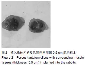
方法:在新西兰大白兔左右两侧竖脊肌内分别植入复合骨形态发生蛋白7的多孔钽片(实验组)和多孔钽片(对照组),植入后2,4,8周,取钽片及其周围0.5 cm肌肉组织,进行扫描电镜、苏木精-伊红染色、Masson染色及硬组织切片观察。
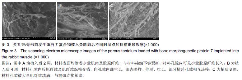
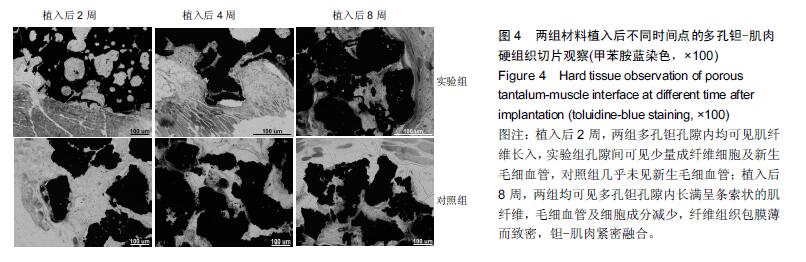
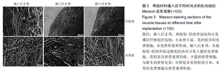
结果与结论:①苏木精-伊红染色:两组材料周围均有纤维性包膜形成,随时间延长,纤维性包膜逐渐由疏松变致密,厚度也逐渐变薄,材料与肌肉交界面无明显炎症反应。两组间纤维包膜厚度比较差异无显著性意义。②扫描电镜:植入2周时,两组多孔钽表面可见少量肌肉及胶原纤维逐渐长入孔隙内部,部分胶原纤维附着于孔壁;植入8周时,多孔钽孔隙内充满了肌腱纤维,纤维与孔壁结合紧密,两组间无明显差异。③硬组织切片:植入2周时,两组多孔钽孔隙均内有少量成纤维细胞及肌纤维长入,实验组材料孔隙内可见有新生小血管长入;植入8周时,两组多孔钽表面和孔隙内均长满呈条索状交 错排列的肌纤维,小血管及细胞成分减少,钽-肌肉紧密融合。④Masson染色:植入8周时,实验组钽-肌肉界面边缘处肌肉内可见大量间充质细胞、骨胶原及软骨基质形成,以及少量新生的软骨化骨,对照组未见软骨化骨。⑤结果表明,复合骨形态发生蛋白7的多孔钽具有良好生物相容性及诱导成骨作用。
中国组织工程研究杂志出版内容重点:生物材料;骨生物材料; 口腔生物材料; 纳米材料; 缓释材料; 材料相容性;组织工程
ORCID: 0000-0002-2962-5069(王志强)
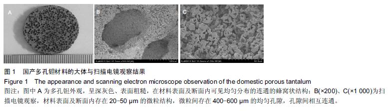
关键词: 生物材料, 材料相容性, 多孔钽, 支架, 骨形态发生蛋白7, 生物相容性, 异位成骨

Abstract:

BACKGROUND: Bone morphogenetic protein 7 (BMP-7) can induce bone and cartilage formation in vivo, and induce chondrogenic and osteogenic differentiation of mesenchymal cells in muscles and around the vessels.

OBJECTIVE: To observe the structure of domestic tantalum-muscle interface fibrous capsule, growth of muscle and small blood vessels into the porous tantalum and the ability of ectopic osteogenesis after implantation of porous tantalum loaded with BMP-7 into the erector spinae of rabbits.
METHODS: Porous tantalum slices loaded with BMP-7 (experimental group) and porous tantalum slices (control group) were implanted into the erector spinae muscle of New Zealand white rabbits. And the porous tantalum slices with surrounding muscle tissues about 0.5 cm thick were removed at 2, 4, 8 weeks after implantation, and observed under scanning electron microscope for hematoxylin eosin staining, Masson staining and hard tissue slice observation.
RESULTS AND CONCLUSION: (1) Hematoxylin-eosin staining: Fibrous capsule formation was observed around the materials in the two groups, and with the extension of time, the fibrous capsules were slightly dense, and thinned. There was no obvious inflammatory reaction in the interface between the material and the muscle. There was no significant difference between the two groups in the fibrous capsules thickness. (2) Scanning electron microscope: 2 weeks after the surgery, a small amount of collagen and muscle fibers were formed in the porous tantalum pores in the two groups, and some of collagen fibers attached to the pore walls. At 8 weeks after the surgery, all the pores of porous tantalum were full of muscle fibers that were combined with the pore wall closely. There was no significant difference between the two groups. (3) Hard tissue slices: 2 weeks after the surgery, a small amount of fibroblast cells and muscle fibers grew into the pores of porous tantalum in the two groups and new capillaries grew into the pores of porous tantalum in the experimental group. At 8 weeks after the surgery, the porous tantalum and all the pores were full of muscle fibers that were combined with the pore wall closely, the number of small blood vessels and cells decreased, and the tantalum and the muscle were fused closely. (4) Masson staining: 8 weeks after the surgery, a large number of mesenchymal cells, ossein and cartilage matrix formed in the muscle gaps and a few cartilage bone tissues were formed in the experimental group, but no cartilage was found in the control group. The study showed that porous tantalum carrying BMP-7 has good biocompatibility and osteogenic induction ability.
中国组织工程研究杂志出版内容重点:生物材料;骨生物材料; 口腔生物材料; 纳米材料; 缓释材料; 材料相容性;组织工程

Key words: Tantalum, Bone Morphogenetic Protein 7, Tissue Engineering