[2] Karkouti K, Wijeysundera DN, Yau TM, et al. Acute kidney injury after cardiac surgery: focus on modifiable risk factors. Circulation. 2009;119(4):495-502.
[3] Sharfuddin AA, Molitoris BA. Pathophysiology of ischemic acute kidney injury. Nat Rev Nephrol. 2011; 7(4):189-200.
[4] Eltzschig HK, Eckle T. Ischemia and reperfusion--from mechanism to translation. Nat Med. 2011;17(11): 1391-1401.
[5] Furuichi K, Shintani H, Sakai Y, et al. Effects of adipose-derived mesenchymal cells on ischemia- reperfusion injury in kidney. Clin Exp Nephrol. 2012; 16(5):679-689.
[6] Reinders ME, Rabelink TJ. Adipose tissue-derived stem cells: can impure cell preparations give pure results. Nephrol Dial Transplant. 2010;25(12):3805- 3807.
[7] Kim J, Kim JI, Na YK, et al. Intra-renal slow cell-cycle cells contribute to the restoration of kidney tubules injured by ischemia/reperfusion. Anat Cell Biol. 2011; 44(3):186-193.
[8] Brodsky SV, Yamamoto T, Tada T, et al. Endothelial dysfunction in ischemic acute renal failure: rescue by transplanted endothelial cells. Am J Physiol Renal Physiol. 2002;282(6):F1140-1149.
[9] Lin F, Cordes K, Li L, et al. Hematopoietic stem cells contribute to the regeneration of renal tubules after renal ischemia-reperfusion injury in mice. J Am Soc Nephrol. 2003;14(5):1188-1199.
[10] Tögel F, Hu Z, Weiss K, et al. Administered mesenchymal stem cells protect against ischemic acute renal failure through differentiation-independent mechanisms. Am J Physiol Renal Physiol. 2005; 289(1):F31-42.
[11] 王志剑,谢平,王共先,等.骨髓干细胞移植对急性缺血性肾损伤大鼠肾小管细胞活性的影响[J].实验与检验医学, 2010,28(2):134-136.
[12] Kale S, Karihaloo A, Clark PR, et al. Bone marrow stem cells contribute to repair of the ischemically injured renal tubule. J Clin Invest. 2003;112(1):42-49.
[13] 保永亮,龚晓燕,黄金玲,等.大黄素对人胃癌MNK-45和MGC8-03细胞增殖的影响[J]. 药物评价研究, 2012, 35(6): 408-411.
[14] 关翠雯,金晶,朱少华,等.大黄素诱导人肾上皮HK-2细胞凋亡及内质网应激的介导作用[J].中草药,2013,44(12): 1621-1627.
[15] 谭力,王联英,向海鹰,等.大黄素对脑缺血再灌注大鼠的保护作用及机制研究[J].中西医结合心脑血管病杂志, 2010, 8(9):1100-1101.
[16] 林胜璋,余耀军,游涛,等.大黄素对大鼠肝脏缺血再灌注损伤的预防作用[J].中国中西医结合外科杂志,2006,12(2): 136-138.
[17] Shigeoka AA, Holscher TD, King AJ, et al. TLR2 is constitutively expressed within the kidney and participates in ischemic renal injury through both MyD88-dependent and -independent pathways. J Immunol. 2007;178(10):6252-6258.
[18] Thurman JM.Triggers of inflammation after renal ischemia/reperfusion.Clin Immunol. 2007;123(1):7-13.
[19] Eltzschig HK, Eckle T. Ischemia and reperfusion--from mechanism to translation. Nat Med. 2011;17(11): 1391-1401.
[20] Friedewald JJ, Rabb H. Inflammatory cells in ischemic acute renal failure. Kidney Int. 2004;66(2):486-491.
[21] Zhu J, Jiang Y, Wu L, et al. Suppression of local inflammation contributes to the neuroprotective effect of ginsenoside Rb1 in rats with cerebral ischemia. Neuroscience. 2012;202:342-351.
[22] Di Paola R, Impellizzeri D, Torre A, et al. Effects of palmitoylethanolamide on intestinal injury and inflammation caused by ischemia-reperfusion in mice. J Leukoc Biol. 2012;91(6):911-920.
[23] Vinten-Johansen J, Jiang R, Reeves JG, et al. Inflammation, proinflammatory mediators and myocardial ischemia-reperfusion Injury. Hematol Oncol Clin North Am. 2007;21(1):123-145.
[24] Grigoryev DN, Liu M, Hassoun HT, et al. The local and systemic inflammatory transcriptome after acute kidney injury. J Am Soc Nephrol. 2008;19(3):547-558.
[25] Hassoun HT, Grigoryev DN, Lie ML, et al. Ischemic acute kidney injury induces a distant organ functional and genomic response distinguishable from bilateral nephrectomy. Am J Physiol Renal Physiol. 2007;293(1): F30-40.
[26] Matsuyama M, Yoshimura R, Hase T, et al. Study of cyclooxygenase-2 in renal ischemia-reperfusion injury. Transplant Proc. 2005;37(1):370-372.
[27] Viñas JL, Sola A, Genescà M, et al. NO and NOS isoforms in the development of apoptosis in renal ischemia/reperfusion. Free Radic Biol Med. 2006; 40(6):992-1003.
[28] Mark LA, Robinson AV, Schulak JA. Inhibition of nitric oxide synthase reduces renal ischemia/reperfusion injury. J Surg Res. 2005;129(2):236-241.
[29] Chong AJ, Shimamoto A, Hampton CR, et al. Toll-like receptor 4 mediates ischemia/reperfusion injury of the heart. J Thorac Cardiovasc Surg. 2004;128(2): 170-179.
[30] Tsung A, Hoffman RA, Izuishi K, et al. Hepatic ischemia/reperfusion injury involves functional TLR4 signaling in nonparenchymal cells. J Immunol. 2005; 175(11):7661-7668.
[31] Leemans JC, Stokman G, Claessen N, et al. Renal-associated TLR2 mediates ischemia/ reperfusion injury in the kidney. J Clin Invest. 2005; 115(10):2894-2903.
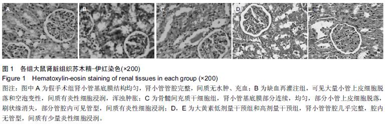
.jpg)


.jpg)
.jpg)