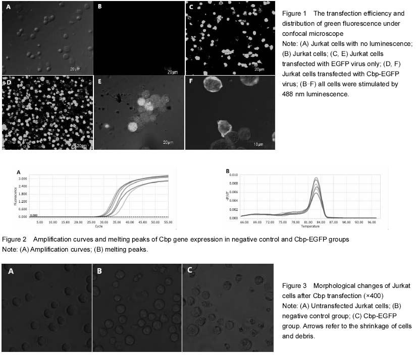
 Transfection rate and distribution of Cbp in Jurkat cells
Jurkat cells were transfected by Cbp-EGFP lentivirus vector and empty lentiviral vector (multiplicity of infection=100) (
Figure 1A), and were detected under the confocal microscope after 96 hours. As shown in Figure 1B, there was no green fluorescence in Jurkat cells untransfected by lentivirus vector. Meanwhile, there was a lot of green fluorescence sustainable expression in negative control and Cbp-EGFP groups (
Figures 1C-D). Under five-fold magnification, the transfection efficiency of lentivirus vector was calculated to be greater than 95%. After further amplification, green fluorescence of the negative control group evenly distributed in the cytoplasm (
Figure 1E), and in the Cbp-EGFP group, green fluorescence focused on the cell membrane on which there was a bit tufted gathering area (
Figure 1F), suggesting it is possibly lipid rafts.
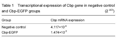
Cbp mRNA expression
The amplification curve and melting curve of negative control and Cbp-EGFP groups showed that gene amplification was a single peak, consistent with the annealing temperature, and no nonspecific amplification (
Figure 2). Cbp mRNA expression was significantly increased in the Cbp-EGFP group, which was 3.5 times as much as the negative control group (
Table 1). It suggested that the mRNA expression of Cbp was indeed at a high level after transfection.
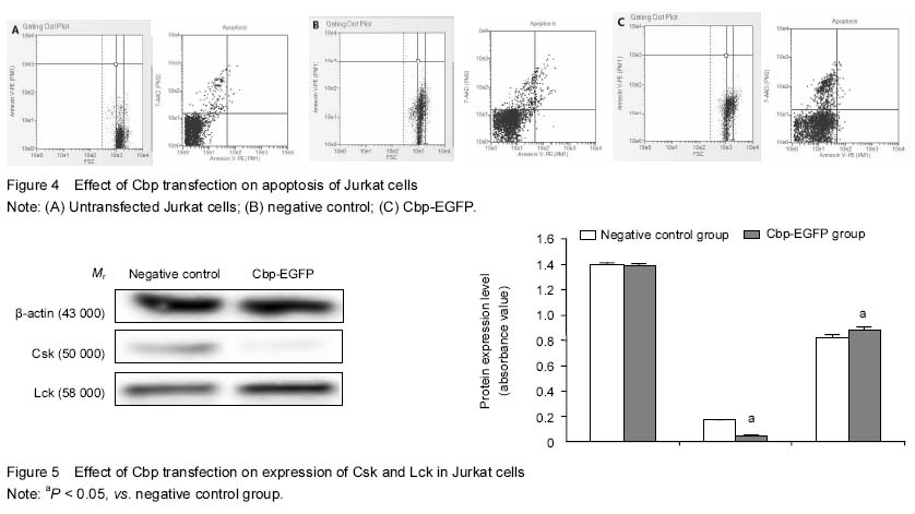
 Cell growth
Jurkat cells is an acute T leukemia cell line and can be immortalized. The growth mode is suspended growth
[16]. As shown in
Figure 3A, the cell state was mellow and bright, liked eggs, and cell morphology was relatively uniform. There were almost no pyknotic cells in the visual field. By comparing Figure 3A and Figure 3B, we found that the cell morphology and size in the negative control and untransfected groups were identical. A small amount of pyknotic cells were seen within the field of vision. But in Figure 3C, the cell growth rate was not as fast as the other groups, the cell size was heterogeneous, and a lot of small cells could be seen. We could easily see the shrinkage of cells and debris under the microscope.
Cell apoptosis
As showed in Figure 4, the cells were concentrated and had the uniform size in the untransfected group; and the same as negative control cells. But there were also a certain amount of small cells in the negative control group which was compared to the untransfected group. In the Cbp-EGFP group, the small-head cells accounted for a large proportion in the whole cells, and the cell size was large. Rates of cell apoptosis and necrosis increased in proper order (untransfected group < negative control group < Cbp-EGFP group) (Figure 4). This suggests that viral vectors are toxic to cells, and Cbp proteins promote apoptosis or necrosis.
Csk and Lck expression
Western blot showed free Lck expression in TCR activated signal pathway was increased, while free inhibitory tyrosine Csk expression was obviously decreased (Figure 5). The statistical results showed the content of Lck increased and Csk protein decreased were both significantly different from those in the Cbp group and negative control group (Figure 5). These suggest that high-expression CBP combined with CSK may inhibit the activity of Src family kinase and play a certain inhibitory role in cell proliferation, which may induce cell apoptosis or cell death.