中国组织工程研究 ›› 2015, Vol. 19 ›› Issue (11): 1668-1672.doi: 10.3969/j.issn.2095-4344.2015.11.006

• 口腔组织构建 oral tissue construction • 上一篇    下一篇

低强度脉冲超声波联合骨形态发生蛋白2促进人牙周膜细胞的成骨分化

刘 俊1,胡 波1,蒋欣益2,孙吉成1,邓 锋1,宋锦璘1   

  1. 1重庆医科大学附属口腔医院,口腔疾病与生物医学重庆市重点实验室,重庆市 401147;2绵阳市中心医院口腔科,四川省绵阳市 621000
  • 修回日期:2014-12-25 出版日期:2015-03-12 发布日期:2015-03-12
  • 通讯作者: 宋锦璘,博士,教授 重庆医科大学附属口腔医院,口腔疾病与生物医学重庆市重点实验室,重庆市 401147
  • 作者简介:刘俊,男,1988年生,四川省金堂县人,汉族,重庆医科大学在读硕士,主要从事牙周组织工程研究。
  • 基金资助:

    国家自然科学基金(30870754),课题名称:基于牙周组织工程技术的低强度脉冲超声波促进牙周病组织修复作用研究

Osteogenic effect of low-intensity pulsed ultrasound combined with bone morphogenetic protein 2 on human periodontal ligament cells  

Liu Jun1, Hu Bo1, Jiang Xin-yi2, Sun Ji-cheng1, Deng Feng1, Song Jin-lin1   

  1. 1The Affiliated Hospital of Stomatology, Chongqing Medical University, Chongqing Research Center for Oral Diseases and Biomedical Science, Chongqing 401147, China; 2Department of Stomatology, the Center Hospital of Mianyang, Mianyang 62100, Sichuan Province, China
  • Revised:2014-12-25 Online:2015-03-12 Published:2015-03-12
  • Contact: Song Jin-lin, M.D., Professor, the Affiliated Hospital of Stomatology, Chongqing Medical University, Chongqing Research Center for Oral Diseases and Biomedical Science, Chongqing 401147, China
  • About author:Liu Jun, Studying for master’s degree, the Affiliated Hospital of Stomatology, Chongqing Medical University, Chongqing Research Center for Oral Diseases and Biomedical Science, Chongqing 401147, China
  • Supported by:

    the National Natural Science Foundation of China, No. 30870754

摘要:

背景:低强度脉冲超声波和骨形态发生蛋白2均可促进人牙周膜细胞成骨分化,但两者联合应用尚未见报道。

目的:验证低强度脉冲超声波和骨形态发生蛋白2联合应用诱导牙周膜细胞成骨分化的生物学效应。
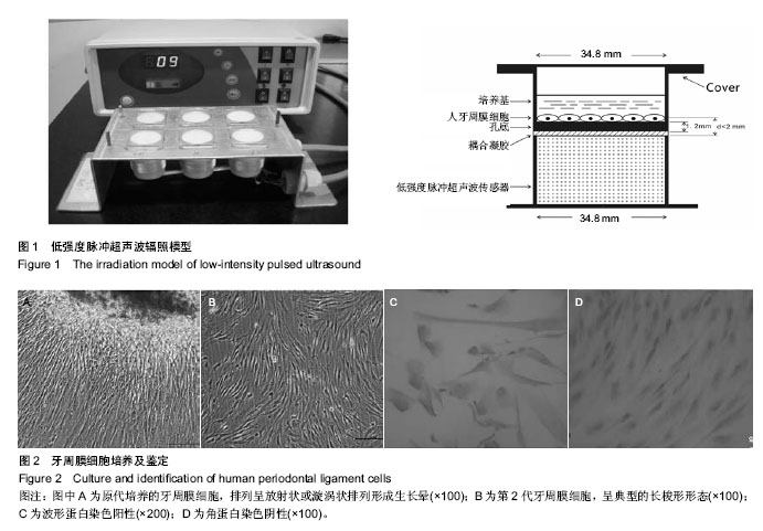
方法:体外分离、原代、传代培养与鉴定牙周膜细胞。取生长良好的第4代牙周膜细胞接种至六孔板中,实验分为4组:对照组、低强度脉冲超声波诱导组、骨形态发生蛋白2诱导组、低强度脉冲超声波联合骨形态发生蛋白2诱导组。采用碱性磷酸酶检测试剂盒检测细胞碱性磷酸酶活性,RT-PCR检测牙周膜细胞成骨相关基因Ⅰ型胶原、骨桥蛋白的表达。
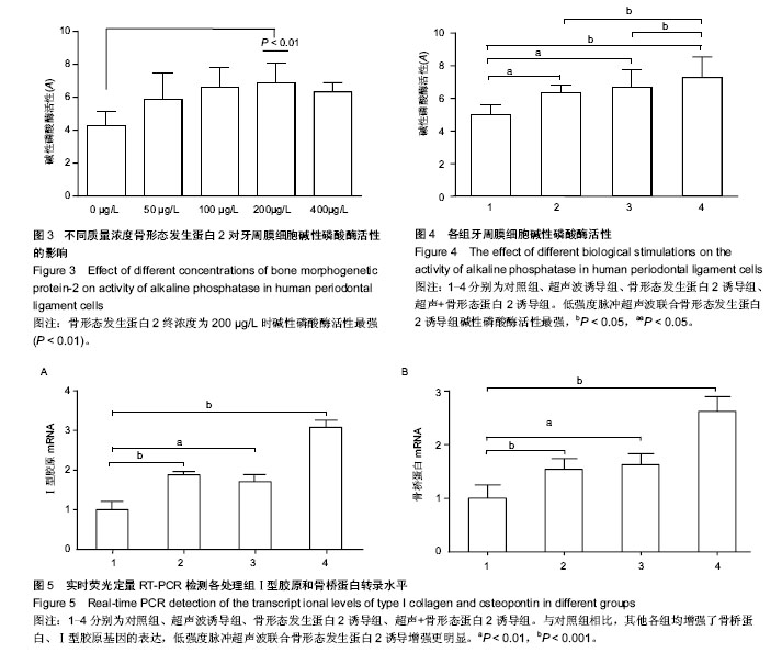
结果与结论通过低强度脉冲超声波、骨形态发生蛋白2和低强度脉冲超声波+骨形态发生蛋白2诱导后,体外培养的牙周膜细胞碱性磷酸酶活性均显著高于对照组(P < 0.05),其中低强度脉冲超声波+骨形态发生蛋白2组碱性磷酸酶活性最强(P < 0.05)。RT-PCR结果分析显示,低强度脉冲超声波与骨形态发生蛋白2都可以上调Ⅰ型胶原、骨桥蛋白基因的表达(P < 0.01),两者联合处理作用更为明显(P < 0.001)。结果初步提示,低强度脉冲超声波和骨形态发生蛋白2均可以增强牙周膜细胞成骨能力,两者联合应用其成骨诱导能力更强。


中国组织工程研究
杂志出版内容重点:组织构建;骨细胞;软骨细胞;细胞培养;成纤维细胞;血管内皮细胞;骨质疏松组织工程


全文链接:

关键词: 组织构建, 骨组织工程, 低强度脉冲超声, 骨形态发生蛋白2, 人牙周膜细胞, 成骨分化, 国家自然科学基金

Abstract:

BACKGROUND: Both of low-intensity pulsed ultrasound (LIPUS) and bone morphogenetic protein-2 (BMP-2) can promote the osteogenic differentiation of human periodontal ligament cells, but there is no report on their combination application.

OBJECTIVE: To evaluate the biological effect of LIPUS combined with BMP-2 on osteogenic differentiation of human periodontal ligament cells.
METHODS: Human periodontal ligament cells were isolated, cultured, passaged and identified. Passage 4 cells were seeded onto 6-well plates and divided into four groups: control group, LIPUS group, BMP-2 group, LIPUS+BMP-2 group. Alkaline phosphatase activity was detected by alkaline phosphatase activity measurement kit, and the expression of type I collagen and osteopontin was detected by RT-PCR.
RESULTS AND CONCLUSION: After LIPUS, BMP-2, and their combination treatment, the activity of alkaline phosphatase was significantly higher than that in the control group (P < 0.05), and the activity of alkaline phosphatase was highest in the LIPUS+BMP-2 group (P < 0.05). RT-PCR results showed that both of LIPUS and BMP-2 could augment the mRNA expression of type I collagen and osteopontin (P < 0.01), but it was more distinct when they were used together (P < 0.001). In a conclusion, the combined utilization of LIPUS and BMP-2 shows stronger ability in promoting the osteogenic differentiation of human periodontal ligament cells.


中国组织工程研究
杂志出版内容重点:组织构建;骨细胞;软骨细胞;细胞培养;成纤维细胞;血管内皮细胞;骨质疏松组织工程


全文链接:

Key words: Sonication, Bone Morphogenetic Proteins, Periodontal Ligament

中图分类号: