| [1] Vail A , Gore SM, Bradley BA, et al. Conclusions of the corneal transplant follow up study. Br J Ophthalmol.1997;81: 631-636.
[2] Eurich D,Neumann UP,Boas-Knoop S,et al.Transforming growth factor-β1-gene polymorphism in the development of kidney diseaseafter liver transplantation.Transplantation. 2012;93(5 ):555-560.
[3] Tanabe T,Kanoh S,Moskowitz WB,et al.Cardiac asthma: transforming growth factor-β from the failing heart leads to squamous metaplasia in human airway cells and in the murine lung.Chest.2012;142(5):1274-1283.
[4] de Menezes Neves PD, Machado JR, dos Reis MA, et al. Distinct expression of interleukin 17,tumor necrosis factor α,transforming growth factor β,and forkhead box P3 in acute rejection after kidneytransplantation. Ann Diagn Pathol.2013; 17(1):75-79.
[5] Huss DJ,Winger RC,Peng H,et al.TGF-beta enhances effector Th1 cell activation but promotes self-regulation via IL-10.Immunol.2010;184(10):5628-5636.
[6] Hu B,He Y,Wu Y,et al.Activated allogeneic NK cells as suppressorsof alloreactive responses. Biol Blood Marrow Transplant.2010;16(6) :772-781.
[7] Han KL,Thomas SV,Koontz SM,et al.Adenosine A-A receptoragonist-mediated increase in donor-derived regulatory T cells suppressesdevelopment of graft-versus- host disease.J Immunol.2013;190(1):458-468.
[8] Wu LM,Zhou L,Xu J, et al.Lack of association between genetic polymorphisms in cytokine genes and tumor recurrence in patients with hepatocellular carcinoma undergoing transplantation.Hepatobiliary Pancreat Dis Int. 2013;12(1):54-59.
[9] Le Texier L,Thebault P,Carvalho-Gaspar M, et al. Immunoregulatory function of IL-27 and TGF-β1 in cardiac allograft transplantation.Transplantation.2012;94(3):226-233.
[10] Williams KA,Coster DJ.Penetrating corneal transplantation in the inbred rat:a new model.Invest Ophthalmol Vis Sci. 1985; 26(1):23-30.
[11] Torres PF, De Vos AF, Van der Gaag R, et al. CytokinemRNA expression during experimental corneal allograft rejection. J Exp Eye Res 2006;63:453-461.
[12] Sano Y, Osawa H, Sotozono C, et al. Cytokine expression during orthotopic corneal allograft rejection in mice.Invest Ophthalmol Vis Sci 2008; 39:1953-1957.
[13] Zhu S, Dekaris I, Duncker G, et al. Early expression of proinflammato rycytokines interleukin and tumor necrosis factor alpha after corneal transplantation. J Interferon Cytokine Res. 2009;19(6):661-669.
[14] 张新华,钟翠平,周播江,等.转化生长因子β1基因修饰树突状细胞延长移植心脏存活时间的研究[J].现代免疫学,2004, 24(6): 464-469
[15] DePaz HA, Oluwole OO, Adeyeri AO, et al. Immature rat myeloid dendritic cells generated in low-dose granulocyte macrophage-colony stimulating factor prolong donor specific rat cardiac allograft survival. Transplantation. 2003; 75(4): 521-552.
[16] 闫峰,蔡莉,惠延年,等.TGFβ2-DC对小鼠同种异体角膜移植排斥反应的抑制作用[J].国际眼科杂志, 2007,7(2):346-349. |
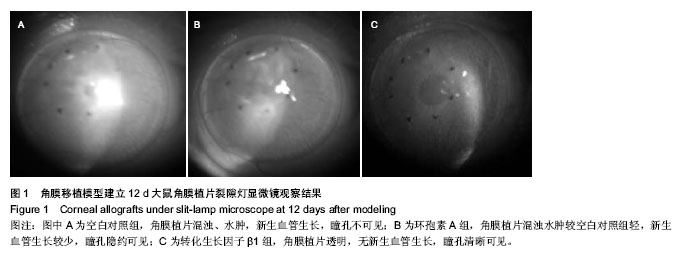

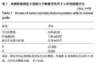
.jpg)