中国组织工程研究 ›› 2015, Vol. 19 ›› Issue (3): 340-346.doi: 10.3969/j.issn.2095-4344.2015.03.003

• 组织工程骨及软骨材料 tissue-engineered bone and cartilage materials • 上一篇    下一篇

骨组织工程诱导性支架材料修复骨缺损

唐俊杰1,2,李文杰2,李 根2,王九娜2,赵 玲2,秦 文2,赵红斌1,2   

  1.   1甘肃农业大学生命科学技术学院,甘肃省兰州市 730070;2解放军兰州军区兰州总医院,甘肃省兰州市 730050
  • 出版日期:2015-01-15 发布日期:2015-01-15
  • 通讯作者: 赵红斌,副主任技师,解放军兰州军区兰州总医院骨科研究所,甘肃省兰州市 730050
  • 作者简介:唐俊杰,女,黑龙江省哈尔滨市人,汉族,甘肃农业大学生命科学技术学院在读硕士,主要从事生物材料研究。
  • 基金资助:

    甘肃省科技重大专项项目(1203FKDA036)

Bone tissue engineering induced composite scaffolds for repair of bone defects

Tang Jun-jie1, 2, Li Wen-jie2, Li Gen2, Wang Jiu-na2, Zhao Ling2, Qin Wen2, Zhao Hong-bin1, 2   

  1. 1College of Life Science and Technology, Gansu Agricultural University, Lanzhou 730070, Gansu Province, China; 2Lanzhou General Hospital of Lanzhou Military Command, Lanzhou 730050, Gansu Province, China
  • Online:2015-01-15 Published:2015-01-15
  • Contact: Zhao Hong-bin, Associate chief technician, College of Life Science and Technology, Gansu Agricultural University, Lanzhou 730070, Gansu Province, China; Lanzhou General Hospital of Lanzhou Military Command, Lanzhou 730050, Gansu Province, China
  • About author:Tang Jun-jie, Studying for master’s degree, College of Life Science and Technology, Gansu Agricultural University, Lanzhou 730070, Gansu Province, China; Lanzhou General Hospital of Lanzhou Military Command, Lanzhou 730050, Gansu Province, China
  • Supported by:

    the Major Science and Technology Project of Gansu Province, No. 1203FKDA036

摘要:

背景:前期实验中发现载淫羊藿苷壳聚糖/胶原/聚己内酯/羟基磷灰石复合支架具有良好的物理和化学性质。


目的:研究载淫羊藿苷壳聚糖/胶原/聚己内酯/羟基磷灰石复合支架修复兔胫骨平台骨缺损的效果。


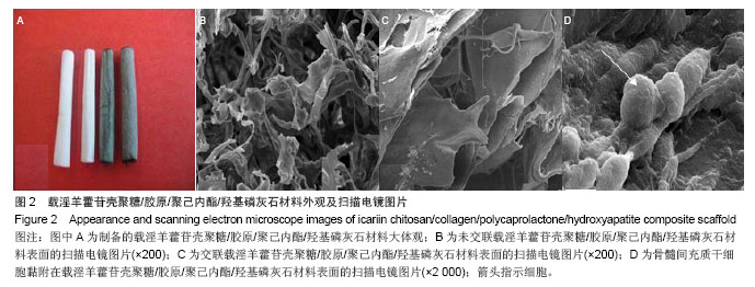
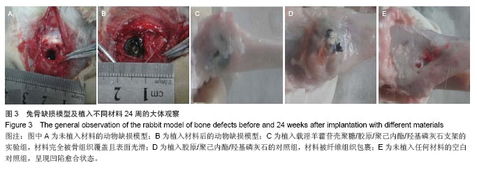
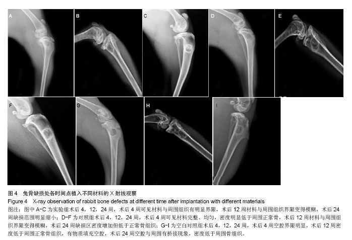
方法:利用静电纺丝技术制备胶原/聚己内酯/羟基磷灰石复合支架壳层,真空干燥法制备载淫羊藿苷壳聚糖微球/胶原支架芯层,将芯层嵌入壳层后利用京尼平交联构建载药复合支架。将载淫羊藿苷壳聚糖微球置入PBS中,观察药物缓释效果。将载药复合支架与大鼠骨髓间充质干细胞共培养7 d,观察细胞黏附效果。取青紫蓝兔15只,复制兔左侧胫骨缺损模型,随机分为3组,实验组于骨缺损处植入载药复合支架,对照组植入胶原/聚己内酯/羟基磷灰石复合支架,空白对照组不植入任何材料。术后4,12,24周行X射线观察、样本大体和组织学观察。


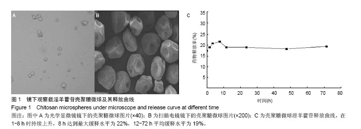
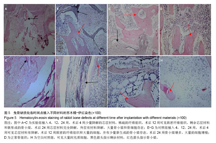
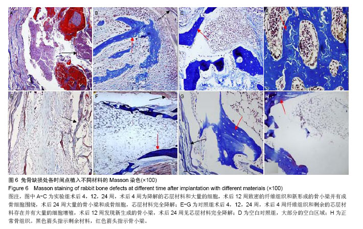
结果与结论:载药复合支架具有疏松多孔结构,利于骨髓间充质干细胞的黏附、增殖;壳聚糖微球体外缓释在72 h内保持19%释放量的良好缓释效果。术后24周,实验组材料完全被新生骨组织覆盖,硬度与正常骨相近,苏木精-伊红染色观察发现有骨小梁、骨细胞和成骨细胞,但缺损区骨密度低于正常骨组织;对照组材料被纤维组织包裹,苏木精-伊红染色显示有骨小梁、骨髓细胞和纤维组织;空白对照组呈凹陷愈合,但硬度低于正常骨组织,苏木精-伊红染色可见大量骨髓细胞和骨小梁。结果表明载药壳聚糖/胶原/聚己内酯/羟基磷灰石复合支架能有效修复兔骨缺损。


中国组织工程研究杂志出版内容重点:生物材料;骨生物材料; 口腔生物材料; 纳米材料; 缓释材料; 材料相容性;组织工程


全文链接:

关键词: 生物材料, 骨生物材料, 壳聚糖, 胶原, 聚己内酯, 羟基磷灰石, 骨缺损, 复合材料, 组织工程

Abstract:

BACKGROUND: In the early experiments, icariin chitosan/collagen/polycaprolactone/hydroxyapatite (CS/Col/PCL/HA) composite scaffold has good physical and chemical properties.

OBJECTIVE: To study the effect of CS/Col/PCL/HA composite scaffolds to repair bone defects of rabbit tibia platform.
METHODS: The outer of Col/PCL/HA composite scaffolds were prepared by electrospinning, and the core containing chitosan microspheres/collagen was made by vacuum freeze-drying process. Then the core was implanted into the outer to construct the drug-loading composite scaffold by genipin crosslinking. The microspheres carrying icariin chitosan were immersed into PBS to observe the slow-release effect. The composite scaffold was used to culture rat bone marrow mesenchymal stem cells for 7 days to observe cell adhesion. Fifteen chinchilla rabbits were taken to make the model of left tibia bone defect, and then randomly divided into three groups: CS/Col/PCL/HA composite scaffold group, Col/PCL/HA composite scaffold group, and blank control group with no materials. X-ray observation, gross observation, and histopathological observation were performed at 4, 12, 24 weeks after the surgery.
RESULTS AND CONCLUSION: The drug-loading composite scaffold had a porous structure that was conducive to cell adhesion and proliferation. Chitosan microspheres kept a 19% drug release within 72 hours in vitro. Twenty-four weeks after surgery, CS/Col/PCL/HA composite scaffolds were completely covered by the newly formed bone tissue, and the scaffold surfaces were smooth and had hardness close to normal bone tissue; hematoxylin-eosin staining showed bone trabecula, bone cells and osteoblasts, but X-ray images showed the bone mineral density in the defect area was slightly lower than that of the normal bone tissue. Col/PCL/HA composite scaffolds were wrapped up by fibrous tissue, and hematoxylin-eosin staining showed bone trabecula, bone marrow cells and dense fibrous tissue. In the blank control group, the hardness of defect areas was significantly lower than that of the normal bone and hematoxylin-eosin staining showed a large number of bone marrow cells and bone trabeculae. The experimental results showed that CS/Col/PCL/HA composite scaffold could repair rabbit bone defects effectively.

中国组织工程研究杂志出版内容重点:生物材料;骨生物材料; 口腔生物材料; 纳米材料; 缓释材料; 材料相容性;组织工程


全文链接:

Key words: Collagen, Chitosan, Hydroxyapatites

中图分类号: