| [1] Chen XQ, Chen LL, Fan L,et al.Stem cells with FGF4-bFGF fused gene enhances the expression of bFGF and improves myocardial repair in rats.Biochem Biophys Res Commun. 2014;447(1):145-151.
[2] Igura K, Okada M, Kim HW,et al.Identification of small juvenile stem cells in aged bone marrow and their therapeutic potential for repair of the ischemic heart.Am J Physiol Heart Circ Physiol. 2013;305(9):H1354-1362.
[3] Valarmathi MT, Goodwin RL, Fuseler JW,et al.A 3-D cardiac muscle construct for exploring adult marrow stem cell based myocardial regeneration.Biomaterials. 2010; 31(12): 3185-200.
[4] 徐信群,王泉兰,应国秋,等.骨髓间充质干细胞移植对心肌梗死大鼠损伤心肌的修复效果[J].南昌大学学报:医学版,2013,53(6): 1-4,21.
[5] 王海萍,张雷,赵静,等.大鼠骨髓间充质干细胞移植治疗心肌梗死的实验研究[J].中国临床解剖学杂志,2012,30(2):209-213.
[6] 田茂,朴海南,陈宇,等. 骨髓间充质干细胞体外构建工程化心肌组织的生存[J].中国组织工程研究,2014,18(20):3133-3138.
[7] 马红芬,张晓刚,史若飞,等.人工脑膜复合大鼠骨髓间充质干细胞修复心肌梗死[J].中国组织工程研究,2013,17(14): 2552-2557.
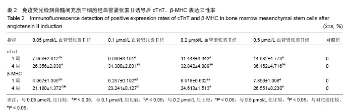
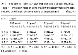
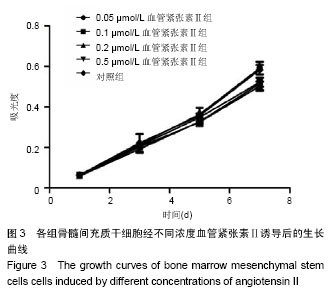
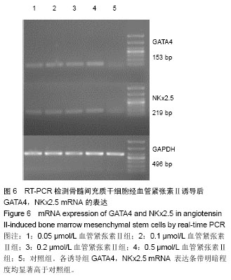
[8] 苑媛,吕安林,陈丹,等.血管紧张素诱导人骨髓间充质干细胞分化为心肌样细胞[J].心脏杂志,2006,18(3):258-261 .
[9] Xing Y, Lv A, Wang L,et al.The combination of angiotensin II and 5-azacytidine promotes cardiomyocyte differentiation of rat bone marrow mesenchymal stem cells.Mol Cell Biochem. 2012;360(1-2):279-287.
[10] 张卫泽,樊艳,陈永清,等.血管紧张素Ⅱ对成人脂肪间充质干细胞向心肌细胞分化的影响[J].第四军医大学学报, 2008,29(8): 692-695.
[11] 顾健,王爱玲,郝玉瑜,等.大鼠骨髓间充质干细胞体外诱导分化为心肌样细胞的研究[J].医学综述,2014,20(6):1109-1111,1116.
[12] 陈玲玲,尹立雪.超声辐照微泡介导5-氮杂胞苷诱导人骨髓间充质干细胞心肌样分化的实验研究[J].中华超声影像学杂志,2013, 22(11):991-996.
[13] 孙庆国,赵文静,王日中,等.体外定向诱导大鼠骨髓间充质干细胞分化为心肌样细胞的实验研究[J].中国实验诊断学,2012,16(12): 2202-2204.
[14] 邸军,李梓菲,陈艳,等.大鼠骨髓间充质干细胞体外向心肌细胞诱导分化的机制[J].中国老年学杂志,2013,33(12):2835-2836.
[15] 王宁,李新华,邢万红,等.细胞代数与5-氮胞苷浓度对体外诱导骨髓间充质干细胞向心肌样细胞定向分化的影响[J].中西医结合心脑血管病杂志,2010, 8(10):1220-1222.
[16] 王宁,邢万红,李新华,等.大鼠骨髓间充质干细胞体外心肌定向诱导及心肌组织构建研究[J].国际生物医学工程杂志,2010,33(3): 163-166.
[17] Makino S, Fukuda K, Miyoshi S,et al.Cardiomyocytes can be generated from marrow stromal cells in vitro.J Clin Invest. 1999;103(5):697-705.
[18] Fukuda K.Development of regenerative cardiomyocytes from mesenchymal stem cells for cardiovascular tissue engineering. Artif Organs. 2001;25(3):187-193.
[19] Liu Y, Song J, Liu W,et al.Growth and differentiation of rat bone marrow stromal cells: does 5-azacytidine trigger their cardiomyogenic differentiation?Cardiovasc Res. 2003;58(2): 460-468.
[20] Xu J, Lin SC, Chen J,et al.CCR2 mediates the uptake of bone marrow-derived fibroblast precursors in angiotensin II-induced cardiac fibrosis.Am J Physiol Heart Circ Physiol. 2011;301(2):H538-547.
[21] Sopel MJ, Rosin NL, Lee TD,et al.Myocardial fibrosis in response to Angiotensin II is preceded by the recruitment of mesenchymal progenitor cells.Lab Invest. 2011;91(4): 565-578.
[22] Qian C, Schoemaker RG, van Gilst WH,et al.The role of the renin-angiotensin-aldosterone system in cardiovascular progenitor cell function.Clin Sci (Lond). 2009;116(4):301-314.
[23] Hakuno D, Fukuda K, Makino S,et al.Bone marrow-derived regenerated cardiomyocytes (CMG Cells) express functional adrenergic and muscarinic receptors.Circulation. 2002;105(3): 380-386.
[24] Wang X, Phillips MI, Mehta JL.LOX-1 and angiotensin receptors, and their interplay.Cardiovasc Drugs Ther. 2011; 25(5):401-417.
[25] Wang X, Khaidakov M, Ding Z,et al.Cross-talk between inflammation and angiotensin II: studies based on direct transfection of cardiomyocytes with AT1R and AT2R cDNA.Exp Biol Med (Maywood). 2012;237(12):1394-1401.
[26] Coyne TM, Marcus AJ, Woodbury D,et al.Marrow stromal cells transplanted to the adult brain are rejected by an inflammatory response and transfer donor labels to host neurons and glia.Stem Cells. 2006;24(11):2483-2492.
[27] Heikinheimo M, Scandrett JM, Wilson DB.Localization of transcription factor GATA-4 to regions of the mouse embryo involved in cardiac development.Dev Biol. 1994;164(2): 361-373.
[28] Xing Y, Lv A, Wang L,et al.Engineered myocardial tissues constructed in vivo using cardiomyocyte-like cells derived from bone marrow mesenchymal stem cells in rats.J Biomed Sci. 2012;19:6.
[29] Liu BW, Lü AL, Hou J,et al.Electrophysiological characteristics of cardiomyocyte-like cells from rat bone marrow derived mesenchymal stem cells by four inductors.Chin Med J (Engl). 2013;126(18):3528-3533.
[30] Li L, Fan D, Wang C,et al.Angiotensin II increases periostin expression via Ras/p38 MAPK/CREB and ERK1/2/TGF-β1 pathways in cardiac fibroblasts.Cardiovasc Res. 2011;91(1): 80-89.
[31] He JG, Chen SL, Huang YY,et al.The nonpeptide AVE0991 attenuates myocardial hypertrophy as induced by angiotensin II through downregulation of transforming growth factor-beta1/Smad2 expression.Heart Vessels. 2010;25(5): 438-443.
[32] Wang N, Ren GD, Zhou Z,et al.Cooperation of myocardin and Smad2 in inducing differentiation of mesenchymal stem cells into smooth muscle cells.IUBMB Life. 2012;64(4):331-339.
[33] Haddad GE, Coleman BR, Zhao A,et al.Regulation of atrial contraction by PKA and PKC during development and regression of eccentric cardiac hypertrophy.Am J Physiol Heart Circ Physiol. 2005;288(2):H695-704.
[34] Li TF, O'Keefe RJ, Chen D.TGF-beta signaling in chondrocytes. Front Biosci. 2005;10:681-688. |