中国组织工程研究 ›› 2013, Vol. 17 ›› Issue (20): 3666-3670.doi: 10.3969/j.issn.2095-4344.2013.20.008

• 口腔组织构建 oral tissue construction • 上一篇    下一篇

线性测量颞下颌关节在咬合重建中的可适位

栾  巍1,石旭旭2   

  1. 1佳木斯大学,黑龙江省佳木斯市  154002
    2大庆油田总医院口腔修复科,黑龙江省大庆市  163001
  • 收稿日期:2012-10-27 修回日期:2013-04-09 出版日期:2013-05-14 发布日期:2013-05-14
  • 通讯作者: 石旭旭,硕士,主任医师,教授,大庆油田总医院口腔修复科,黑龙江省大庆市 163001 shixuxu@tom.com
  • 作者简介:栾巍★,女,1983年生,山东省栖霞市人,2012年佳木斯大学毕业,硕士,主要从事口腔修复种植方面的研究。 12944993@qq.com

Proper position of the temporomandibular joint in the occlusal reconstruction with linear measurement

Luan Wei1, Shi Xu-xu2   

  1. 1 Jiamusi University, Jiamusi  154002, Heilongjiang Province, China
    2 Department of Prosthodontics, Daqing Oilfield General Hospital, Daqing  163001, Heilongjiang Province, China  
  • Received:2012-10-27 Revised:2013-04-09 Online:2013-05-14 Published:2013-05-14
  • Contact: Shi Xu-xu, Master, Chief physician, Professor, Department of Prosthodontics, Daqing Oilfield General Hospital, Daqing 163001, Heilongjiang Province, China shixuxu@tom.com
  • About author:Luan Wei★, Master, Jiamusi University, Jiamusi 154002, Heilongjiang Province, China 12944993@qq.com

摘要:

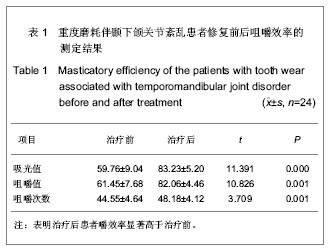
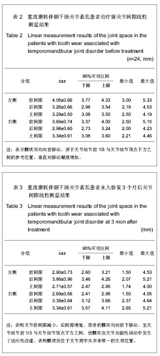
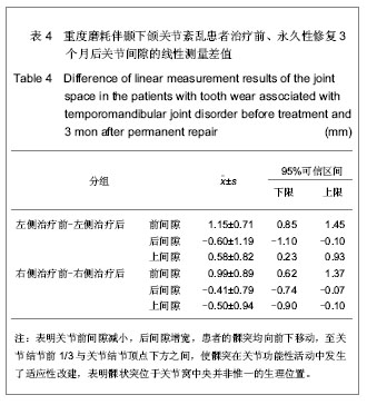
背景:牙合面形态的改变影响咀嚼肌的收缩、咬合力的大小与方向,进而影响包括颞下颌关节在内的整个口颌系统的受力环境,是颞下颌关节生理性和病理性改建的最主要的刺激因素。
目的:测量分析髁状突在咬合重建中的可适位。
方法:对24例牙列重度磨耗伴颞下颌关节紊乱患者采用固定修复方法进行咬合重建治疗,疗程9个月,治疗前后拍摄正中牙合位颞下颌关节CT片利用计算机技术进行线性测量,分析关节间隙的变化。
结果与结论:咬合重建修复治疗后髁突在关节窝内相对参考位置,左侧由-(16.96±2.01)%增至+(14.20±1.24)%;右侧为-(10.64±1.50)%增至(11.51±3.00)%。治疗前的咀嚼效率约为治疗后的62.15%-89.09%,平均为74.88%,差异有显著性意义(P < 0.05)。24例咬合重建后的患者髁状突在关节窝中均向前下移位至关节结节前1/3与关节结节顶点下方之间。提示咬合重建后髁状突向前下移位至关节结节前1/3与关节结节顶点下方之间的可适位改建取得了良好的临床治疗效果,髁状突位于关节窝中央并非惟一的生理位置。

关键词: 组织构建, 口腔组织构建, 骨关节植入物, 咬合重建, 低位咬合, 颞下颌关节紊乱, 牙合垫, 咀嚼效率, 计算机技术, 测量, 关节间隙

Abstract:

BACKGROUND: Morphological changes of occlusal surface will affect the contraction of masticatory muscles, the magnitude and direction of the bite force, thus affecting the force environment of the entire stomatognathic system including the temporomandibular joint, so it is the main stimulus for the physiological and pathological alterations of the temporomandibular joint.
OBJECTIVE: To explore the proper position of the condyle in occlusal reconstruction.
METHODS: Twenty-four cases of tooth wear associated with temporomandibular joint disorder received fixed repair occlusal rehabilitation therapy, and the course was 9 months. The CT image of temporomandibular joint in the median occlusal bit was taken before and after treatment, and computer technology was used for linear measurement to analyze the change of joint space.
RESULTS AND CONCLUSION: The relative reference position of condyle in the glenoid fossa after occlusal reconstruction was changed as follows: the left was increased from -(16.96%±2.01)% to +(14.20%±1.24)%, and the right was increased from -(10.64%±1.50)% to (11.51%±3.00)%. The preoperative masticatory efficiency was 62.15%-89.09% of that after treatment, 74.88% on the average, and the difference was significant (P < 0.05). After occlusal reconstruction, the positions of condyles in the glenoid fossa of 24 patients were moved to the position between the 1/3 of articular eminence and top of articular tubercle. The reconstruction of proper position of condyle to the position between 1/3 of articular eminence and top of articular tubercle has achieved good clinical treatment effect, and indicate that the condyle located in the center of the glenoid fossa is not the only physiological position.

Key words: tissue construction, oral tissue construction, bone and joint implants, occlusal reconstruction, infraocclusion, temporomandibular disorders, occlusal pad, masticatory efficiency, computer technology, measurement, joint space

中图分类号: