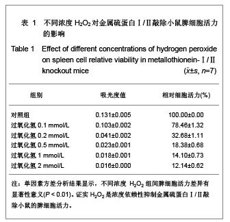
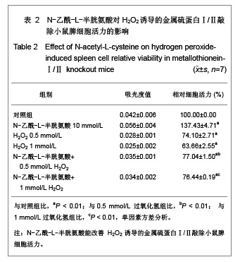
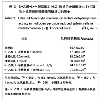
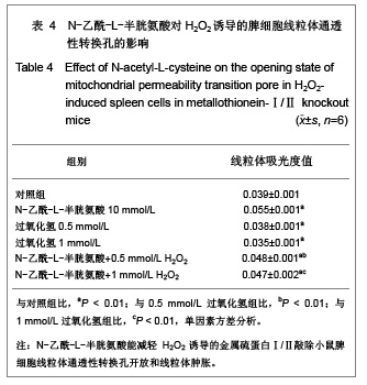
| [1] Thirumoorthy N, Shyam SA, Manisenthil Kumar KT, et al. A review of metallothionein isoforms and their role in pathophysiology. World J Surg Oncol. 2011;9:54.[2] Inoue k, Takano H, Shimada A, et al. Metallothionein as an anti-in?ammatory mediator. Mediators of In?ammation. 2009; 2009:101659.[3] Yao X,Miao W, Li M, et al. Protective effect of albumin on VEGF and brain edema in acute isehemia in rats. Neurosei Lett. 2010;472(3):179-183.[4] Wang M, Wang YM, Li HD, et al. Zhonghua Laonian Xinnaoxueguanbing Zazhi. 2011;13(2):164-167. 王敏,王永明,李海东,等.白蛋白治疗对小鼠脑缺血早期脑组织Toll样受体4和骨髓分化因子88 mRNA表达的影响[J].中华老年心脑血管病杂志,2011,13(2):164-167.[5] Offner H, Vandenbark AA, Hurn PD. Effect of experimental stroke on peripheral immunity: CNS ischemia induces profound immunosuppression. Neuroscience. 2009;158(3): 1098-1111.[6] Kim CH, Vaziri ND, Rodriguez-Iturbe B. Integrin expression and H2O2 production in circulating and splenic leukocytes of obese rats. Obesity (Silver Spring). 2007; 15(9):2209-2216.[7] Liu B, Jian Z, Li Q, et al. Baicalein protects Human melanocytes from H2O2-induced apoptosis via inhibiting mitochondria-dependent caspase activation and the p38 MAPK pathway. Free Radic Biol Med. 2012;53(2):183-193.[8] Li XB, Zhang HC, Guo ZK. Zhongguo Zuzhi Gongcheng Yanjiu. 2012;16(1):22-26. 刘新宾,张红超,郭子宽.骨髓间充质干细胞条件培养液对H2O2损伤大鼠心肌细胞的保护[J].中国组织工程研究, 2012,16(1): 22-26. [9] Gu DS, Li W, Gu WW. Zhongguo Zuzhi Gongcheng Yanjiu. 2011;15(44):8281-8284. 顾东曙,刘闻,顾为望.静脉输注过氧化氢溶液上调小鼠脾脏和外周血CD4+Foxo3+调节性T细胞[J].中国组织工程研究与临床康复,2011,15(44):8281-8284. [10] Yang P, Peairs JJ, Tano R, et al. Oxidant-mediated Akt activation in human RPE cells. Invest Ophthalmol Vis Sci. 2006;47(10):4598-606.[11] Yoshino F, Yoshida A, Okada E, et al. Dental resin curing blue light induced oxidative stress with reactive oxygen. J Photochem Photobiol B. 2012;114:73-78.[12] Nakamura R, Lnoue K, Fujitani Y, et al. In vitro study of the effect of nanoparticle-rch diesel exhaust particles on IL-18 production in splenocytes. J Toxicol Sci. 2011;36(6):823-827.[13] Wu Y, Wang D, Wang X, et al. Caspase 3 is activated through caspase 8 instead of caspase 9 during H2O2-induced apoptosis in HeLa cells. Cell Physiol Biochem. 2011;27(5): 539-546. [14] Ru Y, Yin L, Sun H, et al. A micropreparation of mitochondria from cells using magnetic beads with immounoaf?nity. Anal Biochem. 2012;421(1):219-226.[15] Wang DM, Dong J, Qi ZT, et al. Xian Tiyu Xueyuan Xuebao. 2011;28(4):471-475. 王冬梅,董军,漆正堂,等.动对骨骼肌线粒体通透性转换孔及细胞凋亡的影响[J].西安体育学院学报,2011,28(4):471-475.[16] Petronilli V, Penzo D, Scorrano L, et al. The mitochondrial permeability transition, release of cytochrome c and cell death. J Biol Chem. 2001;267(15):12030-12034. [17] Ajmo CT Jr, Vernon DO, Collier L, et al. The spleen contributes to stroke-induced neurodegeneration. J Neurosci Res. 2008;86(10):2227-2234.[18] Izci Y. Splenectomy may be a prophylactic treatment for cerebral ischemia? Med Hypotheses. 2010;75(4):347-349. [19] Yu X, Guo J, Fang H, et al. Basal metallothionein-Ⅰ/Ⅱ protects against NMDA-mediated oxidative injury in cortical neuron/astrocyte cultures. Toxicology. 2011;282(1-2):16-22.[20] Ghanizadeh A. Gold implants and increased expression of metallothionein-Ⅰ/Ⅱ as a novel hypothesized therapeutic approach for autism. Toxicology. 2011; 283(1):63-64.[21] Hamdi Y, Kaddour H, Vaudry D, et al. The octadecaneuropeptide ODN protects astrocytes against hydrogen peroxide-induced apoptosis via a PKA/MAPK- dependent mechanism. PLoS One. 2012;7(8):e42498. [22] Baumeister P, Huebner T, Reiter M, et al. Reduction of oxidative DNA fragmentation by ascorbic acid, zinc and N-acetylcysteine in nasal mucosa tissue cultures. Anticancer Res. 2009;29(11):4571-4574.[23] Khouri R, Novais F, Santana G, et al. DETC induces Leishmania parasite killing in human in vitro and murine in vivo models: a promising therapeutic alternative in Leishmaniasis. PLoS One. 2010;5(12):e14394.[24] Xie M, Roy R. Increased levels of hydrogen peroxide induce a HIF-1-dependent modi?cation of lipid metabolism in AMPK compromised C. elegans Dauer Larvae. Cell Metab. 2012; 16(3):322-335.[25] Wu JP, Chen HC, Li MH. Bioaccumulation and toxicodynamics of cadmiumt of reshwater planarian and the protective effect of N-Acetylcysteine. Arch Environ Contam Toxicol. 2012;63(2):220-229.[26] Yao X, Li M, He J, et al. Effect of early acute high concentrations of iodide exposure on mitochondrial superoxide production in FRTL cells. Free Radic Biol Med. 2012; 52(8):1343-1352.[27] Li M, Li LY, Chen ZP, et al. Tianjin Yiyao. 2010;38(3): 212-215. 李敏,李兰英,陈组培,等.过氧化氢对FRTL细胞线粒体膜电位和超氧化物生成的影响[J].天津医药,2010,38(3):212-215.[28] Kinnally KW, Peixoto PM, Ryu SY, et al. Is mPTP the gate keeper for necrosis, apoptosis, or both? Biochim Biophys Acta. 2011;1813(4):616-622.[29] Drahota Z, Milerova M, Endlicher R, et al. Developmental changes of the sensitivity of cardiac and liver mitochondrial permeability transition pore to calcium load and oxidative stress. Physiol Res. 2012;61(Suppl 1):S165-172.[30] Loor G, Kondapalli J, Lwase H, et al. Mitochondrial oxidant stress triggers cell death in simulated ischemia-reperfusion. Biochim Biophys Acta. 2011;1813(7):1382-1394.[31] The Ministry of Science and Technology of the People’s Republic of China. Guidance Suggestions for the Care and Use of Laboratory Animals. 2006-09-30. |
.jpg)